

TEAD-1 overexpression in the mouse heart promotes an age-dependent heart dysfunction
- PMID: 20194497
- PMCID: PMC2859535
- DOI: 10.1074/jbc.M109.063057
TEAD-1 overexpression in the mouse heart promotes an age-dependent heart dysfunction
Abstract
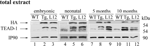
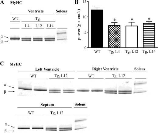
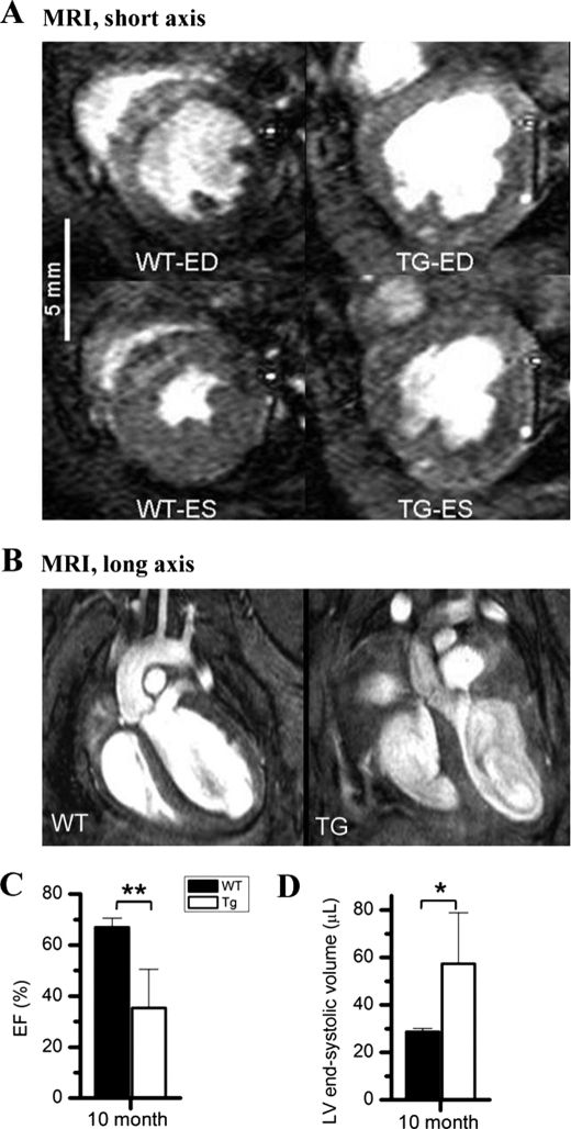
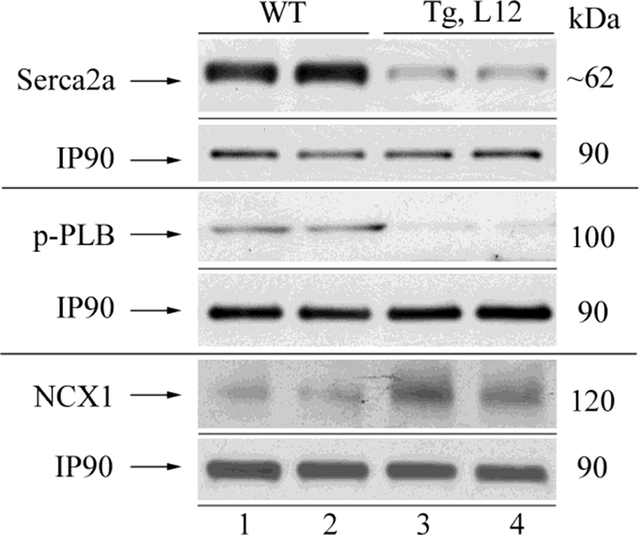
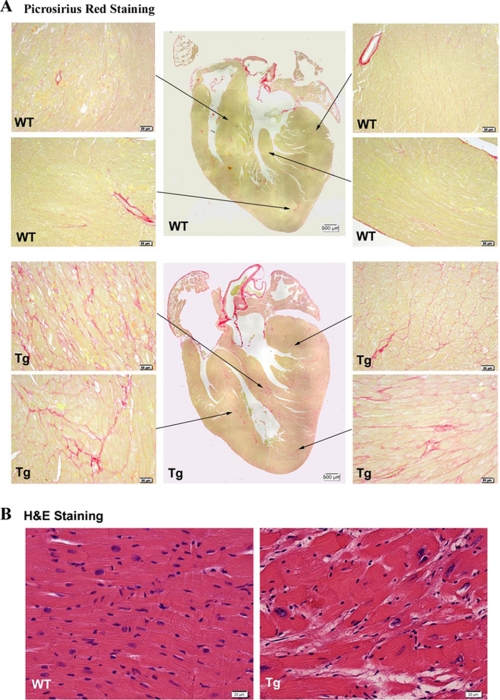
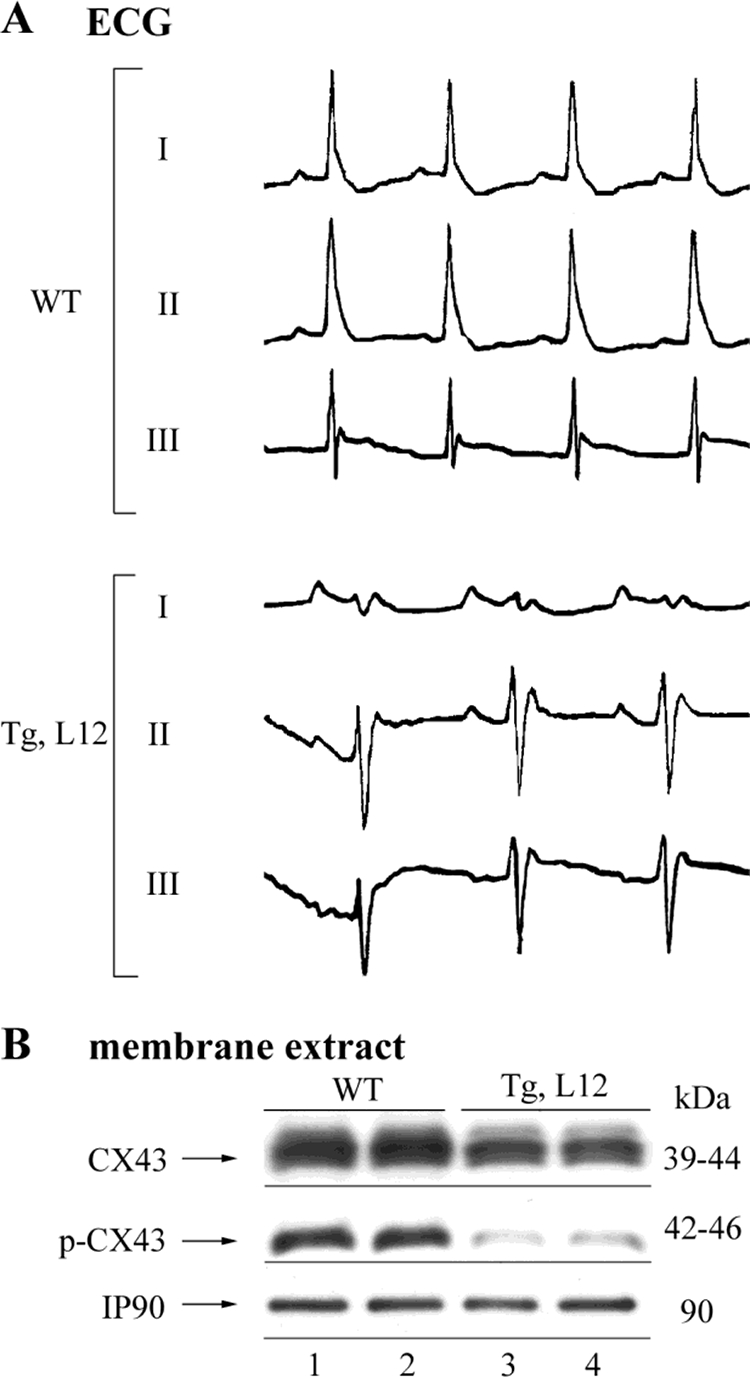
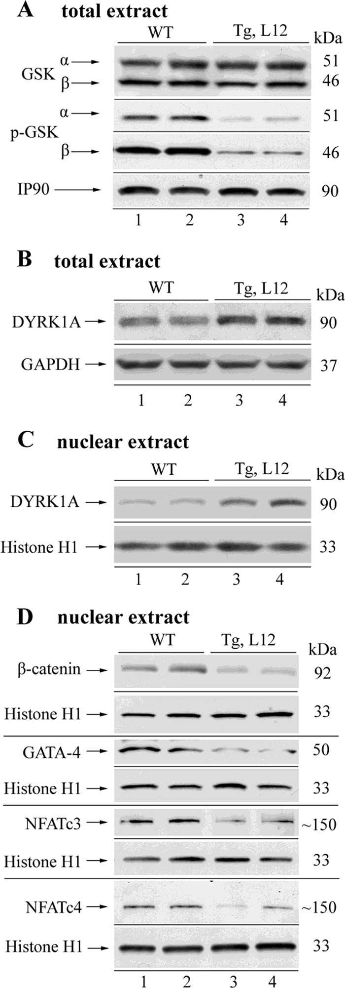
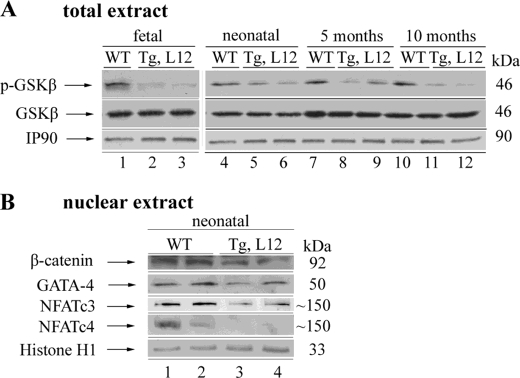
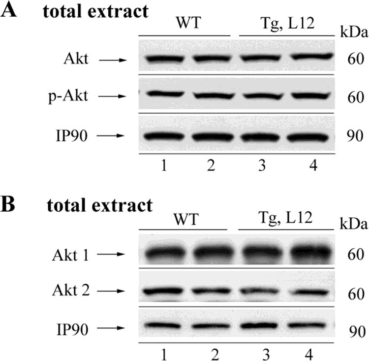
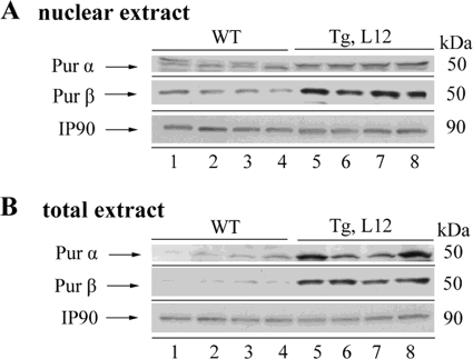
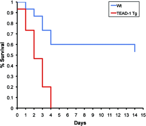
TEA domain transcription factor-1 (TEAD-1) is essential for proper heart development and is implicated in cardiac specific gene expression and the hypertrophic response of primary cardiomyocytes to hormonal and mechanical stimuli, and its activity increases in the pressure-overloaded hypertrophied rat heart. To investigate whether TEAD-1 is an in vivo modulator of cardiac specific gene expression and hypertrophy, we developed transgenic mice expressing hemagglutinin-tagged TEAD-1 under the control of the muscle creatine kinase promoter. We show that a sustained increase in TEAD-1 protein leads to an age-dependent dysfunction. Magnetic resonance imaging revealed decreases in cardiac output, stroke volume, ejection fraction, and fractional shortening. Isolated TEAD-1 hearts revealed decreased left ventricular power output that correlated with increased betaMyHC protein. Histological analysis showed altered alignment of cardiomyocytes, septal wall thickening, and fibrosis, although electrocardiography displayed a left axis shift of mean electrical axis. Transcripts representing most members of the fetal heart gene program remained elevated from fetal to adult life. Western blot analyses revealed decreases in p-phospholamban, SERCA2a, p-CX43, p-GSK-3alpha/beta, nuclear beta-catenin, GATA4, NFATc3/c4, and increased NCX1, nuclear DYKR1A, and Pur alpha/beta protein. TEAD-1 mice did not display cardiac hypertrophy. TEAD-1 mice do not tolerate stress as they die over a 4-day period after surgical induction of pressure overload. These data provide the first in vivo evidence that increased TEAD-1 can induce characteristics of cardiac remodeling associated with cardiomyopathy and heart failure.
Figures
Similar articles
-
Targeted disruption of Hspa4 gene leads to cardiac hypertrophy and fibrosis.J Mol Cell Cardiol. 2012 Oct;53(4):459-68. doi: 10.1016/j.yjmcc.2012.07.014. Epub 2012 Aug 1. J Mol Cell Cardiol. 2012. PMID: 22884543
-
The chromatin-binding protein Smyd1 restricts adult mammalian heart growth.Am J Physiol Heart Circ Physiol. 2016 Nov 1;311(5):H1234-H1247. doi: 10.1152/ajpheart.00235.2016. Epub 2016 Sep 23. Am J Physiol Heart Circ Physiol. 2016. PMID: 27663768 Free PMC article.
-
Cardiac-restricted Overexpression of TRAF3 Interacting Protein 2 (TRAF3IP2) Results in Spontaneous Development of Myocardial Hypertrophy, Fibrosis, and Dysfunction.J Biol Chem. 2016 Sep 9;291(37):19425-36. doi: 10.1074/jbc.M116.724138. Epub 2016 Jul 27. J Biol Chem. 2016. PMID: 27466370 Free PMC article.
-
Transcriptional pathways and potential therapeutic targets in the regulation of Ncx1 expression in cardiac hypertrophy and failure.Adv Exp Med Biol. 2013;961:125-35. doi: 10.1007/978-1-4614-4756-6_11. Adv Exp Med Biol. 2013. PMID: 23224875 Free PMC article. Review.
-
Mechanotransduction in cardiac hypertrophy and failure.Circ Res. 2015 Apr 10;116(8):1462-1476. doi: 10.1161/CIRCRESAHA.116.304937. Circ Res. 2015. PMID: 25858069 Free PMC article. Review.
Cited by
-
Sequencing of mRNA identifies re-expression of fetal splice variants in cardiac hypertrophy.J Mol Cell Cardiol. 2013 Sep;62:99-107. doi: 10.1016/j.yjmcc.2013.05.004. Epub 2013 May 17. J Mol Cell Cardiol. 2013. PMID: 23688780 Free PMC article.
-
Potential Role of Exercise in Regulating YAP and TAZ During Cardiomyocytes Aging.Curr Cardiol Rev. 2022;18(5):24-33. doi: 10.2174/1573403X18666220404152924. Curr Cardiol Rev. 2022. PMID: 35379136 Free PMC article. Review.
-
Discovery of a subtype-selective, covalent inhibitor against palmitoylation pocket of TEAD3.Acta Pharm Sin B. 2021 Oct;11(10):3206-3219. doi: 10.1016/j.apsb.2021.04.015. Epub 2021 May 1. Acta Pharm Sin B. 2021. PMID: 34729310 Free PMC article.
-
TEA domain transcription factor 1(TEAD1) induces cardiac fibroblasts cells remodeling through BRD4/Wnt4 pathway.Signal Transduct Target Ther. 2024 Feb 19;9(1):45. doi: 10.1038/s41392-023-01732-w. Signal Transduct Target Ther. 2024. PMID: 38374140 Free PMC article.
-
YAP and TAZ Regulate Cc2d1b and Purβ in Schwann Cells.Front Mol Neurosci. 2019 Jul 17;12:177. doi: 10.3389/fnmol.2019.00177. eCollection 2019. Front Mol Neurosci. 2019. PMID: 31379499 Free PMC article.
References
-
- Dorn G. W., 2nd, Robbins J., Sugden P. H. (2003) Circ. Res. 92, 1171–1175 - PubMed
-
- Heineke J., Molkentin J. D. (2006) Nat. Rev. 7, 589–600 - PubMed
-
- Swynghedauw B. (2006) J. Exp. Biol. 209, 2320–2327 - PubMed
-
- Opie L. H., Commerford P. J., Gersh B. J., Pfeffer M. A. (2006) Lancet 367, 356–367 - PubMed
-
- Akazawa H., Komuro I. (2003) Circ. Res. 92, 1079–1088 - PubMed
Publication types
MeSH terms
Substances
Grants and funding
LinkOut - more resources
Full Text Sources
Medical
Molecular Biology Databases
Miscellaneous
