. 2010 May 28;285(22):16748-56.
doi: 10.1074/jbc.M109.092999. Epub 2010 Mar 1.

Human GLUD2 glutamate dehydrogenase is expressed in neural and testicular supporting cells

Affiliations

Human GLUD2 glutamate dehydrogenase is expressed in neural and testicular supporting cells

Cleanthe Spanaki et al. J Biol Chem. .

Abstract

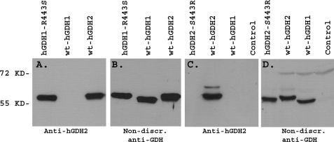
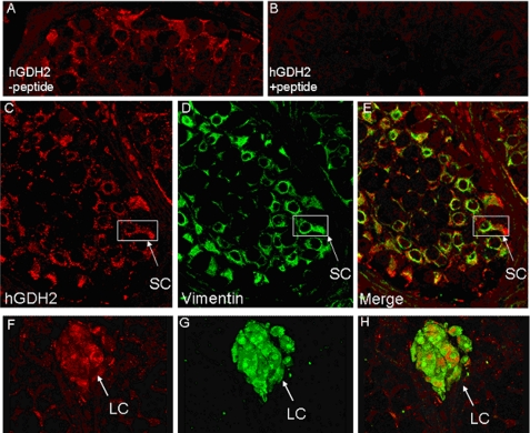
Mammalian glutamate dehydrogenase (GDH) is an allosterically regulated enzyme that is expressed widely. Its activity is potently inhibited by GTP and thought to be controlled by the need of the cell for ATP. In addition to this housekeeping human (h) GDH1, humans have acquired (via a duplication event) a highly homologous isoenzyme (hGDH2) that is resistant to GTP. Although transcripts of GLUD2, the gene encoding hGDH2, have been detected in human neural and testicular tissues, data on the endogenous protein are lacking. Here, we developed an antibody specific for hGDH2 and used it to study human tissues. Western blot analyses revealed, to our surprise, that endogenous hGDH2 is more densely expressed in testis than in brain. At the subcellular level, hGDH2 localized to mitochondria. Study of testicular tissue using immunocytochemical and immunofluorescence methods revealed that the Sertoli cells were strongly labeled by our anti-hGDH2 antibody. In human cerebral cortex, a robust labeling of astrocytes was detected, with neurons showing faint hGDH2 immunoreactivity. Astrocytes and Sertoli cells are known to support neurons and germ cells, respectively, providing them with lactate that largely derives from the tricarboxylic acid cycle via conversion of glutamate to alpha-ketoglutarate (GDH reaction). As hGDH2 is not subject to GTP control, the enzyme is able to metabolize glutamate even when the tricarboxylic acid cycle generates GTP amounts sufficient to inactivate the housekeeping hGDH1 protein. Hence, the selective expression of hGDH2 by astrocytes and Sertoli cells may provide a significant biological advantage by facilitating metabolic recycling processes essential to the supportive role of these cells.


Figures

FIGURE 1.
FIGURE 1.
Western blot analysis of purified hGDH1 and hGDH2 proteins. Recombinant GLUD1 and GLUD2 cDNAs were expressed in Sf21 cells, producing hGDH1 and hGDH2 proteins, respectively. These were purified to homogeneity as described under “Experimental Procedures.” Increasing concentrations of the purified proteins were electrophoresed on an 8.5% SDS-polyacrylamide gel and blotted with either our anti-hGDH2 antibody (left panel) or a commercially available antibody raised against bovine liver GDH (right panel). The amount of purified hGDH1 and hGDH2 loaded is shown (in nanograms) above the corresponding lane. Under these conditions, the anti-hGDH2 antibody recognized only purified hGDH2, whereas the commercially available anti-GDH antibody recognized both human isoproteins (non-discriminating). hGDH2 migrated somewhat higher than hGDH1 (estimated molecular masses of 58 and 56 kDa, respectively), as shown previously for purified hGDH1 and hGDH2 separated by SDS-PAGE and visualized with Coomassie Blue (9).
FIGURE 2.
FIGURE 2.
Effect of mutations at residue 443 on the specificity of the anti-hGDH2 antibody. Mutant and wild-type (wt) hGDH1 and hGDH2 were produced in Sf21 cells using the baculovirus expression system. Recombinant human expressed proteins purified to homogeneity (A and B) or crude Sf21 cell extracts (C and D) were electrophoresed on an 8.5% gel and blotted with our anti-hGDH2 antibody (A and C) or the commercially available non-discriminating (non-discr.) anti-GDH antibody (B and D). The anti-hGDH2 antibody, raised against a 12-amino acid-long peptide that corresponds to residues 436–447 of the mature hGDH2 protein, identified the R443S hGDH1 mutant and wild-type hGDH2, both of which carry Ser443 instead of Arg443, which occurs in wild-type hGDH1. In contrast, when Ser443 of hGDH2 was replaced with Arg, the antibody no longer recognized the mutant hGDH2 enzyme.
FIGURE 3.
FIGURE 3.
Immunoblots of human testis using the anti-hGDH2 and non-discriminating anti-GDH antibodies. Crude extracts and partially purified GDH preparations from human testis were run on 8.5% gels. Expressed recombinant hGDH1 and hGDH2 were used as controls. The anti-hGDH2 antibody visualized a single hGDH2-specific band in both the purified (Pur.) and crude testis extracts (A). In contrast, the non-discriminating (Non-discr.) antibody revealed two bands of nearly equal amounts, representing hGDH1 and hGDH2 (B).
FIGURE 4.
FIGURE 4.
Western blot analysis of human brain, testis, and liver. A, crude extracts from human parietal, temporal, and frontal lobe (60 μg each) and human testis and liver (20 μg each) were electrophoresed on an 8.5% SDS-polyacrylamide gel and transferred to a nitrocellulose membrane. Blotting was performed using either the anti-hGDH2 antibody (upper panel) or the anti-GDH non-discriminating (non discr.) antibody (lower panel). B, shown are the results from Western blot analyses of human frontal lobe, testis, and liver using different amounts of crude tissue extracts. The total protein (micrograms) loaded is shown above each lane. Blotting was performed using the anti-hGDH2 antibody. These blots show that human testis contains substantially higher levels of hGDH2 than human brain and that human liver has none. This differential expression of hGDH2 contrasts with the fact that human liver extracts contain higher levels of GDH activity (1.301 ± 0.41 μmol of NADPH oxidized per min/mg of protein) than human brain extracts (0.356 ± 0.021, 0.501 ± 0.027, and 0.467 ± 0.014 μmol/min/mg of protein for parietal, temporal, and frontal lobe, respectively), and the crude human testis extracts had even lower GDH activity (0.078 ± 0.003 μmol of NADPH oxidized per min/mg of protein, mean ± S.D.). These enzyme assays measured the sum of hGDH1 and hGDH2 activity.
FIGURE 5.
FIGURE 5.
Distribution of hGDH2 and hGDH1 in mitochondrial and cytosolic fractions prepared from human testis. The mitochondrial fraction was isolated using the Ficoll method (see “Experimental Procedures”). Equal protein amounts of whole lysate and mitochondrial and cytosolic fractions were electrophoresed on an 8.5% SDS-polyacrylamide gel, and immunoblotting was performed using the anti-hGDH2 antibody (A) and the non-discriminating (Non-discr.) anti-GDH antibody (B). Actin (C) and manganese superoxide dismutase (MnSOD; D), visualized by the respective antibodies, served as cytosolic and mitochondrial markers, respectively. Both hGDH1 and hGDH2 were enriched in the mitochondrial fraction, whereas the cytosolic fraction was essentially devoid of GDH immunoreactivity. These immunoblotting data agree with the results of GDH activity assays using these fractions (data not shown).
FIGURE 6.
FIGURE 6.
Localization of hGDH2 in human testis. Shown are images of paraffin-embedded fixed sections of human testis stained with rabbit antiserum against hGDH2 and visualized with AEC+ chromogen (red pigment). A, punctate immunoreactivity for hGDH2 was found in the cytoplasm of Sertoli cells (SC) inside the seminiferous tubules, as well as in the cytoplasm of Leydig cells (LC) in the intermediate space. Spermatocytes (SPC) were devoid of hGDH2 immunoreactivity. B, preincubation with the immunogenic peptide markedly attenuated the staining. Some residual staining can be observed in the cytoplasm of Leydig cells. C, incubation of the tissue with the rabbit preimmune serum (PIS) revealed mild staining of the Leydig cell cytoplasm, whereas the seminiferous tubules had no hGDH2 immunoreactivity. D, omitting the primary antibody revealed only nonspecific staining attributed to the secondary antibody (Sec Ab).
FIGURE 7.
FIGURE 7.
Localization of hGDH2 in the cytoplasm of Sertoli and Leydig cells in human testis by double immunofluorescence studies. Shown are images of paraffin-embedded formalin-fixed sections of human testis stained with 1) rabbit antiserum against hGDH2 (red staining), 2) mouse monoclonal antibody against vimentin (green staining), and 3) mouse monoclonal antibody against calretinin (green staining). In A, the rabbit antiserum was not preincubated, whereas in B, it was preincubated with the immunogenic peptide prior to staining. There was specific punctate immunoreactivity for hGDH2 in the cytoplasm of Sertoli cells (SC; A and C), which were labeled also with vimentin (D and E). This reactivity was almost completely abolished after preincubation of the rabbit antiserum with the immunogenic peptide (B). There was also hGDH2-specific staining in the cytoplasm of Leydig cells (LC) (F and H), which were also labeled with calretinin (G and H).
FIGURE 8.
FIGURE 8.
Localization of hGDH2 mostly in astrocytes in human brain. Shown are images of unfixed human brain gray (A–F) and white (G–I) matter, double-stained with monoclonal antibodies (red staining) to GFAP (marker for astrocytes), NeuN (neuronal marker (n)), and MOG (marker for oligodendrocytes (o) and myelin (m)), in combination with rabbit antiserum against hGDH2 (green staining). Cell nuclei were visualized with 4′,6-diamidino-2-phenylindole (blue staining). Merged images are shown in the right panels. Strong punctate immunoreactivity for hGDH2 was found in the cytoplasm (closed arrowheads in A, D, and G) and proximal processes (open arrowheads) of cells that were identified as astrocytes (a) by their strong GFAP reactivity (B). Much weaker and rather diffuse hGDH2 reactivity was present in the cytoplasm of NeuN-positive neurons (D–F). There was no overlap between MOG and hGDH2 localization (G–I), indicating a lack of hGDH2 expression in oligodendrocytes.
FIGURE 9.
FIGURE 9.
Specific labeling of hGDH2 in human brain. Shown are images of unfixed human brain cortex immunostained with mouse monoclonal antibody against GFAP (red staining) and rabbit antiserum against hGDH2 (green staining) in the absence (A) and presence (B) of antigen (immunogenic peptide). Cell nuclei were visualized with 4′,6-diamidino-2-phenylindole (DAPI; blue staining). There was specific punctate immunoreactivity for hGDH2 in the cytoplasm (arrowheads) of astrocytes (a), which were labeled with GFAP (arrows). hGDH2 immunoreactivity was almost completely abolished after preincubation of the rabbit antiserum with the immunogenic peptide (B).

Similar articles

Cited by

References

    1. Smith E. (1979) Proc. Am. Philos. Soc. 123, 73–84
    1. Hudson R. C., Daniel R. M. (1993) Comp. Biochem. Physiol. B 106, 767–792 - PubMed
    1. Plaitakis A., Berl S., Yahr M. D. (1984) Ann. Neurol. 15, 144–153 - PubMed
    1. Mavrothalassitis G., Tzimagiorgis G., Mitsialis A., Zannis V., Plaitakis A., Papamatheakis J., Moschonas N. (1988) Proc. Natl. Acad. Sci. 85, 3494–3498 - PMC - PubMed
    1. Shashidharan P., Michaelidis T. M., Robakis N. K., Kresovali A., Papamatheakis J., Plaitakis A. (1994) J. Biol. Chem. 269, 16971–16976 - PubMed

Publication types