. 2011 Jan;6(1):24-37.
doi: 10.1093/scan/nsq003. Epub 2010 Mar 1.

Neural correlates of recognition memory for emotional faces and scenes

Affiliations

Neural correlates of recognition memory for emotional faces and scenes

Michelle L Keightley et al. Soc Cogn Affect Neurosci. 2011 Jan.

Abstract

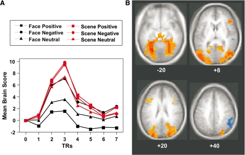
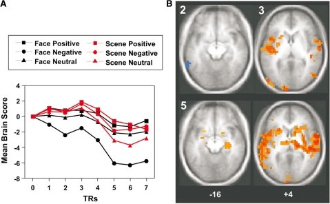
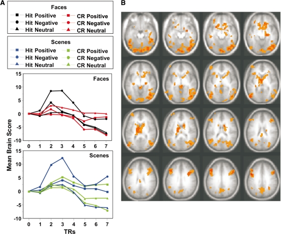
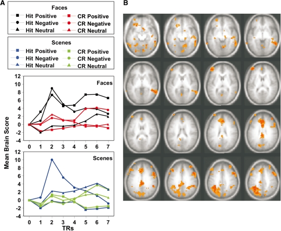
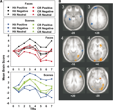
We examined the influence of emotional valence and type of item to be remembered on brain activity during recognition, using faces and scenes. We used multivariate analyses of event-related fMRI data to identify whole-brain patterns, or networks of activity. Participants demonstrated better recognition for scenes vs faces and for negative vs neutral and positive items. Activity was increased in extrastriate cortex and inferior frontal gyri for emotional scenes, relative to neutral scenes and all face types. Increased activity in these regions also was seen for negative faces relative to positive faces. Correct recognition of negative faces and scenes (hits vs correct rejections) was associated with increased activity in amygdala, hippocampus, extrastriate, frontal and parietal cortices. Activity specific to correctly recognized emotional faces, but not scenes, was found in sensorimotor areas and rostral prefrontal cortex. These results suggest that emotional valence and type of visual stimulus both modulate brain activity at recognition, and influence multiple networks mediating visual, memory and emotion processing. The contextual information in emotional scenes may facilitate memory via additional visual processing, whereas memory for emotional faces may rely more on cognitive control mediated by rostrolateral prefrontal regions.


Figures

Fig.1
Fig.1
Results of the analysis of stimulus and valence, LV1. (A) Temporal brain scores (averaged over participants) for each face and scene condition are plotted across the seven TRs in the analysis period. Brain scores are summary scores of activity across the entire brain and the graph shows how the pattern of activity across the brain is expressed over the 16 s period. These time courses may be thought of as whole-brain hemodynamic response functions. (B) Areas making a significant contribution to this pattern are shown on the average MRI of the 18 participants in MNI space (the same average MRI also is used in subsequent figures). Data shown are from TR2 and the Z level of each slice is shown beneath it. Activity in the red areas was greater for scenes and negative faces. Blue areas showed less activity for scenes and negative faces.
Fig. 2
Fig. 2
Results of the analysis of stimulus and valence, LV2. (A) Temporal brain scores (averaged over participants) for each face and scene condition are plotted across the analysis period. (B) Areas making a significant contribution to this pattern. Data shown are from TR2, TR3 and TR5, and the Z-level of each slice is shown beneath it. Activity across the TRs distinguished negative faces from the other conditions, with red areas showing less activity for negative faces and blue areas more activity for negative faces.
Fig. 3
Fig. 3
Results of the contrast of hits vs correct rejections for faces and scenes—LV1. (A) Temporal brain scores (averaged over participants) are shown for faces and scenes separately for clarity, although these scores come from the same analysis. (B) Regions contributing to the pattern (from TR3) are shown (images are from Z = −24 to Z = +36 in 4 mm steps). This LV differentiated negative hits from correct rejections and was characterized by increased activity in orange/red areas for correctly recognized negative faces and scenes.
Fig. 4
Fig. 4
Results of the contrast of hits vs correct rejections for faces and scenes—LV2. (A) Temporal brain scores (averaged over participants) are shown for faces and scenes separately for clarity, although these scores come from the same analysis. (B) Regions contributing to the pattern (from TR 2) are shown (images are from Z = −20 to Z = +40 in 4 mm steps). This LV-differentiated emotional hits for faces from correct rejections and was characterized by increased activity in orange/red areas for correctly recognized emotional faces.
Fig. 5
Fig. 5
Results of the contrast of hits vs correct rejections for faces and scenes—LV3. (A) Temporal brain scores (averaged over participants) are shown for faces and scenes separately for clarity, although these scores come from the same analysis. (B) Regions contributing to the pattern are shown. Data shown are from TR3, TR4 and TR5, and the Z-level of each slice is shown beneath it. This LV differentiated positive hits from correct rejections and was characterized by increased activity in orange/red areas and decreased activity in blue areas for correctly recognized positive faces and scenes.

References

    1. Addis DR, Moscovitch M, Crawley AP, McAndrews MP. Recollective qualities modulate hippocampal activation during autobiographical memory retrieval. Hippocampus. 2004;14:752–62. - PubMed
    1. Adolphs R, Cahill L, Schul R, Babinsky R. Impaired declarative memory for emotional material following bilateral amygdala damage in humans. Learning and Memory. 1997;4:291–300. - PubMed
    1. Adolphs R, Damasio H, Tranel D, Cooper G, Damasio AR. A role for somatosensory cortices in the visual recognition of emotion as revealed by three-dimensional lesion mapping. Journal of Neuroscience. 2000;20:2683–90. - PMC - PubMed
    1. Adolphs R, Tranel D, Damasio H, Damasio A. Impaired recognition of emotion in facial expressions following bilateral damage to the human amygdala. Nature. 1994;372:669–72. - PubMed
    1. Adolphs R, Tranel D, Denburg N. Impaired emotional declarative memory following unilateral amygdala damage. Learning & Memory. 2000;7:180–6. - PMC - PubMed

Publication types