. 2010 May;78(5):2079-88.
doi: 10.1128/IAI.01450-09. Epub 2010 Mar 1.

Indispensable role for the eukaryotic-like ankyrin domains of the ankyrin B effector of Legionella pneumophila within macrophages and amoebae

Affiliations

Indispensable role for the eukaryotic-like ankyrin domains of the ankyrin B effector of Legionella pneumophila within macrophages and amoebae

Christopher T D Price et al. Infect Immun. 2010 May.

Erratum in

  • Infect Immun. 2013 Jul;81(7):2660

Abstract

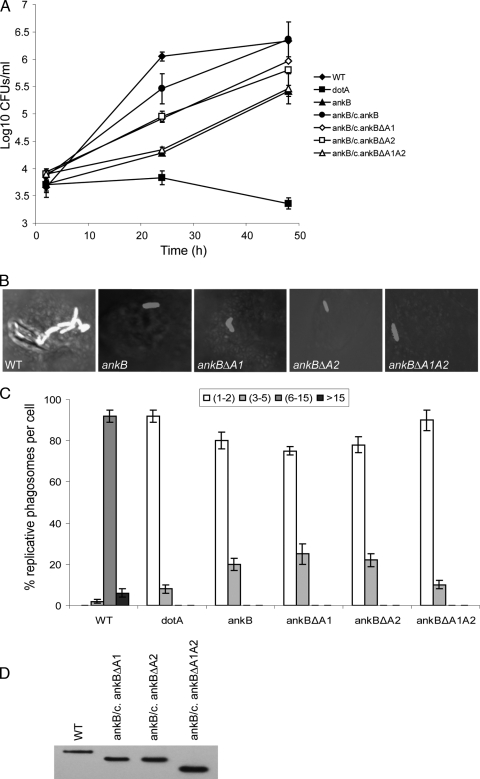
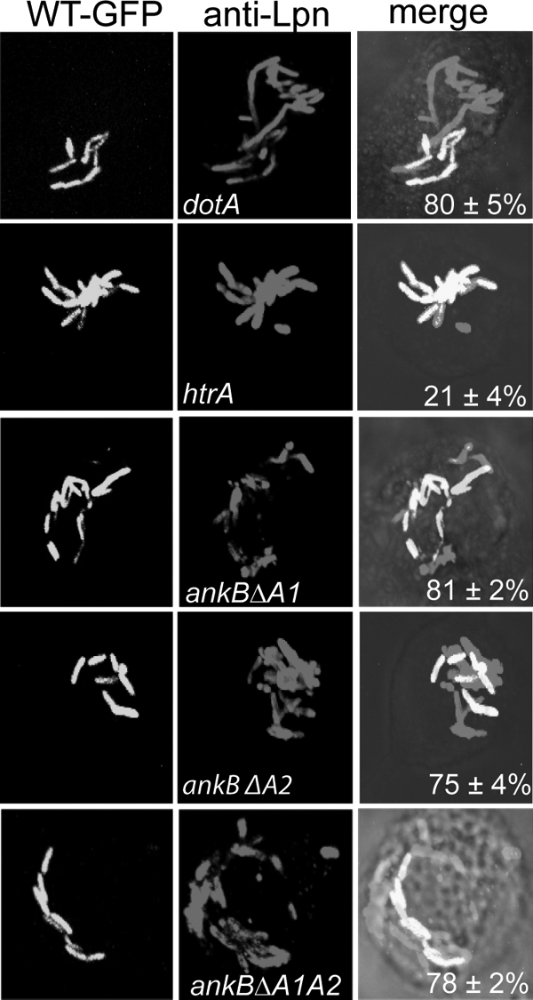
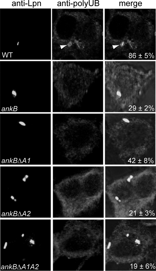
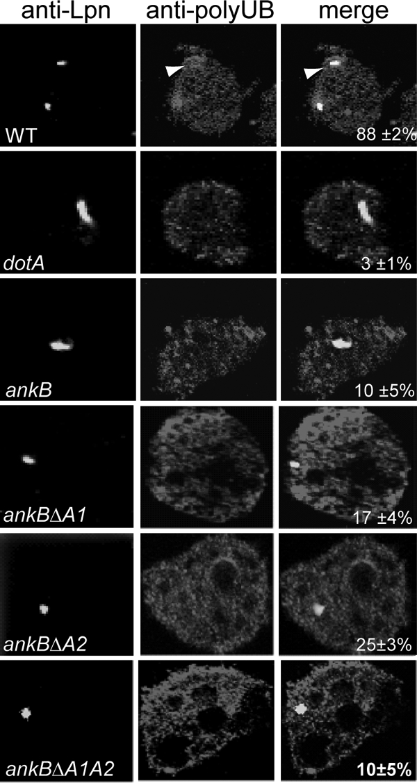
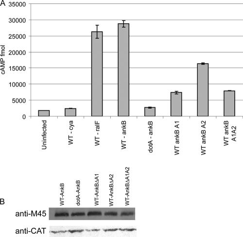
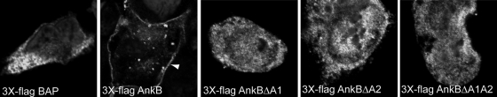
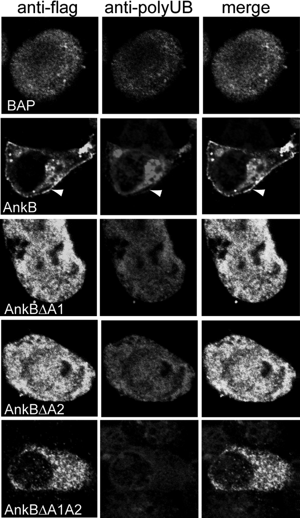
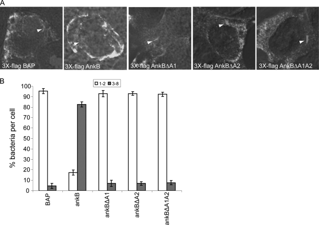
The Dot/Icm-translocated ankyrin B (AnkB) effector of Legionella pneumophila exhibits molecular mimicry of eukaryotic F-box proteins and is essential for intracellular replication in macrophages and protozoa. In addition to two eukaryotic-like ankyrin (ANK) domains, AnkB harbors a conserved eukaryotic F-box domain, which is involved in polyubiquitination of proteins throughout the eukaryotic kingdom. We have recently shown that the F-box domain of the AnkB effector is essential for decoration of the Legionella-containing vacuole (LCV) with polyubiquitinated proteins within macrophages and protozoan hosts. To decipher the role of the two ANK domains in the function of AnkB, we have constructed in-frame deletion of either or both of the ANK domain-encoding regions (ankB Delta A1, ankB Delta A2, and ankB Delta A1A2) to trans-complement the ankB null mutant. Deletion of the ANK domains results in defects in intracellular proliferation and decoration of the LCV with polyubiquitinated proteins. Export of the truncated variants of AnkB was reduced, and this may account for the observed defects. However, while full-length AnkB ectopically expressed in mammalian cells trans-rescues the ankB null mutant for intracellular proliferation, ectopic expression of AnkB Delta A1, AnkB Delta A2, and AnkB Delta A1A2 fails to trans-rescue the ankB null mutant. Importantly, ectopically expressed full-length AnkB is targeted to the host cell plasma membrane, where it recruits polyubiquitinated proteins. In contrast, AnkB Delta A1, AnkB Delta A2, and AnkB Delta A1A2 are diffusely distributed throughout the cytosol and fail to recruit polyubiquitinated proteins. We conclude that the two eukaryotic-like ANK domains of AnkB are essential for intracellular proliferation, for targeting AnkB to the host membranes, and for decoration of the LCV with polyubiquitinated proteins.


Figures

FIG. 1.
FIG. 1.
The two ANK domains of AnkB are essential for intracellular growth of L. pneumophila in macrophages. (A) Monolayers of U937 macrophages were infected with the WT strain and the isogenic dotA or ankB mutants or the ankB mutant complemented with either WT ankB (c/ankB) or one of the ankB mutant alleles. The infection was carried out in triplicate with an MOI of 10 for 1 h followed by 1 h of gentamicin treatment to kill extracellular bacteria. The infected monolayers were lysed at different time points and plated onto agar plates for colony enumeration. The results are representative of three independent experiments performed in triplicate. Error bars represent standard deviations. (B and C) Single cell analyses of L. pneumophila replicative phagosomes. At 10 h postinfection, 100 infected cells were analyzed by laser scanning confocal microscopy for formation of replicative phagosomes, and representative images are shown in panel B. L. pneumophila was stained by a polyclonal anti-L. pneumophila antibody and Alexa Fluor 555-conjugated anti-rabbit IgG (red). Quantification of the number of bacteria/cell at 10 h is shown in panel C. The dotA mutant was used as a negative control. Infected cells from multiple coverslips were examined in each experiment. The results are representative of three independent experiments performed in triplicate. Error bars represent standard deviations. (D) Immunoblot analysis shows no detectable differences in the expression levels of the AnkB variants in L. pneumophila. Total bacterial proteins equivalent to 1 × 10⁸ bacteria were loaded onto SDS-polyacrylamide gels and immunoblotted using an anti-AnkB rabbit antiserum (1:60,000 dilution).
FIG. 2.
FIG. 2.
L. pneumophila bacteria expressing mutant alleles of AnkB are rescued within cells harboring the wild-type bacteria. Coinfections were performed using the wild-type L. pneumophila strain AA100 and the ankB mutant or using the ankB mutant harboring one of the mutant alleles ankBΔA1, ankBΔA2, and ankBΔA1A2. Infected U937 cells were gentamicin treated, washed, and fixed at 10 h after infection. Bacterial phagosomes were scored for all infected cells in randomly selected fields by confocal microscopy. Cells harboring phagosomes containing both strains (WT-GFP [green fluorescent protein] and a mutant) were scored. The htrA mutant was used as a negative control while the dotA mutant was used as a positive control. Representative confocal images are shown in panel A, while quantitative analyses are shown in panel B. Rescue was calculated by quantifying the percentage of the cells harboring the ankB mutant replicating within cells harboring the WT strain, and the numbers are shown in the merged images. The results are representative of three independent experiments performed in triplicate by analyses of 100 infected cells. Error bars represent standard deviations.
FIG. 3.
FIG. 3.
The two ANK domains of AnkB are essential for acquisition of polyubiquitinated proteins by the LCV within macrophages. Representative confocal images of infected macrophages for colocalization of phagosomes with polyubiquitinated proteins at 2 h post-infection of U937 cells by the wild-type strain of L. pneumophila, the isogenic ankB mutant, or the ankB mutant harboring one of the mutant ankB alleles on a plasmid. The cells were labeled with anti-Lpn antibody (green) and antipolyubiquitin (red). The arrowheads indicate heavy colocalization of polyubiquitin with the bacterium. Quantification of colocalization of the LCVs with polyubiquitin at 2 h postinfection has been determined by analysis of 100 infected cells and is shown in the merged images. The data are representative of three independent experiments.
FIG. 4.
FIG. 4.
The two ANK domains of AnkB are essential for acquisition of polyubiquitinated proteins by the LCV within A. polyphaga. Representative confocal images of infected A. polyphaga for colocalization of phagosomes with polyubiquitinated proteins at 2 h post-infection of A. polyphaga by the wild-type strain of L. pneumophila, the ankB isogenic mutant, or the ankB mutant harboring one of the mutant ankB alleles on a plasmid. The cells were labeled with anti-Lpn antibody (green) and antipolyubiquitin (red). The arrowheads indicate heavy colocalization of polyubiquitin with the bacterium. Quantification of colocalization of the LCVs with polyubiquitin at 2 h postinfection is shown in the merged images and has been determined by analysis of 100 infected cells. The data are representative of three independent experiments.
FIG. 5.
FIG. 5.
Translocation of the AnkB variants into host cells. (A) Translocation of AnkB encoded by mutant alleles into U937 cells was determined at 1 h postinfection using adenylate cyclase assays and cyclic AMP (cAMP) levels as readout. Data points are the average cyclic AMP concentrations per well for a representative experiment performed three times in triplicate. Error bars represent standard deviations. (B) Equivalent expression of the AnkB-Cya fusion alleles in L. pneumophila was determined by immunoblot assays of Cya-AnkB fusions expressed in L. pneumophila. Proteins derived from equivalent numbers of bacteria (1 × 10⁸) were loaded onto an SDS-polyacrylamide gel, and Cya fusion proteins were detected on Western blots probed with an anti-M45 antibody, recognizing the N-terminal M45 epitope on all Cya fusions. The blots were reprobed with anti-CAT antibodies, which showed equivalent expression of another protein encoded on the same reporter plasmid.
FIG. 6.
FIG. 6.
Ectopic expression of 3×Flag AnkBΔA1, AnkBΔA2, or AnkBΔA1A2 in HEK293 cells shows altered cellular distribution compared with WT 3×Flag AnkB. Representative confocal images of HEK293 cells transiently transfected with plasmid DNA encoding 3×Flag AnkB, AnkBΔA1, AnkBΔA2, or AnkBΔA1A2 or the BAP control. At 24 h posttransfection, fixed and permeabilized HEK293 cells were labeled with mouse anti-Flag M2 antibody and an Alexa Fluor 488 secondary antibody (green). Nuclei were stained with DAPI (4′,6-diamidino-2-phenylindole) (blue). The white arrowhead indicates localization of 3×Flag AnkB to the cell plasma membrane.
FIG. 7.
FIG. 7.
Ectopically expressed 3×Flag AnkBΔA1, AnkBΔA2, and AnkBΔA1A2 do not recruit polyubiquitinated proteins to the cell plasma membrane. At 24 h following transfection, fixed and permeabilized cells were labeled using a rabbit anti-Flag antibody (green) and a mouse antipolyubiquitin antibody (red). Nuclei were stained with DAPI (4′,6-diamidino-2-phenylindole) (blue). The white arrowheads indicate colocalization of 3×Flag AnkB with polyubiquitinated proteins at the cell periphery.
FIG. 8.
FIG. 8.
Ectopic expression of 3×Flag AnkBΔA1, AnkBΔA2, or AnkBΔA1A2 fails to rescue intravacuolar replication of the ankB mutant. HEK293 cells were transiently transfected with plasmids encoding 3×Flag BAP, 3×Flag AnkB, 3×Flag AnkBΔA1, 3×Flag AnkBΔA2, or 3×Flag AnkBΔA1A2 for 24 h prior to infection. Transfected HEK293 cells were then infected with the WT strain or the ankB or dotA mutant strain, and after 2 and 12 h, 100 infected cells were analyzed by confocal microscopy for formation of replicative phagosomes. (A) Representative confocal microscopy images of transfected HEK293 cells at 12 h postinfection. Ectopically expressed 3×Flag proteins were detected using antibody (green) while bacteria were detected using a rabbit anti-L. pneumophila antibody (red). Nuclei were stained with DAPI (blue). White arrowheads indicate bacteria inside cells. (B) Quantitation of single cell analysis of ankB mutant replicative phagosomes in transfected HEK293 cells 12 h postinfection. At 2 h and 12 h post-infection of HEK293 cells, 100 infected cells were analyzed by confocal microscopy for formation of replicative phagosomes. The WT strain and the dotA mutant bacteria were used as positive and negative controls, respectively. Infected cells from multiple coverslips were examined in each experiment. All the results are representative of three independent experiments performed in triplicate. Error bars represent standard deviations.

Similar articles

Cited by

References

    1. Abu Kwaik, Y. 1996. The phagosome containing Legionella pneumophila within the protozoan Hartmannella vermiformis is surrounded by the rough endoplasmic reticulum. Appl. Environ. Microbiol. 62:2022-2028. - PMC - PubMed
    1. Abu-Zant, A., R. Asare, J. E. Graham, and Y. Abu Kwaik. 2006. Role for RpoS but not RelA of Legionella pneumophila in modulation of phagosome biogenesis and adaptation to the phagosomal microenvironment. Infect. Immun. 74:3021-3026. - PMC - PubMed
    1. Al-Khodor, S., T. Al-Quadan, and Y. Abu Kwaik. Temporal and differential regulation of expression of the eukaryotic-like ankyrin effectors of L. pneumophila. Environ. Microbiol., in press. - PubMed
    1. Al-Khodor, S., C. T. Price, F. Habyarimana, A. Kalia, and Y. Abu Kwaik. 2008. A Dot/Icm-translocated ankyrin protein of Legionella pneumophila is required for intracellular proliferation within human macrophages and protozoa. Mol. Microbiol. 70:908-923. - PMC - PubMed
    1. Al-Khodor, S., C. T. D. Price, A. Kalia, and Y. Abu Kwaik. Functional diversity of ankyrin repeats in microbial proteins. Trends Microbiol. 18:132-139. - PMC - PubMed

Publication types

LinkOut - more resources