. 2010 May;78(5):2342-52.
doi: 10.1128/IAI.01140-09. Epub 2010 Mar 1.

Role of host sphingosine kinase 1 in the lung response against Cryptococcosis

Affiliations

Role of host sphingosine kinase 1 in the lung response against Cryptococcosis

Travis McQuiston et al. Infect Immun. 2010 May.

Abstract

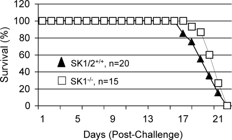
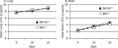
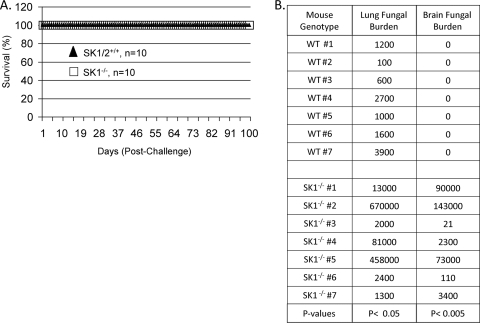
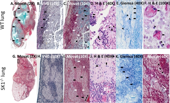
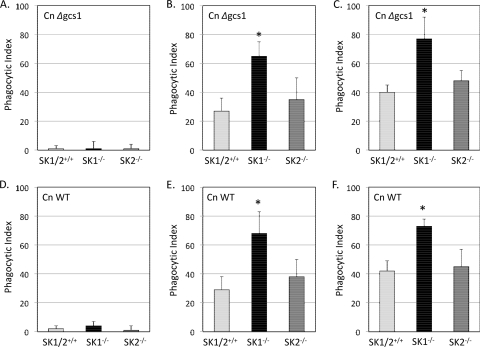
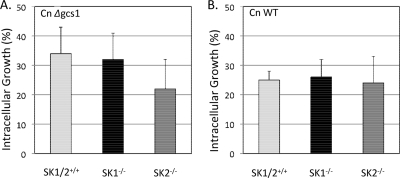
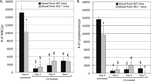
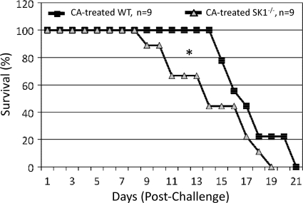
Cryptococcus neoformans is a fungal pathogen causing pulmonary infection and a life-threatening meningoencephalitis in human hosts. The fungus infects the host through inhalation, and thus, the host response in the lung environment is crucial for containment or dissemination of C. neoformans to other organs. In the lung, alveolar macrophages (AMs) are key players in the host lung immune response, and upon phagocytosis, they can kill C. neoformans by evoking an effective immune response through a variety of signaling molecules. On the other hand, under conditions not yet fully defined, the fungus is able to survive and proliferate within macrophages. Since the host sphingosine kinase 1 (SK1) regulates many signaling functions of immune cells, particularly in macrophages, in this study we determined the role of SK1 in the host response to C. neoformans infection. Using wild-type (SK1/2(+/+)) and SK1-deficient (SK1(-/-)) mice, we found that SK1 is dispensable during infection with a facultative intracellular wild-type C. neoformans strain. However, SK1 is required to form a host lung granuloma and to prevent brain infection by a C. neoformans mutant strain lacking the cell wall-associated glycosphingolipid glucosylceramide (Delta gcs1), previously characterized as a mutant able to replicate only intracellularly. Specifically, in contrast to those from SK1/2(+/+) mice, lungs from SK1(-/-) mice have no collagen deposition upon infection with C. neoformans Delta gcs1, and AMs from these mice contain significantly more C. neoformans cells than AMs from SK1/2(+/+) mice, suggesting that under conditions in which C. neoformans is more internalized by AMs, SK1 may become important to control C. neoformans infection. Indeed, when we induced immunosuppression, a host condition in which wild-type C. neoformans cells are increasingly found intracellularly, SK1(-/-) survived significantly less than SK1/2(+/+) mice infected with a facultative intracellular wild-type strain, suggesting that SK1 has an important role in controlling C. neoformans infection under conditions in which the fungus is predominantly found intracellularly.


Figures

FIG. 1.
FIG. 1.
SK1 is dispensable for virulence of a facultative intracellular C. neoformans wild-type strain. C57BL/6J wild-type mice (SK1/2+/+) and sphingosine kinase 1 knockout mice (SK1−/−) were challenged intranasally with 5 × 10⁵ C. neoformans wild-type strain H99 cells and monitored daily. Data are representative of three individual experiments. There was no significant difference in survival between SK1/2+/+ and SK1−/− mice (19.6 ± 1.4 days versus 20.9 ± 1.0 days).
FIG. 2.
FIG. 2.
SK1 deficiency does not affect fungal organ burden in mice challenged with a facultative intracellular C. neoformans wild-type strain. C57BL/6J wild-type mice (SK1/2+/+, n = 9) and sphingosine kinase 1 knockout mice (SK1−/−, n = 9) were challenged intranasally with 5 × 10⁵ C. neoformans wild-type strain H99 cells and monitored daily. Three mice from each group were sacrificed at days 5, 10, and 15 after C. neoformans challenge. Lungs (A) and brains (B) from these mice were processed and analyzed for CFU. There were no significant differences in fungal burden in the lungs or brains of SK1/2+/+ and SK1−/− mice at any of the time points examined.
FIG. 3.
FIG. 3.
SK1 prevents dissemination of an obligately intracellular C. neoformans strain for replication. C57BL/6J wild-type mice (SK1/2+/+) and sphingosine kinase 1 knockout mice (SK1−/−) were infected intranasally with 5 × 10⁵ C. neoformans strain Δgcs1 cells and monitored daily. (A) All mice survived until 100 days postchallenge, at which time the fungal burden in the lungs, livers, spleens, kidneys, and brain of 7 mice from each group was determined. (B) Lungs and brains of SK1−/− mice contained statistically significantly greater numbers of C. neoformans cells than lungs and brains from SK1/2+/+ mice.
FIG. 4.
FIG. 4.
SK1 modulates the granulomatous inflammatory response to an obligately intracellular C. neoformans strain for replication. Histology of lungs of C57BL/6J wild-type (SK1/2+/+) mice (A to F) and sphingosine kinase 1 knockout (SK1−/−) mice (G to L) challenged intranasally with 5 × 10⁵ C. neoformans Δgcs1 cells was analyzed after 100 days postchallenge using Movat staining (A, C, G, I, and L), Verhoeff-van Gieson (VVG) staining (B and H), hematoxylin and eosin (H&E) staining (D, F, and J), and Giemsa staining (E and K). Boxed areas in A and G are magnified in the other representative images. (A) White arrows indicate Δgcs1 cells stained with Alcian blue in necrotic regions; black arrows indicate the fibrotic tissue encapsulating the well-developed granulomas (magnification, ×2). (B) Black arrows indicate the collagen (stained pink/purple) deposited in the fibrotic tissues. White arrows indicate recruited leukocytes inside the fibrotic tissue and within the necrotic core of the granuloma (magnification, ×20). (C) White arrows indicate leukocytes (stained deep red) which reside between the fibrotic tissues and within the necrotic region; black arrows indicate intracellular C. neoformans cells (Alcian blue-stained), whereas black arrowheads indicate extracellular C. neoformans cells (magnification, ×20). (D) H&E staining reveals a mixed population of leukocytes (stained dark purple) representing the host inflammatory response. Neutrophils (black arrowheads) are the most represented granulocyte cell type at 100 days after C. neoformans challenge and are often proximal to AMs containing the intracellular Δgcs1 strain (black arrowheads). Note the absence of eosinophils, which are easily identified using H&E by their bilobed nuclei and reddish-stained cytosol. Crystal-like structures (white arrows; stained pink), which are mostly Ym1 and/or Ym2 derived, suggest eosinophil involvement during some junction of the host immune response (magnification, ×40). (E) Giemsa staining shows leukocytes, which are stained dark blue-purple, located on the periphery of the granuloma (black arrows). Neutrophils (black arrowheads) are the predominant leukocytes present in the leukocyte infiltration. (F) The necrotic core of well-developed granulomas contained giant macrophages (black arrows) often containing the crystal-like structures, which are stained pink. Mixed with cellular debris are numerous extracellular Δgcs1 cells (white arrowheads) and some intact neutrophils (black arrowheads) (magnification, ×100). (G to L) Histology of the lungs of Δgcs1 strain-challenged SK1−/− mice. (G) Δgcs1 cells are found throughout the lung section and are stained lightly with Alcian blue (magnification, ×2). Note that in both panels G and H, there is no fibrotic tissue with collagen deposition, as observed in panels A and B. The bright red structure in the lower right corner of H is a blood vessel. Note the lack of fibrotic tissue and the giant macrophages containing intracellular Δgcs1 cells (black arrowheads) (magnification, ×20). (I) Intracellular and abundant amounts of capsule are stained lightly with Alcian blue (black arrows). The deep red sections correspond to large numbers of leukocytes (white arrows), which are dispersed throughout the section. (J) Foamy histiocytes and giant macrophages (upper) containing intracellular Δgcs1 cells (black arrows) comprise much of the infected lung tissue from Δgcs1 strain-challenged SK1−/− mice. Large numbers of leukocytes (lower), specifically neutrophils (white arrows) and lymphocytes (white arrowheads), are located near phagocytes containing intracellular fungal cells (magnification, ×40). (K) Giemsa staining, which stains the nuclei of leukocytes dark blue-purple, shows the presence of increased leukocyte infiltration (compare panel E). This influx of leukocytes comprised predominately neutrophils and lymphocytes. More lymphocytes (black arrows), which have purple-stained cytosols (K), were observed in the host inflammatory response of Δgcs1 strain-challenged SK1−/− mice than in Δgcs1 strain-challenged SK1/2+/+ mice (E). (L) Macrophages are filled with Alcian blue-stained intracellular Δgcs1 cells (white arrows) with large numbers of leukocytes bordering them (black arrowheads).
FIG. 5.
FIG. 5.
SK1 deficiency in AMs increases the phagocytosis of C. neoformans. AMs from C57BL/6J wild-type (SK1/2+/+) and sphingosine kinase 1 (SK1−/−) and 2 (SK2−/−) knockout mice were incubated with either the C. neoformans Δgcs1 strain (A, B, and C) or the wild-type (WT) strain H99 (D, E, and F) at an MOI of 1:1. Cocultures were stopped after 2 h by removal of the medium and fixation with MeOH. Cells were stained with Giemsa, and internalization of C. neoformans was determined using light microscopy. A minimum of 500 AMs was examined per coculture to determine and calculate the phagocytic index. Data are averages ± standard deviations from five separate experiments. Student's t test was conducted to determine significance (*, P < 0.05).
FIG. 6.
FIG. 6.
SK1 deficiency in AMs does not affect intracellular growth of C. neoformans. AMs from C57BL/6J wild-type (SK1/2+/+) and sphingosine kinase 1 (SK1−/−) and 2 (SK2−/−) knockout mice were incubated with either the C. neoformans Δgcs1 strain (A) or the wild-type (WT) strain H99 (B) at an MOI of 1:1. Medium from cocultures was removed after 2 h, and cocultures were washed to remove extracellular C. neoformans. Fresh medium was then added for an additional 2 h. After a total of 4 h, the medium was removed and MeOH was added for fixation. Cells were stained with Giemsa, and internalization of C. neoformans was determined using light microscopy. A minimum of 100 internalized C. neoformans cells for each coincubation were examined per coculture to determine the number of buds. Data are averages ± standard deviations from five separate experiments. Student's t test was conducted to determine significance (*, P < 0.05).
FIG. 7.
FIG. 7.
Corticosteroid significantly decreases total white blood cells and lymphocytes in WT and SK1−/− mice. C57BL/6J wild-type mice (SK1/2+/+, n = 9) and sphingosine kinase knockout mice (SK1−/−, n = 9) received intraperitoneal injections of 125 mg/kg of cortisone acetate (CA) 24 h prior to, immediately prior to, and at days 1, 4, and 7 after intranasal challenge with 5 × 10⁵ C. neoformans cells. Blood was collected from the saphenous vein immediately prior to injection with CA. Data are averages ± standard deviations from three different pools of blood from three mice. Student's t test was conducted to determine significance (*, P < 0.05; §, P < 0.01; €, P < 0.001; $, P < 0.0001; ¢, P < 0.00001).
FIG. 8.
FIG. 8.
Role of SK1 during immunosuppression in host susceptibility to a facultative intracellular C. neoformans wild-type strain. C57BL/6J wild-type mice (SK1/2+/+) and sphingosine kinase 1 knockout mice (SK1−/−) were administered a regimen of cortisone acetate (CA) to induce immunosuppression and challenged intranasally with 5 × 10⁵ C. neoformans strain H99 cells, as described in Materials and Methods. The Wilcoxon rank sum test was used to determine significance (*, P < 0.05).

Similar articles

Cited by

References

    1. Allende, M. L., T. Sasaki, H. Kawai, A. Olivera, Y. Mi, G. van Echten-Deckert, R. Hajdu, M. Rosenbach, C. A. Keohane, S. Mandala, S. Spiegel, and R. L. Proia. 2004. Mice deficient in sphingosine kinase 1 are rendered lymphopenic by FTY720. J. Biol. Chem. 279:52487-52492. - PubMed
    1. Ammit, A. J., A. T. Hastie, L. C. Edsall, R. K. Hoffman, Y. Amrani, V. P. Krymskaya, S. A. Kane, S. P. Peters, R. B. Penn, S. Spiegel, and R. A. Panettieri, Jr. 2001. Sphingosine 1-phosphate modulates human airway smooth muscle cell functions that promote inflammation and airway remodeling in asthma. FASEB J. 15:1212-1214. - PubMed
    1. Argraves, K. M., P. J. Gazzolo, E. M. Groh, B. A. Wilkerson, B. S. Matsuura, W. O. Twal, S. M. Hammad, and W. S. Argraves. 2008. High density lipoprotein-associated sphingosine 1-phosphate promotes endothelial barrier function. J. Biol. Chem. 283:25074-25081. - PMC - PubMed
    1. Billich, A., F. Bornancin, P. Devay, D. Mechtcheriakova, N. Urtz, and T. Baumruker. 2003. Phosphorylation of the immunomodulatory drug FTY720 by sphingosine kinases. J. Biol. Chem. 278:47408-47415. - PubMed
    1. Clancy, C. J., M. H. Nguyen, R. Alandoerffer, S. Cheng, K. Iczkowski, M. Richardson, and J. R. Graybill. 2006. Cryptococcus neoformans var. grubii isolates recovered from persons with AIDS demonstrate a wide range of virulence during murine meningoencephalitis that correlates with the expression of certain virulence factors. Microbiology 152:2247-2255. - PubMed

Publication types

MeSH terms

Substances

LinkOut - more resources