

The proline-rich region of pneumococcal surface proteins A and C contains surface-accessible epitopes common to all pneumococci and elicits antibody-mediated protection against sepsis
- PMID: 20194601
- PMCID: PMC2863538
- DOI: 10.1128/IAI.01199-09
The proline-rich region of pneumococcal surface proteins A and C contains surface-accessible epitopes common to all pneumococci and elicits antibody-mediated protection against sepsis
Abstract
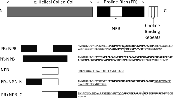
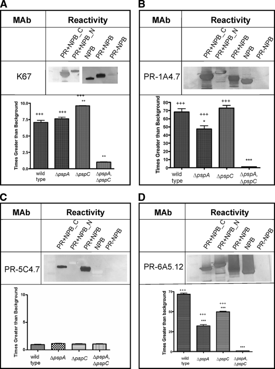
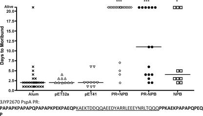
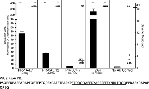
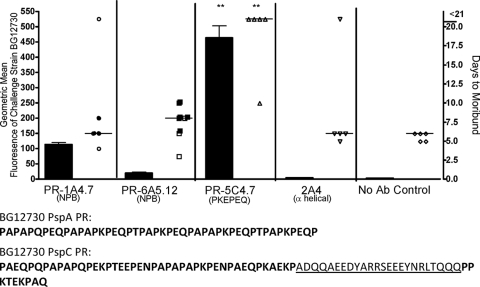
Pneumococcal surface protein A (PspA) and PspC of Streptococcus pneumoniae are surface virulence proteins that interfere with complement deposition and elicit protective immune responses. The C-terminal halves of PspA and PspC have some structural similarity and contain highly cross-reactive proline-rich (PR) regions. In many PR regions of PspA and PspC, there exists an almost invariant nonproline block (NPB) of about 33 amino acids. Neither the PR regions nor their NPB exhibit the alpha-helical structure characteristic of much of the protection-eliciting N-terminal portions of PspA and PspC. Prior studies of PspA and PspC as immunogens focused primarily on the alpha-helical regions of these molecules that lack the PR and NPB regions. This report shows that immunization with recombinant PR (rPR) molecules and passive immunization with monoclonal antibodies reactive with either NPB or PR epitopes are protective against infection in mice. PR regions of both PspA and PspC were antibody accessible on the pneumococcal surface. Our results indicate that while PspA could serve as a target of these protective antibodies in invasive infections, PspC might not. When antibody responses to rPR immunogens were evaluated by using flow cytometry to measure antibody binding to live pneumococci, it was observed that the mice that survived subsequent challenge produced significantly higher levels of antibodies reactive with exposed PR epitopes than the mice that became moribund. Due to their conservation and cross-reactivity, the PR regions and NPB regions represent potential vaccine targets capable of eliciting cross-protection immunity against pneumococcal infection.
Figures
Similar articles
-
The pspC gene of Streptococcus pneumoniae encodes a polymorphic protein, PspC, which elicits cross-reactive antibodies to PspA and provides immunity to pneumococcal bacteremia.Infect Immun. 1999 Dec;67(12):6533-42. doi: 10.1128/IAI.67.12.6533-6542.1999. Infect Immun. 1999. PMID: 10569772 Free PMC article.
-
Mapping of epitopes recognized by antibodies induced by immunization of mice with PspA and PspC.Clin Vaccine Immunol. 2014 Jul;21(7):940-8. doi: 10.1128/CVI.00239-14. Epub 2014 May 7. Clin Vaccine Immunol. 2014. PMID: 24807052 Free PMC article.
-
Streptococcus pneumoniae Binds to Host Lactate Dehydrogenase via PspA and PspC To Enhance Virulence.mBio. 2021 May 4;12(3):e00673-21. doi: 10.1128/mBio.00673-21. mBio. 2021. PMID: 33947761 Free PMC article.
-
Systemic and mucosal protective immunity to pneumococcal surface protein A.Ann N Y Acad Sci. 1996 Oct 25;797:118-26. doi: 10.1111/j.1749-6632.1996.tb52954.x. Ann N Y Acad Sci. 1996. PMID: 8993356 Review.
-
A Jack of All Trades: The Role of Pneumococcal Surface Protein A in the Pathogenesis of Streptococcus pneumoniae.Front Cell Infect Microbiol. 2022 Feb 2;12:826264. doi: 10.3389/fcimb.2022.826264. eCollection 2022. Front Cell Infect Microbiol. 2022. PMID: 35186799 Free PMC article. Review.
Cited by
-
Impaired function of antibodies to pneumococcal surface protein A but not to capsular polysaccharide in Mexican American adults with type 2 diabetes mellitus.Clin Vaccine Immunol. 2012 Sep;19(9):1360-9. doi: 10.1128/CVI.00268-12. Epub 2012 Jul 3. Clin Vaccine Immunol. 2012. PMID: 22761295 Free PMC article.
-
Evaluation of a vaccine formulation against Streptococcus pneumoniae based on choline-binding proteins.Clin Vaccine Immunol. 2015 Feb;22(2):213-20. doi: 10.1128/CVI.00692-14. Epub 2014 Dec 17. Clin Vaccine Immunol. 2015. PMID: 25520146 Free PMC article.
-
Pneumococcal surface protein A inhibits complement deposition on the pneumococcal surface by competing with the binding of C-reactive protein to cell-surface phosphocholine.J Immunol. 2012 Dec 1;189(11):5327-35. doi: 10.4049/jimmunol.1201967. Epub 2012 Oct 26. J Immunol. 2012. PMID: 23105137 Free PMC article.
-
A new candidate epitope-based vaccine against PspA PhtD of Streptococcus pneumoniae: a computational experimental approach.Front Cell Infect Microbiol. 2023 Nov 15;13:1271143. doi: 10.3389/fcimb.2023.1271143. eCollection 2023. Front Cell Infect Microbiol. 2023. PMID: 38035337 Free PMC article.
-
Serotype-independent pneumococcal vaccines.Cell Mol Life Sci. 2013 Sep;70(18):3303-26. doi: 10.1007/s00018-012-1234-8. Epub 2012 Dec 27. Cell Mol Life Sci. 2013. PMID: 23269437 Free PMC article. Review.
References
-
- Balachandran, P., A. Brooks-Walter, A. Virolainen-Julkunen, S. K. Hollingshead, and D. E. Briles. 2002. Role of pneumococcal surface protein C in nasopharyngeal carriage and pneumonia and its ability to elicit protection against carriage of Streptococcus pneumoniae. Infect. Immun. 70:2526-2534. - PMC - PubMed
-
- Barocchi, M. A., S. Censini, and R. Rappuoli. 2007. Vaccines in the era of genomics: the pneumococcal challenge. Vaccine 25:2963-2973. - PubMed
-
- Bast, D. J., M. Yue, X. Chen, D. Bell, L. Dresser, R. Saskin, L. A. Mandell, D. E. Low, and J. C. de Azavedo. 2004. Novel murine model of pneumococcal pneumonia: use of temperature as a measure of disease severity to compare the efficacies of moxifloxacin and levofloxacin. Antimicrob. Agents Chemother. 48:3343-3348. - PMC - PubMed
Publication types
MeSH terms
Substances
Grants and funding
LinkOut - more resources
Full Text Sources
Other Literature Sources
Medical
Molecular Biology Databases
Research Materials
