. 2010 May;30(9):2206-19.
doi: 10.1128/MCB.01181-09. Epub 2010 Mar 1.

Par1b/MARK2 phosphorylates kinesin-like motor protein GAKIN/KIF13B to regulate axon formation

Affiliations

Par1b/MARK2 phosphorylates kinesin-like motor protein GAKIN/KIF13B to regulate axon formation

Yuta Yoshimura et al. Mol Cell Biol. 2010 May.

Abstract

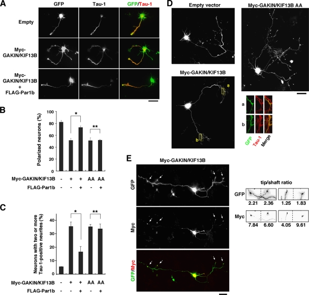
Here we report that Par1b/MARK2 regulates axon formation via phosphorylation of a kinesin superfamily protein GAKIN/KIF13B. Accumulating evidence indicated the importance of the evolutionarily conserved kinase Par1b in the regulation of cell polarity. Using hippocampal neurons in culture, it has been shown that Par1b regulates axon specification, but the underlying mechanism remains uncharacterized. We identify GAKIN/KIF13B as a novel Par1b-binding protein and reveal that GAKIN/KIF13B is a physiological substrate for Par1b, and the phosphorylation sites are conserved from Drosophila. In hippocampal neurons, GAKIN/KIF13B accumulates at the distal part of the microtubules in the tips of axons, but not of dendrites. Overexpression of GAKIN/KIF13B by itself can induce the formation of extra axons, which is inhibited by the coexpression of Par1b in a manner dependent on its kinase activity. In contrast, small interfering RNA (siRNA)-mediated knockdown of GAKIN/KIF13B severely retards neurite extension and promotes the axonless phenotype. The extra axon phenotype caused by Par1b siRNA is suppressed by cointroduction of GAKIN/KIF13B siRNA, thus placing the GAKIN/KIF13B function downstream of Par1b. We also find that GAKIN/KIF13B acts downstream of the phosphatidylinositol 3-kinase (PI3K) signaling via Par1b phosphorylation. These results reveal that GAKIN/KIF13B is a key intermediate linking Par1b to the regulation of axon formation.


Figures

FIG. 1.
FIG. 1.
Binding of Par1b to GAKIN/KIF13B. (A) Anti-FLAG immunoprecipitates (IP) from lysates of MDCK clones stably expressing FLAG-Par1b or GFP as a control were subjected to SDS-PAGE and visualized by silver staining. The positions of FLAG-Par1b and GAKIN/KIF13B are indicated to the right of the gel. The relative molecular weights (in thousands) [Mr(K)] are indicated to the left of the gel. (B) Equal amounts of lysates of MDCK, SH-SY5Y cells, or cortical neurons at 2 DIV were subjected to immunoprecipitation (IP) with antibody against Par1b or GAKIN/KIF13B. The precipitates were analyzed by immunoblotting (IB) with the indicated antibodies. Immunoprecipitations with control rabbit IgG and without lysates (− Lysates) were also performed as controls. The multiple bands for Par1b represent alternative splicing isoforms (indicated by the black arrowheads to the right of the gels). (C) Domain map of GAKIN/KIF13B and its deletion constructs (the numbers are the positions of the amino acid residues). (D) COS-7 cells were transfected with the indicated constructs, and the lysates were subjected to anti-FLAG immunoprecipitation. The lysates and precipitates were analyzed by immunoblotting with the indicated antibodies. The positions of nonspecific bands (*) and IgG (**) are indicated. (E) Recombinant His-Par1b was incubated with GST-Stalk2 or GST immobilized on beads as a control. Proteins were subjected to SDS-PAGE and visualized by staining with Coomassie brilliant blue. Ten percent of His-Par1b was loaded as input. (F) His-GAKIN/KIF13B and His-Par1b were mixed and immunoprecipitated with anti-Par1b antibody. The precipitates were analyzed by immunoblotting with the indicated antibodies.
FIG. 2.
FIG. 2.
Phosphorylation of GAKIN/KIF13B by Par1b. (A) FLAG-GAKIN/KIF13B was expressed in COS-7 cells and then immunoprecipitated. The precipitates were subjected to kinase assays for His-Par1b (+). The control experiments (−) were performed with samples that had been obtained by subjecting E. coli lysate (not expressing His-Par1b) to the same purification procedure as His-Par1b. Autoradiography represents the phosphorylation level of FLAG-GAKIN/KIF13B. (B) The indicated deletion constructs of GAKIN/KIF13B were expressed and subjected to kinase assays as in panel A. The positions of IgG (*) and autophosphorylated His-Par1b (**) are indicated. (C) Alignment of the amino acid sequence of the region of human GAKIN/KIF13B containing Ser1381 and Ser1410 with the corresponding sequences. The human (Homo sapiens), mouse (Mus musculus), zebrafish (Danio rerio), and fruit fly (Drosophila melanogaster) sequences are shown. The evolutionarily conserved amino acids surrounding Ser1381 and Ser1410 are shown on a gray background. The 14-3-3 binding motif is also indicated. (D) The indicated mutagenized constructs of FLAG-Stalk2 were subjected to kinase assays as in panel A.
FIG. 3.
FIG. 3.
Par1b phosphorylation induces 14-3-3 binding with GAKIN/KIF13B. (A) COS-7 cells were transfected with the indicated constructs. The lysates and anti-FLAG immunoprecipitates were subjected to immunoblotting with the indicated antibodies. (B) Lysates of MDCK cells or hippocampal neurons at 2 DIV were subjected to immunoprecipitation with antibody against GAKIN/KIF13B. The precipitates were analyzed by immunoblotting with the indicated antibodies. Immunoprecipitations with rabbit IgG and without lysates were performed as controls. (C) MDCK, SH-SY5Y cells, or hippocampal neurons at 2 DIV were transfected with the indicated siRNAs. The lysates and anti-GAKIN/KIF13B immunoprecipitates were subjected to immunoblotting with the indicated antibodies. The numbers below the column indicate the ratio of band intensity normalized to actin (means of three independent experiments). (D) MDCK cells were transfected with the indicated constructs. The cells were cultured for an additional day and stained for FLAG and Myc. Bar, 20 μm. (E) Myc-GAKIN/KIF13B accumulation in MDCK cells. Myc-GAKIN/KIF13B accumulation is defined as large dot-like signals observed exclusively at the tips of protrusions. Three independent experiments were performed, and >60 cells were counted in each experiment. The values show the means plus SDs (error bars). Values that are significantly different (P < 0.001) are indicated by an asterisk.
FIG. 4.
FIG. 4.
Accumulation of GAKIN/KIF13B at axon tips. (A) Representative GAKIN/KIF13B-stained images of stage 1, stage 2, and stage 3 hippocampal neurons from E18 rats are indicated. The accumulation of GAKIN/KIF13B at the tips of neurites is indicated by the small white arrows. Bars, 20 μm. (B) Percentage of hippocampal neurons that have a single neurite (gray) or two or more neurites (black) containing GAKIN/KIF13B accumulation at stage 2 (n = 100) or stage 3 (n = 96). GAKIN/KIF13B accumulation is defined here as a level greater than 4 times the shaft intensity level. (C) Neurons at 3 DIV were stained for GAKIN/KIF13B and Tau-1 or MAP2. Merged images are also presented (GAKIN/KIF13B shown in green; Tau-1 and MAP2 shown in red). Bar, 20 μm. (D) (Top) Neurons were transfected with GFP and then stained for GAKIN/KIF13B at 3 DIV (green, GFP; red, GAKIN/KIF13B). Bar, 20 μm. (Bottom) The fluorescence intensity in the axon tip, shaft, and cell body was measured and is shown in the graph plot using line scan imaging (purple, GFP; blue, GAKIN/KIF13B). (E) Anti-GAKIN/KIF13B antibody was preincubated with GST (control) or GST-CAP-Gly immobilized on beads for 2 h. Neurons at 3 DIV were stained with absorbed anti-GAKIN/KIF13B antibody or IgG (control) (green) together with anti-Tau-1 antibody (red). Green and red signals at the axon tips are also shown separately. Bar, 20 μm. (F) Neurons at 3 DIV were stained for β-tubulin and GAKIN/KIF13B, and the growth cone area is magnified and indicated. A merged image is also shown (GAKIN/KIF13B [red]; β-tubulin [green]). Bar, 5 μm.
FIG. 5.
FIG. 5.
GAKIN/KIF13B plays a positive role in axon formation. (A) Hippocampal neurons from E18 rats were transfected with empty vector, Myc-GAKIN/KIF13B, or Myc-GAKIN/KIF13B plus FLAG-Par1b together with GFP and then stained for Tau-1 at 3 DIV. Merged images are also presented (GFP shown in green and Tau-1 in red). Bar, 20 μm. (B) Neurons were transfected with the indicated constructs, and the percentage of polarized neurons was determined at 3 DIV. Three independent experiments were performed, and >80 cells were counted in each experiment. The values in panels B and C are means plus SDs (error bars). Values that are significantly different are indicated as follows: *, P < 0.01; **, P = 0.33. (C) Percentage of neurons that have two or more axons positive for Tau-1. Three independent experiments were performed, and >80 cells were counted in each experiment. Values that are significantly different are indicated as follows: *, P < 0.01; **, P = 0.55. (D) Neurons were transfected with empty vector, Myc-GAKIN/KIF13B, or Myc-GAKIN/KIF13B AA together with GFP at 48 h after plating by lipofection and observed for GFP fluorescence at 3.5 DIV. Magnified views of the two areas surrounded by yellow dotted lines (a and b) in neurons transfected with Myc-GAKIN/KIF13B and labeled by GFP fluorescence (green), Tau-1 staining (red), and both GFP fluorescence and Tau-1 staining (merged images) are shown. Bar, 20 μm. (E) Neurons were transfected with Myc-GAKIN/KIF13B together with GFP as in panel D. A merged image is also shown (green, GFP; red, Myc). Arrows indicate the tips of the axon-like neurites are indicated by the small white arrows. Bar, 20 μm. The tip/shaft ratios of the GFP and Myc signal intensities are shown to the right of the photomicrographs.
FIG. 6.
FIG. 6.
GAKIN/KIF13B functions downstream of Par1b in axon specification. (A) Hippocampal neurons from E18 rats were transfected with the indicated siRNAs and GFP by electroporation. Lysates of the neurons at 2 DIV were then subjected to immunoblotting with anti-GAKIN/KIF13B antibody. The numbers below the top blot indicate the ratios of band intensity normalized to actin. (B) Neurons transfected as in panel A were visualized with GFP at 3 DIV. Bar, 50 μm. (C) Neurons transfected as in panel A were stained for Tau-1 at 3 DIV. Bar, 20 μm. (D) Percentage of axonless neurons at 3 DIV. Three independent experiments were performed, and >80 cells were counted in each experiment. The values show the means plus SDs (error bars). Values that are significantly different (P < 0.001) are indicated by an asterisk. The axons were determined here to be neurites positive for Tau-1 staining and longer than 100 μm. (E) Neurons transfected with the indicated siRNAs were stained for Tau-1 at 3 DIV. Bar, 50 μm. (F) Percentage of neurons with none, one (single), or two or more axons at 3 DIV. Three independent experiments were performed, and >80 cells were counted in each experiment.
FIG. 7.
FIG. 7.
PI3K regulates GAKIN/KIF13B localization. (A) Hippocampal neurons from E18 rats were transfected with the indicated constructs together with GFP and then stained for GAKIN/KIF13B at 2 DIV. Bar, 10 μm. (B) Neurons at 1 DIV were treated with LY294002 (20 μM), PKCζ inhibitor (10 μM), KT5720 (200 nM), or DMSO (control) for an additional 24 h and stained for β-tubulin and GAKIN/KIF13B (β-tubulin [green]; GAKIN/KIF13B [red]). The tip/shaft ratios of the GAKIN/KIF13B signal intensities are also shown. Bar, 10 μm. (C) Percentage of neurites with GAKIN/KIF13B accumulation at the tip of the neurite at 2 DIV. Three independent experiments were performed, and >80 cells were counted in each experiment. The values show the means plus SDs (error bars). Values that are significantly different are indicated as follows: *, P < 0.001; **, P = 0.50. (D) Lysates of neurons at 2 DIV treated as in panel B were subjected to immunoblotting with anti-GAKIN/KIF13B antibody. The numbers below the top blot indicate the ratios of band intensity normalized to actin. (E) Neurons were transfected with Myc-GAKIN/KIF13B and Myc-GAKIN/KIF13B AA, plated, and cultured for 24 h. LY294002 or DMSO was added, and the cells were incubated for 24 h. Neurons at 2 DIV were stained for β-tubulin and Myc. Merged images (green, β-tubulin; red, Myc) are also presented. The areas outlined by yellow dotted lines in the Tubulin/Myc column are magnified. Bar, 10 μm. (F) Percentage of neurons with accumulation of Myc-GAKIN/KIF13B or Myc-GAKIN/KIF13B AA at the tip of the neurite at 2 DIV in the presence or absence of LY294002. Three independent experiments were performed, and >80 cells were counted in each experiment. The values show the means plus SDs (error bars). Values that are significantly different are indicated as follows: *, P < 0.001; **, P = 0.09. (G) MDCK cells were transfected with Myc-GAKIN/KIF13B or Myc-GAKIN/KIF13B AA together with GFP. LY294002 or DMSO was added, and the cells were incubated for 20 h. The cells were stained for Myc. Bar, 20 μm. The accumulation of Myc-GAKIN/KIF13B AA in cells treated with LY294002 is indicated by white arrowheads.
FIG. 8.
FIG. 8.
Schematic diagram of a model for positive-feedback signaling mediated by Par1/GAKIN/KIF13B. PIP3 is generated from PIP2 by PI3K and activates aPKC via PDK1. Activated aPKC forms the complex with Par3/Par6 and phosphorylates Par1. Phosphorylated Par1 is excluded from the membrane and functionally inactivated. Unphosphorylated Par1 phosphorylates GAKIN/KIF13B and inhibits microtubule-dependent accumulation of GAKIN/KIF13B cooperatively with 14-3-3. Unphosphorylated (14-3-3-free) GAKIN/KIF13B accumulates at the protrusion tips with PIP3-containing vesicles. Such a spatial positive-feedback mechanism may assemble PIP3 at specific sites in cells to regulate cell polarity.

Similar articles

Cited by

References

    1. Akhmanova, A., C. C. Hoogenraad, K. Drabek, T. Stepanova, B. Dortland, T. Verkerk, W. Vermeulen, B. M. Burgering, C. I. De Zeeuw, F. Grosveld, and N. Galjart. 2001. Clasps are CLIP-115 and -170 associating proteins involved in the regional regulation of microtubule dynamics in motile fibroblasts. Cell 104:923-935. - PubMed
    1. Altschuler, S. J., S. B. Angenent, Y. Wang, and L. F. Wu. 2008. On the spontaneous emergence of cell polarity. Nature 454:886-889. - PMC - PubMed
    1. Arimura, N., and K. Kaibuchi. 2007. Neuronal polarity: from extracellular signals to intracellular mechanisms. Nat. Rev. Neurosci. 8:194-205. - PubMed
    1. Asaba, N., T. Hanada, A. Takeuchi, and A. H. Chisti. 2003. Direct interaction with a kinesin-related motor mediates transport of mammalian discs large tumor suppressor homologue in epithelial cells. J. Biol. Chem. 278:8395-8400. - PubMed
    1. Benton, R., and D. S. Johnston. 2003. Drosophila PAR-1 and 14-3-3 inhibit Bazooka/PAR-3 to establish complementary cortical domains in polarized cells. Cell 115:691-704. - PubMed

Publication types