. 2010 Mar 15;207(3):465-74.
doi: 10.1084/jem.20091513. Epub 2010 Mar 1.

Essential roles of sphingosine-1-phosphate receptor 2 in human mast cell activation, anaphylaxis, and pulmonary edema

Affiliations

Essential roles of sphingosine-1-phosphate receptor 2 in human mast cell activation, anaphylaxis, and pulmonary edema

Carole A Oskeritzian et al. J Exp Med. .

Abstract

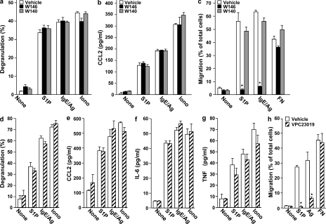
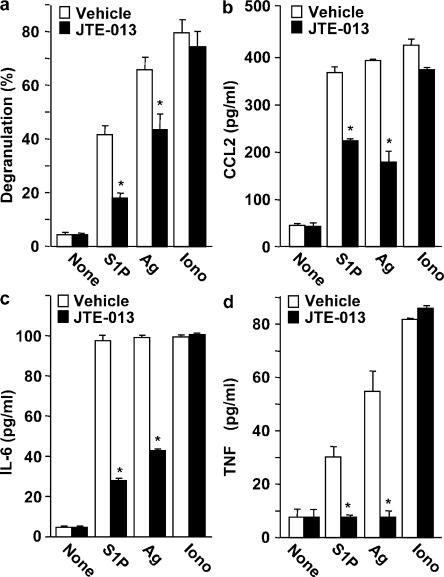
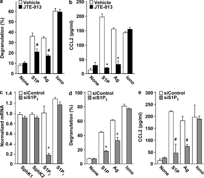
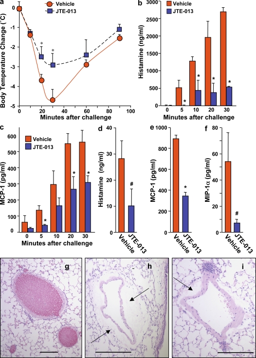
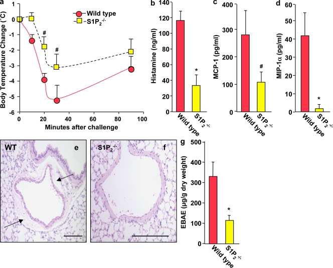
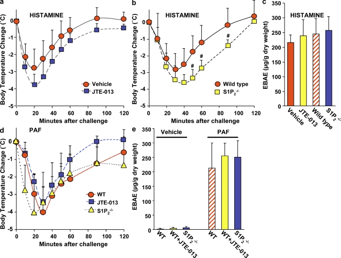
Systemic exacerbation of allergic responses, in which mast cells play a critical role, results in life-threatening anaphylactic shock. Sphingosine-1-phosphate (S1P), a ligand for a family of G protein-coupled receptors, is a new addition to the repertoire of bioactive lipids secreted by activated mast cells. Yet little is known of its role in human mast cell functions and in anaphylaxis. We show that S1P(2) receptors play a critical role in regulating human mast cell functions, including degranulation and cytokine and chemokine release. Immunoglobulin E-triggered anaphylactic responses, including elevation of circulating histamine and associated pulmonary edema in mice, were significantly attenuated by the S1P(2) antagonist JTE-013 and in S1P(2)-deficient mice, in contrast to anaphylaxis induced by administration of histamine or platelet-activating factor. Hence, S1P and S1P(2) on mast cells are determinants of systemic anaphylaxis and associated pulmonary edema and might be beneficial targets for anaphylaxis attenuation and prophylaxis.


Figures

Figure 1.
Figure 1.
S1P1 is not involved in degranulation or cytokine and chemokine secretion from human CB-MCs and Sk-MCs but is essential for human mast cell motility. (a–c) Purified CB-MCs were stimulated with vehicle, 100 nM S1P, or 1 µM ionomycin, or sensitized overnight with IgE, and then stimulated with 30 ng/ml DNP-HSA (Ag), for 2 h. Where indicated, CB-MCs were pretreated for 30 min with vehicle, 1 µM W146, or 1 µM W140 before stimulation. Degranulation was assessed by the percentage of β-hexosaminidase release (a). Secretion of CCL2 (b) was measured by ELISA. (c) Duplicate cultures pretreated for 30 min with vehicle, 1 µM W146, or 1 µM W140 were allowed to migrate in transwell chambers toward vehicle, 100 nM S1P, 30 ng/ml Ag, or 20 µg/ml fibronectin (FN) for 24 h. Similar results were obtained in three independent experiments using CB-MC from three different donors. (d–h) Sk-MCs were stimulated with vehicle, 100 nM S1P, or 1 µM ionomycin, or sensitized overnight with IgE, and then stimulated with 30 ng/ml Ag for 2 h. Where indicated, Sk-MCs were pretreated for 30 min with vehicle or 1 µM VPC23019 before stimulation. Degranulation (d) and secretion of CCL2 (e), IL-6 (f), and TNF (g) was measured. (h) Migration of duplicate cultures toward vehicle, 100 nM S1P, 30 ng/ml Ag, or 20 µg/ml fibronectin was determined in transwell chambers. Data are expressed as the percentage of migrating cells and are the means ± SD of triplicate determinations. Similar results were obtained in three independent experiments using CB-MC and Sk-MC from three different donors. *, P < 0.01, compared with vehicle treatment.
Figure 2.
Figure 2.
S1P2 antagonism impairs degranulation and cytokine and chemokine secretion from human Sk-MCs. Sk-MCs were stimulated with vehicle, 100 nM S1P, or 1 µM ionomycin, or sensitized overnight with IgE, and then stimulated with 30 ng/ml Ag for 2 h. Sk-MCs were pretreated for 30 min with vehicle or 1 µM JTE013 before stimulation, as indicated. Degranulation was assessed by β-hexosaminidase release (a). Secretion of CCL2 (b), IL-6 (c), and TNF (d) was measured by ELISA. Data are the means ± SD of triplicate determinations. Similar results were obtained in three independent experiments using Sk-MC from three different donors. *, P < 0.01, compared with vehicle treatment.
Figure 3.
Figure 3.
S1P2 antagonism or down-regulation of S1P2 decreases S1P- and Ag-induced degranulation and chemokine secretion from CB-MCs. (a and b) Purified CB-MCs were stimulated with vehicle, 100 nM S1P, or 1 µM ionomycin, or sensitized overnight with IgE, and then stimulated with 30 ng/ml Ag for 2 h. CB-MCs were pretreated for 30 min with vehicle or 1 µM JTE013 before stimulation, as indicated. Degranulation (a) and secretion of CCL2 (b) were measured. *, P ≤ 0.01; #, P ≤ 0.05, compared with vehicle treatment. (c–e) CB-MCs transfected with control siRNA or S1P2 siRNA were stimulated with vehicle, 100 nM S1P, or 1 µM ionomycin, or sensitized overnight with IgE, and then stimulated with 30 ng/ml Ag for 2 h. (c) RNA was isolated and mRNA levels of S1P1, S1P2, SphK1, SphK2, and GAPDH were determined by quantitative real-time PCR. Degranulation (d) and secretion of CCL2 (e) were measured. *, P ≤ 0.01; #, P ≤ 0.05, compared with scrambled siRNA. Data are the means ± SD of triplicate determinations. Similar results were obtained in three independent experiments with CB-MC from three different donors.
Figure 4.
Figure 4.
S1P2 antagonist attenuates PSA. C57BL/6 mice were injected i.p. with IgE anti-DNP mAb. 12 h later, mice were injected i.p. with vehicle (DMSO; n = 6) or JTE013 (20 µg/mouse; n = 6) and, 30 min later, systemic anaphylaxis was induced by i.p. injection of DNP-HSA. Body temperature was monitored at the indicated times (a). Serum levels of histamine at the indicated times (b) or at 120 min (d), MCP-1/CCL2 at the indicated times (c) or at 120 min (e), and MIP-1α/CCL3 at 120 min (f) were determined by ELISA. Data are the means ± SD. (g–i) Representative micrographs of hematoxylin and eosin (H&E)–stained lung tissues from control mice (g), Ag-challenged mice (h), and mice treated with JTE013 before Ag challenge (i). Arrows indicate pulmonary vascular edema. Note the extensive vascular edema in vehicle-treated Ag-challenged lung (h) compared with JTE013-treated Ag-challenged lung (i). Bars, 100 µm. *, P ≤ 0.01; #, P ≤ 0.05, compared with vehicle treated mice. Similar results were obtained in three independent experiments.
Figure 5.
Figure 5.
PSA is markedly reduced in S1P2 knockout mice. S1P2−/− mice (n = 6) and wild-type mice (n = 6) were injected i.p. with IgE anti-DNP mAb, and systemic anaphylaxis was elicited 12 h later by injection of DNP-HSA. Body temperature was monitored at the indicated times (a). Serum levels of histamine (b), MCP-1/CCL2 (c), and MIP-1α/CCL3 (d) at 120 min were determined by ELISA. Data are means ± SD. (e and f) Representative micrographs of H&E-stained lung tissues showing extensive pulmonary perivascular edema in Ag-challenged wild-type mice (e), which was nearly absent around lung blood vessels of Ag-challenged S1P2−/− mice (f). Arrows indicate pulmonary vascular edema. Lung vascular permeability was determined by quantifying Evans blue extravasation (EBAE; g). Similar results were obtained in three independent experiments. *, P ≤ 0.01; #, P ≤ 0.05, compared with wild-type mice. Bars, 100 µm.
Figure 6.
Figure 6.
Blockade of S1P2 or S1P2 deficiency does not mitigate anaphylaxis triggered by histamine or PAF. (a) C57BL/6 mice were injected i.v. with histamine (3 mg/mouse) together with vehicle (n = 6) or JTE-013 (20 µg/mouse; n = 6), and body temperature was monitored at the indicated times. (b) C57BL/6 × 129sv wild-type (n = 6) and S1P2 knockout mice (n = 9) were injected i.v. with histamine (3 mg/mouse), and body temperature was monitored. (c) Lung vascular permeability was determined by quantifying Evans blue extravasation. (d) C57BL/6 × 129sv wild-type mice were injected i.v. with PAF (500 ng/mouse) together with vehicle (n = 9) or JTE-013 (n = 9). S1P2 null mice were injected with PAF (500 ng/mouse; n = 9). Body temperature was monitored at the indicated times. (e) Lung vascular permeability was determined by quantifying Evans blue extravasation in C57BL/6 × 129sv wild-type mice and in S1P2 null mice injected i.v. with vehicle, PAF, or JTE-013, as indicated. Data are the means ± SD of triplicate determinations. Similar results were obtained in two independent experiments.

Similar articles

Cited by

References

    1. Ammit A.J., Hastie A.T., Edsall L.C., Hoffman R.K., Amrani Y., Krymskaya V.P., Kane S.A., Peters S.P., Penn R.B., Spiegel S., Panettieri R.A., Jr 2001. Sphingosine 1-phosphate modulates human airway smooth muscle cell functions that promote inflammation and airway remodeling in asthma. FASEB J. 15:1212–1214 - PubMed
    1. Camerer E., Regard J.B., Cornelissen I., Srinivasan Y., Duong D.N., Palmer D., Pham T.H., Wong J.S., Pappu R., Coughlin S.R. 2009. Sphingosine-1-phosphate in the plasma compartment regulates basal and inflammation-induced vascular leak in mice. J. Clin. Invest. 119:1871–1879 - PMC - PubMed
    1. Finkelman F.D. 2007. Anaphylaxis: lessons from mouse models. J. Allergy Clin. Immunol. 120:506–515 10.1016/j.jaci.2007.07.033 - DOI - PubMed
    1. Galli S.J., Grimbaldeston M., Tsai M. 2008. Immunomodulatory mast cells: negative, as well as positive, regulators of immunity. Nat. Rev. Immunol. 8:478–486 10.1038/nri2327 - DOI - PMC - PubMed
    1. Garcia J.G., Liu F., Verin A.D., Birukova A., Dechert M.A., Gerthoffer W.T., Bamberg J.R., English D. 2001. Sphingosine 1-phosphate promotes endothelial cell barrier integrity by Edg-dependent cytoskeletal rearrangement. J. Clin. Invest. 108:689–701 - PMC - PubMed

Publication types

MeSH terms