. 2010 Mar 15;207(3):455-63.
doi: 10.1084/jem.20091725. Epub 2010 Mar 1.

Evidence that vitamin D(3) promotes mast cell-dependent reduction of chronic UVB-induced skin pathology in mice

Affiliations

Evidence that vitamin D(3) promotes mast cell-dependent reduction of chronic UVB-induced skin pathology in mice

Lisa Biggs et al. J Exp Med. .

Abstract

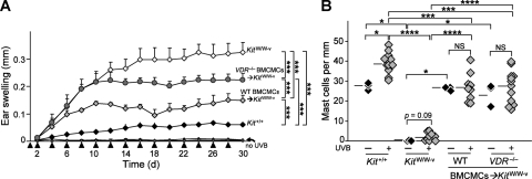
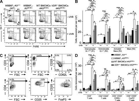
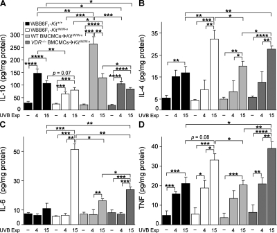
Mast cell production of interleukin-10 (IL-10) can limit the skin pathology induced by chronic low-dose ultraviolet (UV)-B irradiation. Although the mechanism that promotes mast cell IL-10 production in this setting is unknown, exposure of the skin to UVB irradiation induces increased production of the immune modifying agent 1alpha,25-dihydroxyvitamin D(3) (1alpha,25[OH](2)D(3)). We now show that 1alpha,25(OH)(2)D(3) can up-regulate IL-10 mRNA expression and induce IL-10 secretion in mouse mast cells in vitro. To investigate the roles of 1alpha,25(OH)(2)D(3) and mast cell vitamin D receptor (VDR) expression in chronically UVB-irradiated skin in vivo, we engrafted the skin of genetically mast cell-deficient WBB6F(1)-Kit(W/W-v) mice with bone marrow-derived cultured mast cells derived from C57BL/6 wild-type or VDR(-/-) mice. Optimal mast cell-dependent suppression of the inflammation, local production of proinflammatory cytokines, epidermal hyperplasia, and epidermal ulceration associated with chronic UVB irradiation of the skin in Kit(W/W-v) mice required expression of VDR by the adoptively transferred mast cells. Our findings suggest that 1alpha,25(OH)(2)D(3)/VDR-dependent induction of IL-10 production by cutaneous mast cells can contribute to the mast cell's ability to suppress inflammation and skin pathology at sites of chronic UVB irradiation.


Figures

Figure 1.
Figure 1.
1α,25(OH)2D3-induced IL-10 production by BMCMCs. (A) FACS analysis of WT (B6) BMCMCs and VDR−/− BMCMCs for VDR expression. (B) IL-10 in supernatants of 5-wk-old (B6J) BMCMCs cultured for 24 h with 0.1–1,000 nM 1α,25(OH)2D3. Data (n = 5 per group) are of mean values obtained in five different experiments (all measurements for each experiment were performed in duplicate). *, P < 0.05 or **, P < 0.01 for the indicated comparisons. See Fig. S1 B for data using VDR−/− BMCMCs.
Figure 2.
Figure 2.
Mast cell VDR expression helps to limit tissue swelling associated with chronic low-dose UVB irradiation. (A) Ear swelling after 15 exposures to 2 kJ/m2 of UVB (each arrowhead indicates a single exposure) in WT WBB6F1-Kit+/+ mice (black diamond, Kit+/+; n = 11), mast cell–deficient WBB6F1-KitW/W-v mice (open diamond, KitW/W-v; n = 9), and WBB6F1-KitW/W-v mice engrafted in the ear pinnae with WT C57BL/6-VDR+/+ BMCMCs (light gray diamond, WT BMCMC→KitW/W-v, n = 9) or VDR−/− BMCMCs (dark gray circle, VDR−/− BMCMC→KitW/W-v; n = 12). ***, P < 0.0001 for the indicated comparisons between groups of UVB-irradiated mice. Data (n = 9–12 per group) are from the three independent experiments performed (each with three to four mice per group), each of which gave similar results. Ear swelling measurements in ears not irradiated with UVB (“no UVB”) are highly overlapping for the four groups of mice; for each group, responses in UVB-irradiated ears were significantly different from those in the non-UVB–irradiated ears at P < 0.0001. (B) Numbers of dermal mast cells in ear pinnae after 15 exposures to 2 kJ/m2 of UVB. *, P < 0.05; **, P < 0.01; ***, P < 0.001; or ****, P < 0.0001, for the indicated comparisons. Data (3–12 mice per group) are from the three experiments performed, each of which gave similar results; each experiment analyzed one ear from a mouse not treated with UVB (– UVB) and single ears from three to four UVB-treated (+ UVB) mice.
Figure 3.
Figure 3.
Mast cells and mast cell VDR expression are required to limit pathology induced by chronic low-dose UVB irradiation of ear skin. Cross sections of ears obtained from WBB6F1-Kit+/+ (WT) mice (A–C), WBB6F1-KitW/W-v (KitW/W-v) mice (D–F), WT BMCMC→KitW/W-v mice (G–I), or VDR−/− BMCMC→KitW/W-v mice (J–L) at 24 h after the final exposure to 2 kJ/m2 UVB (15 exposures, 2 d apart; B, C, E, F, H, I, K, and L); sections stained with H&E (A, B, D, E, G, H, J, and K), or Toluidine blue (C, F, I, and L). Mice that did not receive UVB irradiation were also killed for analysis of skin histology at the end of the experiment (A, D, G, and J). C*, cartilage. Double-headed arrows indicate thickness of dermis (D) or epidermis (Ep). Red arrowheads indicate mast cells (C, F, I, and L). Bars: 100 µm (inset in A; for A–L); 1,000 µm (A; for insets in A, B, D, E, G, H, J, and L); 10 µm (C and I insets). Results are representative of those obtained in the three different experiments performed, each with three to four mice analyzed per group.
Figure 4.
Figure 4.
Mast cell VDR expression helps to limit leukocyte numbers in ear skin exposed to chronic low-dose UVB irradiation. Flow cytometric analysis of granulocytes and macrophages (A and B) and CD4+ and CD4+CD25+ T cells (C and D) 24 h after the last of 15 exposures to 2 kJ/m2 UVB irradiation (+ UVB) or at control sites not treated with UVB irradiation (– UVB) in ear skin of WBB6F1-Kit+/+ (WT) mice, mast cell–deficient WBB6F1-KitW/W-v (KitW/W-v) mice, WT BMCMC→KitW/W-v mice, and VDR−/− BMCMC→KitW/W-v mice. Percentage values in A and C refer to percentage of total viable cells present in the depicted section of the plot. (A) Representative FACS plots of granulocytes and macrophages in ears derived from individual mice. (B) Granulocytes (gated population R1, Gr-1hi; R2, Gr-1+) and macrophages (macs; R3 [“activated macrophages”], F4/80+ Gr-1hi; R4, F4/80+) recovered per ear. (C) Representative FACS plots of T cell populations in ears derived from individual mice, where live populations of CD3+ T cells were first gated and then CD4+CD25+CD62L T cells were selected, and where 4% paraformaldehyde-fixed CD4+ T cells were gated, and then CD25+ FoxP3+ T cells were selected. Percentages of gated T cell populations indicated are the percentage of the total cell population in that ear. (D) CD4+ T cell populations recovered per ear based on flow cytometric analysis of fixed cells (for CD4+CD25+FoxP3+ T cells) or live cells (the other three groups). (B and D) Data (n = 6–12 per group) are of mean values from 3 different experiments, each of which gave similar results, in which individual ears of three to four mice per group were analyzed in each experiment. *, P < 0.05; **, P < 0.01; ***, P < 0.001 for the indicated comparisons.
Figure 5.
Figure 5.
Ear skin cytokine levels 24 h after 4 or 15 UVB exposures. Levels of IL-10 (A), IL-4 (B), IL-6 (C), and TNF (D) in lysates of control ears not treated with UVB irradiation (– UVB) or ears treated with 4 or 15 exposures to 2 kJ/m2 UVB irradiation in WBB6F1-Kit+/+ (WT) mice, mast cell–deficient WBB6F1-KitW/W-v (KitW/W-v) mice, WT BMCMC→KitW/W-v mice, and VDR−/− BMCMC→KitW/W-v mice. Data (n = 6–8 per group) are from the two experiments performed, each of which gave similar results, in which three to four mice/group were tested per experiment. *, P < 0.05; **, P < 0.01; ***, P < 0.001; ****, P < 0.0001 for the indicated comparisons.

Similar articles

Cited by

References

    1. Alard P., Niizeki H., Hanninen L., Streilein J.W. 1999. Local ultraviolet B irradiation impairs contact hypersensitivity induction by triggering release of tumor necrosis factor-alpha from mast cells. Involvement of mast cells and Langerhans cells in susceptibility to ultraviolet B. J. Invest. Dermatol. 113:983–990 10.1046/j.1523-1747.1999.00772.x - DOI - PubMed
    1. Baroni E., Biffi M., Benigni F., Monno A., Carlucci D., Carmeliet G., Bouillon R., D’Ambrosio D. 2007. VDR-dependent regulation of mast cell maturation mediated by 1,25-dihydroxyvitamin D3. J. Leukoc. Biol. 81:250–262 10.1189/jlb.0506322 - DOI - PubMed
    1. Beissert S., Schwarz A., Schwarz T. 2006. Regulatory T cells. J. Invest. Dermatol. 126:15–24 10.1038/sj.jid.5700004 - DOI - PubMed
    1. Boonstra A., Barrat F.J., Crain C., Heath V.L., Savelkoul H.F., O’Garra A. 2001. 1α,25-Dihydroxyvitamin D3 has a direct effect on naive CD4+ T cells to enhance the development of Th2 cells. J. Immunol. 167:4974–4980 - PubMed
    1. Bouillon R., Carmeliet G., Verlinden L., van Etten E., Verstuyf A., Luderer H.F., Lieben L., Mathieu C., Demay M. 2008. Vitamin D and human health: lessons from vitamin D receptor null mice. Endocr. Rev. 29:726–776 10.1210/er.2008-0004 - DOI - PMC - PubMed

Publication types

MeSH terms