. 2010 Nov;20(11):2749-59.
doi: 10.1093/cercor/bhq023. Epub 2010 Mar 1.

Abnormal activation of the primary somatosensory cortex in spasmodic dysphonia: an fMRI study

Affiliations

Abnormal activation of the primary somatosensory cortex in spasmodic dysphonia: an fMRI study

Kristina Simonyan et al. Cereb Cortex. 2010 Nov.

Abstract

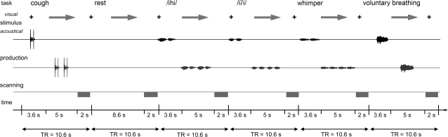
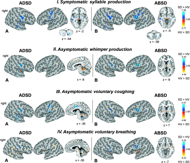
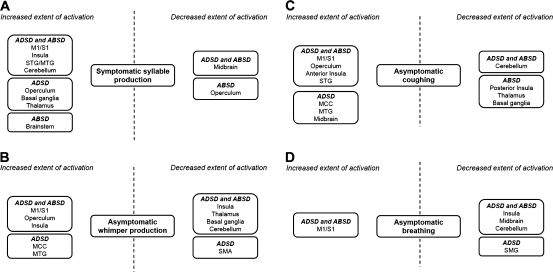
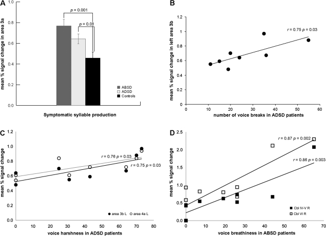
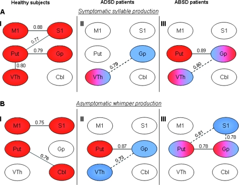
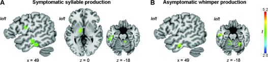
Spasmodic dysphonia (SD) is a task-specific focal dystonia of unknown pathophysiology, characterized by involuntary spasms in the laryngeal muscles during speaking. Our aim was to identify symptom-specific functional brain activation abnormalities in adductor spasmodic dysphonia (ADSD) and abductor spasmodic dysphonia (ABSD). Both SD groups showed increased activation extent in the primary sensorimotor cortex, insula, and superior temporal gyrus during symptomatic and asymptomatic tasks and decreased activation extent in the basal ganglia, thalamus, and cerebellum during asymptomatic tasks. Increased activation intensity in SD patients was found only in the primary somatosensory cortex during symptomatic voice production, which showed a tendency for correlation with ADSD symptoms. Both SD groups had lower correlation of activation intensities between the primary motor and sensory cortices and additional correlations between the basal ganglia, thalamus, and cerebellum during symptomatic and asymptomatic tasks. Compared with ADSD patients, ABSD patients had larger activation extent in the primary sensorimotor cortex and ventral thalamus during symptomatic task and in the inferior temporal cortex and cerebellum during symptomatic and asymptomatic voice production. The primary somatosensory cortex shows consistent abnormalities in activation extent, intensity, correlation with other brain regions, and symptom severity in SD patients and, therefore, may be involved in the pathophysiology of SD.


Figures

Figure 1.
Figure 1.
Schematic illustration of the experimental design. The subject fixated on the black cross and listened to the acoustically presented sample task for a 3.6-s period. Samples were pseudorandomized and presented as pairs of voluntary coughing, syllable and whimper production, and a single voluntary breathing. No stimulus was presented for resting control condition, during which the subject maintained normally paced breathing. A green arrow cued the subject to initiate the task production within a 5-s period, which was followed by a 2-s period of image acquisition.
Figure 2.
Figure 2.
(A,B) Significant differences in brain activation extent during production of symptomatic voice and asymptomatic laryngeal tasks in ADSD (IA, IIA, IIIA, and IVA) and ABSD (IB, IIB, IIIB, and IVB) patient groups compared with HV are presented on the inflated cortical surfaces; differences in the activation in the subcortical regions and cerebellum are shown in the series of axial or sagittal brain images of a single subject in the Talairach–Tournoux standard space (cluster size threshold ≥ 450 mm3 at a voxelwise threshold of P = 0.005). The color bar represents t-values (10 degrees of freedom) and reflects the significance of greater activation extent in SD patients compared with HV (SD > HV; dark blue to light blue) and the significance of greater activation extent in HV compared with SD patients (HV > SD; red to yellow). The coordinates are given in the RAI (right-anterior-inferior) conversion.
Figure 3.
Figure 3.
Block diagrams depicting abnormalities in brain activation extent that are common and distinct in ADSD and ABSD patients compared with healthy subjects during production of symptomatic syllable (A) and asymptomatic whimper (B), coughing (C), and breathing (D). M1/S1, ventral primary sensorimotor cortex; STG/MTG, superior and middle temporal gyrus; MCC, middle cingulate cortex; SMG, supramarginal gyrus.
Figure 4.
Figure 4.
(A) Bar graphs depicting significant differences in brain activation intensity in the primary somatosensory cortex (area 3a) during symptomatic syllable production between controls and patients with ADSD and ABSD. No significant differences in the intensity of activation between groups were observed during production of asymptomatic tasks. Error bars show standard error. (B) A trend toward a positive correlation between the mean percent signal change in the left primary somatosensory cortex (area 3b) and symptom severity (i.e., number of voice breaks) in ADSD patients. (C) Trends toward a positive correlation between voice harshness during symptomatic speech production and the mean percent signal change in the left primary somatosensory cortex (area 3b) and left primary motor cortex (area 4a) in ADSD patients. (D) Significant positive relationship between the mean percent signal change in the right cerebellum (IV–V and VI) and voice breathiness during symptomatic speech production in ABSD patients. R, right; L, left; Cbl, cerebellum.
Figure 5.
Figure 5.
Functional correlations within the sensorimotor cortical and subcortical regions involved in the control of voluntary voice production during symptomatic syllable and asymptomatic whimper production in healthy subjects (AI and BI), ADSD (AII and BII) and ABSD (AIII and BIII) patients. Red, significant correlations; blue, correlations not found in healthy subjects but significant in the patient groups. Solid lines show the statistically significant correlations present in all groups; dotted lines show statistically significant correlations present only in patients. M1, primary motor cortex; S1, primary somatosensory cortex; Put, putamen; VTh, ventral thalamus; Gp, globus pallidus; Cbl, cerebellum.
Figure 6.
Figure 6.
Significant differences in brain activation extent during production of symptomatic syllable (A) and asymptomatic whimper (B) in ABSD patients compared with ADSD patients are shown in the series of axial or sagittal brain images of a single subject in the Talairach–Tournoux standard space (cluster size threshold ≥ 450 mm3 at a voxelwise threshold of P = 0.005). The color bar indicates the t-values (10 degrees of freedom). The coordinates are given in the RAI (right-anterior-inferior) conversion.

Similar articles

Cited by

References

    1. Ali SO, Thomassen M, Schulz GM, Hosey LA, Varga M, Ludlow CL, Braun AR. Alterations in CNS activity induced by botulinum toxin treatment in spasmodic dysphonia: an H215O PET study. J Speech Lang Hear Res. 2006;49:1127–1146. - PubMed
    1. Andrew J, Fowler CJ, Harrison MJ. Stereotaxic thalamotomy in 55 cases of dystonia. Brain. 1983;106(Pt 4):981–1000. - PubMed
    1. Arbib M. Preceptual structures and distributed motor control. In: Brookhart JMMV, editor. Handbook of physiology. Motor control. Bethesda, MD: Americal Physiological Society; 1981. pp. 1449–1480.
    1. Barkmeier JM, Case JL, Ludlow CL. Identification of symptoms for spasmodic dysphonia and vocal tremor: a comparison of expert and nonexpert judges. J Commun Disord. 2001;34:21–37. - PubMed
    1. Birn RM, Bandettini PA, Cox RW, Shaker R. Event-related fMRI of tasks involving brief motion. Hum Brain Mapp. 1999;7:106–114. - PMC - PubMed

Publication types