. 2010 Apr 1;184(7):3450-60.
doi: 10.4049/jimmunol.0903831. Epub 2010 Mar 1.

CXC chemokine ligand (CXCL) 9 and CXCL10 are antagonistic costimulation molecules during the priming of alloreactive T cell effectors

Affiliations

CXC chemokine ligand (CXCL) 9 and CXCL10 are antagonistic costimulation molecules during the priming of alloreactive T cell effectors

Joshua M Rosenblum et al. J Immunol. .

Abstract

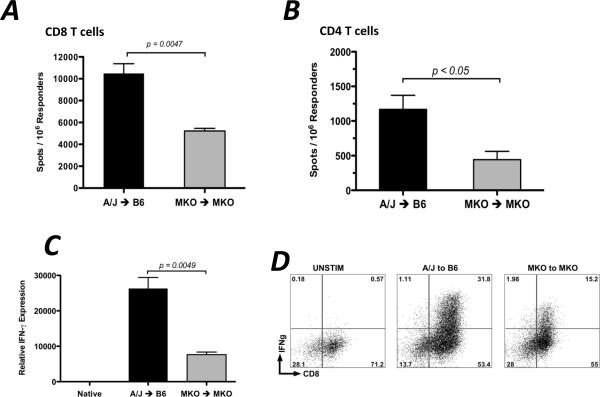
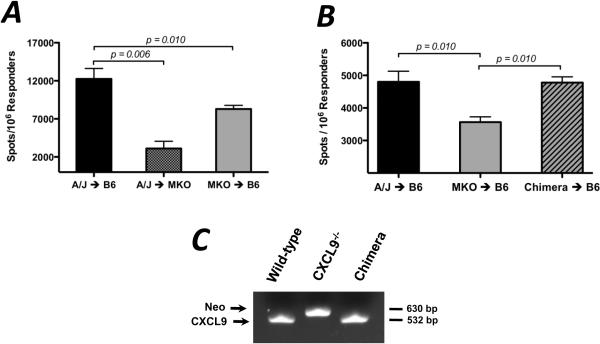
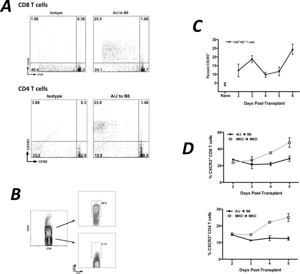
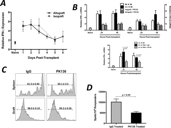
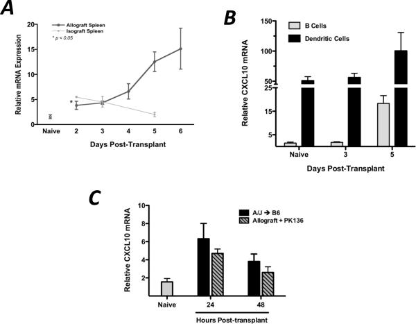
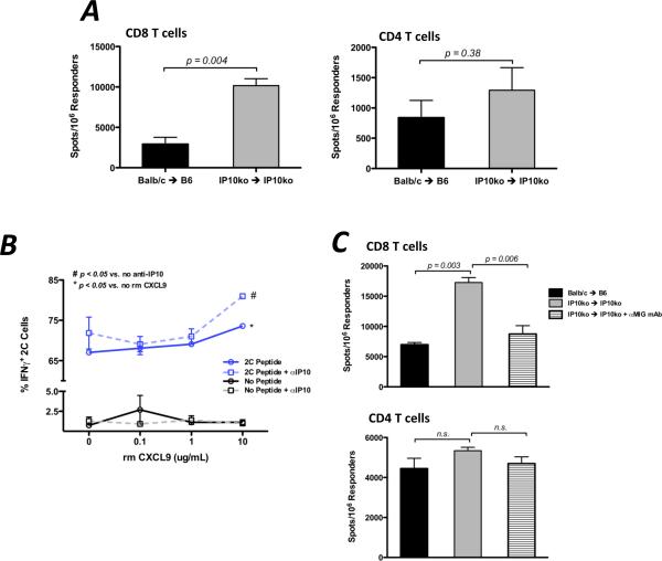
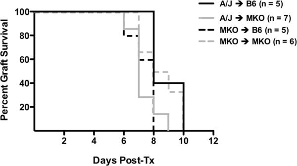
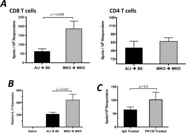
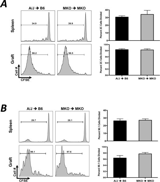
Donor Ag-reactive CD4 and CD8 T cell production of IFN-gamma is a principal effector mechanism promoting tissue injury during allograft rejection. The CXCR3-binding chemokines CXCL9 and CXCL10 recruit donor-reactive T cells to the allograft, but their role during the priming of donor-reactive T cells to effector function is unknown. Using a murine model of MHC-mismatched cardiac transplantation, we investigated the influence of CXCL9 and CXCL10 during donor-reactive T cell priming. In allograft recipient spleens, CXCL9 and CXCL10 were expressed as early as 24 h posttransplant and increased with similar kinetics, concurrently with CXCR3 expression on T cells. CXCL9, but not CXCL10, expression required NK cell production of IFN-gamma. The absence of CXCL9 in donor allografts, recipients, or both significantly decreased the frequency of donor-reactive CD8 T cells producing IFN-gamma and increased the frequency of donor-reactive CD8 T cells producing IL-17A. In contrast, the absence of CXCL10 increased the frequency of IFN-gamma-producing CD8 T cells in a CXCL9-dependent manner. These data provide novel evidence that donor-reactive CD8 T cells use the CXCR3 chemokine axis as a costimulation pathway during priming to allografts where CXCL9 promotes the development of IFN-gamma-producing CD8 T cells, and CXCL10 antagonizes this skewing.


Figures

Figure 1
Figure 1. CXCL9 is required for maximum generation of donor-reactive, IFN-γ-producing CD8 T cells
Wild-type or CXCL9−/− B6 mice received wild-type or CXCL9−/− A/J heart allografts. Recipient spleens were harvested on day 7 post-transplant and purified CD8 (A) or CD4 (B) T cells producing IFN-γ were enumerated by ELISPOT assay. Data is representative of at least 3 independent experiments. (C) mRNA was purified from total graft homogenates on day 7 post-transplant. Quantitative RT-PCR analysis was performed on 5–6 samples/group. Expression of Mrpl32 was used as the endogenous control, and expression of IFN-γ in each sample was normalized to the expression in a random native heart sample. (D) Graft-infiltrating cells were purified on day 7 post-transplant and were re-stimulated in vitro with PMA/Ionomycin for 4 hr with monensin for the last 2 hr. Intracellular cytokine staining was performed using standard techniques and reagents. Numbers in each plot are percentages of total lymphocytes, and plots represent 4–5 samples per group. (E) Allografts were harvested at the times indicated and the number of graft-infiltrating CD8 T cells was quantitated using flow cytometry. Bars in each panel represent mean ± SEM; n = 5–6/group, * p < 0.05, ** p < 0.01.
Figure 2
Figure 2. Donor- and recipient-derived CXCL9 influence the development of donor-reactive IFN-γ-producing CD8 T cells
(A) Wild-type or CXCL9−/− B6 mice received wild-type or CXCL9−/− A/J heart allografts as shown. Recipient spleens were harvested on day 7 post-transplant and purified CD8 T cells producing IFN-γ were enumerated by ELISPOT assay. Data is representative of at least 3 independent experiments; n = 4–5/group, * p < 0.05, ** p < 0.01. (B) Chimerism of donor grafts in B was confirmed by PCR analysis of DNA from PBMCs collected from mice just prior to transplant. Data is representative of four mice per group. (C) CXCL9−/− A/J mice were irradiated and wild-type A/J bone marrow was adoptively transferred; mice were then rested for 8 weeks. These chimeric mice were then used as heart donors to wild-type B6 mice. CD8 T cells purified from recipient spleens on day 7 post-transplant, and IFN-γ-producing cells were enumerated using ELISPOT. Data is representative of 2 independent experiments; n = 3–4/group, ** p < 0.01.
Figure 3
Figure 3. CXCL9 is produced in the graft recipient spleen as early as 48 hr post-transplant, primarily by CD11c+ DCs
(A) Wild-type isograft and allograft recipient spleens were harvested at the times post-transplant shown. Total mRNA was purified from spleen homogenates, and expression of IFN-γ mRNA was normalized to a random naïve spleen sample. n = 5–6/group, representative of three independent experiments. (B) B cells (CD19+CD11c) or dendritic cells (CD19CD11c+) were flow sorted from recipient spleens at various times post-transplant. mRNA was prepared from the purified cell populations, and IFN-γ expression was normalized to a random naïve B cell sample. n = 5–6/group; mean ± SEM. (C) Total recipient spleen homogenates were analyzed for CXCL9 mRNA expression by qRT-PCR. Bars represent mean ± SEM; n = 4–5/group, * p < 0.05.
Figure 4
Figure 4. CXCR3 is expressed in the recipient spleen by proliferating, activated T cells with kinetics similar to CXCL9 expression
(A) 2.5×10⁶ 2C CD8 T cells were adoptively transferred to wild-type B6 recipients of A/J heart allografts one day prior to transplant. Recipient spleens were harvested on the days indicated and CXCR3 expression on 2C CD8 T cells was measured by flow cytometry. Data represents percent of total 2C CD8 T cells expressing CXCR3; n = 3–4/time point; mean ± SEM. (B) The percentage of endogenous polyclonal CD8 (top panel) or CD4 (bottom panel) T cells expressing CXCR3 was determined at the times post transplant shown. Data are representative of two independent experiments; n = 3–4/time point; mean ± SEM. (C) 2.5×10⁶ CD8 or CD4 T cells were CFSE labeled and were transferred to wild-type B6 recipients of A/J heart allografts one day prior to transplant. Recipient spleens were harvested on day 5 post-transplant, and CXCR3 expression on CFSE labeled proliferating cells was measured by flow cytometry. Numbers represent percentage of total transferred cells in each quadrant; plots are representative of three mice per group. (D) Spleens from wild-type B6 recipients of wild-type A/J cardiac allografts were harvested on day 5 post-transplant, and cells were processed using standard techniques for flow cytometry. Numbers represent percentage of total CD8 T cells expressing CXCR3; plots are representative of 4 individual mice.
Figure 5
Figure 5. Recipient NK cells produce IFN-γ during the first 48hr post-transplant in the recipient spleen
(A) Wild-type isograft or allograft recipient spleens were harvested at the times post-transplant shown. Total mRNA was purified from spleen homogenates and IFN-γ mRNA expression was determined by qRT-PCR; n = 4–6/group, mean ± SEM. (B and C) Wild-type (B) or RAG1−/− (C) isograft or allograft recipients were treated with control IgG or with NK-depleting mAb (PK136, 250ug d −3, −2, −1, 1, i.p.). Total mRNA was purified from spleen homogenates at the times shown. qRT-PCR analysis was performed as in (A); n = 4–5/group, mean ± SEM, * p < 0.05, ** p < 0.01. (D) Wild-type B6 allograft recipients (n = 3/group) were treated with control IgG or PK136 (250ug d −3, −2, −1, 1, 3, 5; i.p.). Recipient spleens were harvested on day 7 post-transplant and the number of purified CD8 T cells producing IFN-γ were enumerated by ELISPOT assay. Data is representative of two independent experiments, mean ± SEM, * p < 0.05.
Figure 6
Figure 6. CXCL10/IP10 is produced in the allograft recipient spleen by DCs and B cells, independent of NK cells
(A) mRNA expression of CXCL10 was determined in allograft and isograft recipient spleens at various times post-transplant (n = 3–4/group). Data was analyzed as in 3A. (B) B cells and DCs were flow-sort purified as in 3B and CXCL10 mRNA expression was determined as described. (C) Wild-type allograft recipients were depleted of NK cells as described, and total spleen homogenates were taken at the times indicated for qRT-PCR analysis of CXCL10 expression. Data represent two individual experiments, n = 3–4/group; mean ± SEM.
Figure 7
Figure 7. CXCL10/IP10 antagonizes the action of CXCL9 during priming of donor-reactive, IFN-γ-producing CD8 T cells
(A) Spleens from wild-type or CXCL10−/− B6 recipients of wild-type or CXCL10−/− Balb/c heart allografts were harvested on day 7 post-transplant. CD8 or CD4 T cells were purified and the number of donor-specific cells producing IFN-γ was quantified using ELISPOT. (B) Flow sort purified naïve 2C CD8 T cells were cocultured with B6 spleen cells pulsed or not pulsed with 2C peptide for 3 days in the presence of anti-CXCL10 mAb (5 ug/mL) or control Ab and increasing amounts of recombinant CXCL9 as shown. 2C cells producing IFN-γ were then determined by intracellular cytokine staining. Data represents two independent experiments, 3 replicates/group; mean ± SEM. (C) CXCL10−/− allograft combinations were treated with control IgG or with anti-CXCL9 mAb (150ug, i.p., d1, 3, 5). The number of IFN-γ-producing CD4 or CD8 T cells was enumerated by ELISPOT on day 7 post-transplant. Data represents two independent experiments, n = 3/group; mean ± SEM, * p < 0.05, ** p < 0.01.
Figure 8
Figure 8. CXCL9 does not influence the proliferation of donor-reactive CD8 or CD4 T cells in vivo
(A) 5×10⁶ Ld-reactive naïve 2C transgenic CD8 T cells were labeled with CFSE and were transferred to wild-type or CXCL9−/− B6 mice one day prior to transplant with wild-type or CXCL9−/− A/J heart grafts. Grafts and recipient spleens were harvested on day 5 post-transplant, and CFSE dilution was measured by standard flow cytometry techniques. Histograms are representative of 4–6 mice per group, and graphs represent percent of 2C CD8 T cells that have divided. (B) 10×106 CD90.1 CD4 T cells were labeled with CFSE and were adoptively transferred to CD90.2 mice one day prior to transplant. Proliferation of CD4 T cells was determined at day 5 post-transplant as in (A). (C) 5×10⁶ CFSE labeled 2C CD8 T cells were adoptively transferred to wild-type B6 allograft recipients treated with control IgG or PK136 one day prior to transplant. Proliferation of 2C cells in the recipient spleens and grafts was determined by flow cytometry on day 5 post-transplant. Histograms are representative of 3 mice per group; numbers represent mean ± SEM percent of total 2C cells that have divided.
Figure 9
Figure 9. In the absence of CXCL9, donor-specific production of IL-17 by CD8 T cells is increased
(A) Wild-type or CXCL9−/− B6 mice received wild-type or CXCL9−/− A/J heart allografts, and CD8 or CD4 T cells were purified from recipient spleens on day 7 post-transplant. The number of donor-specific cells producing IL-17 was enumerated using ELISPOT. Data represents 3 independent experiments. n = 5–6/group, mean ± SEM, * p < 0.05. (B) Total mRNA was purified from graft homogenates at day 7 post-transplant. IL-17 mRNA expression was normalized to endogenous Mrpl32 expression using a random native heart sample as the baseline. Data represents 2 independent experiments.
Figure 10
Figure 10

Similar articles

Cited by

References

    1. Heeger PS. T-cell allorecognition and transplant rejection: a summary and update. Am J Transplant. 2003;3:525–533. - PubMed
    1. Hall BM, Dorsch SE. Cells mediating allograft rejection. Immunol Rev. 1984;77:31–59. - PubMed
    1. Hidalgo LG, Halloran PF. Role of IFN-gamma in allograft rejection. Crit Rev Immunol. 2002;22:317–349. - PubMed
    1. Larsen CP, Morris PJ, Austyn JM. Migration of dendritic leukocytes from cardiac allografts into host spleens. A novel pathway for initiation of rejection. J Exp Med. 1990;171:307–314. - PMC - PubMed
    1. Trinchieri G. Interleukin-12: a proinflammatory cytokine with immunoregulatory functions that bridge innate resistance and antigen-specific adaptive immunity. Annu Rev Immunol. 1995;13:251–276. - PubMed

Publication types

MeSH terms