. 2010 Mar 16;107(11):5018-23.
doi: 10.1073/pnas.0913485107. Epub 2010 Mar 1.

Drosophila parkin requires PINK1 for mitochondrial translocation and ubiquitinates mitofusin

Affiliations

Drosophila parkin requires PINK1 for mitochondrial translocation and ubiquitinates mitofusin

Elena Ziviani et al. Proc Natl Acad Sci U S A. .

Abstract

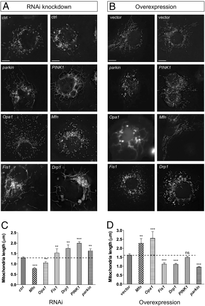
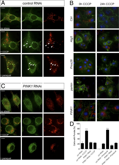
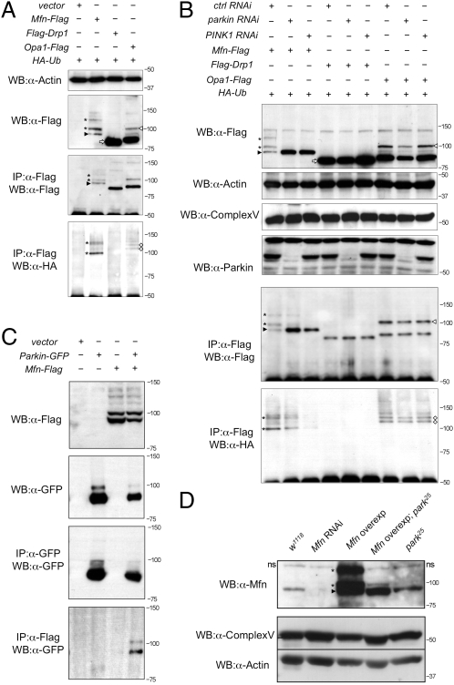
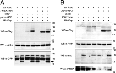
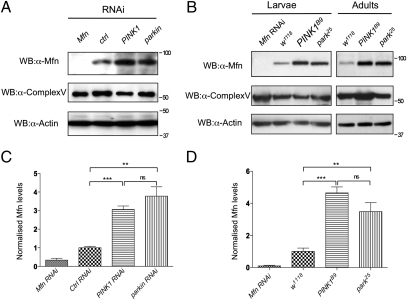
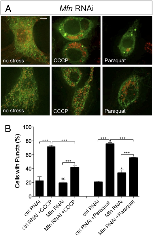
Loss of the E3 ubiquitin ligase Parkin causes early onset Parkinson's disease, a neurodegenerative disorder of unknown etiology. Parkin has been linked to multiple cellular processes including protein degradation, mitochondrial homeostasis, and autophagy; however, its precise role in pathogenesis is unclear. Recent evidence suggests that Parkin is recruited to damaged mitochondria, possibly affecting mitochondrial fission and/or fusion, to mediate their autophagic turnover. The precise mechanism of recruitment and the ubiquitination target are unclear. Here we show in Drosophila cells that PINK1 is required to recruit Parkin to dysfunctional mitochondria and promote their degradation. Furthermore, PINK1 and Parkin mediate the ubiquitination of the profusion factor Mfn on the outer surface of mitochondria. Loss of Drosophila PINK1 or parkin causes an increase in Mfn abundance in vivo and concomitant elongation of mitochondria. These findings provide a molecular mechanism by which the PINK1/Parkin pathway affects mitochondrial fission/fusion as suggested by previous genetic interaction studies. We hypothesize that Mfn ubiquitination may provide a mechanism by which terminally damaged mitochondria are labeled and sequestered for degradation by autophagy.


Conflict of interest statement

The authors declare no conflict of interest.

Figures

Fig. 1.
Fig. 1.
Drosophila PINK1 and parkin activity promotes mitochondrial fission and/or inhibits fusion. (A) S2R+ cells were treated with the indicated RNAi probe for 3 days. Rhodamine 123 was used to visualize mitochondrial morphology in live cells. (B) S2R+ cells were transfected with plasmids to express indicated genes and cotransfected with mitoGFP to visualize mitochondria. (C and D) Quantification of mitochondrial length as imaged in A and B, respectively. Two control (ctrl) RNAi and vector-only treatments are shown to demonstrate phenotype variability. (Scale bar: 5 μm.) The bar graphs represent the mean and sem for at least three independent experiments. Significance was determined by one-way ANOVA with Bonferroni correction (**P < 0.01, ***P < 0.001).
Fig. 2.
Fig. 2.
PINK1 is required for Parkin translocation and mitophagy. (A) S2R+ cells cotransfected with parkin-GFP and mitoDsRed, treated with control dsRNA and then treated with CCCP or paraquat and fixed for imaging. Most Parkin-GFP puncta colocalize with (arrow) or abut (arrowhead) mitochondria. (B) Cells were treated with indicated dsRNAs and exposed to CCCP for 24 h. Samples were fixed and stained to label mitochondria (anti-Complex Vα, red), actin (phalloidin-488, green) or nuclei (Hoechst, blue). (C) S2R+ cells transfected as in A and treated with PINK1 dsRNA before exposure to CCCP or paraquat. Comparison of Parkin-GFP distribution under different image analysis methods is shown in Fig. S5. (D) Percentage of cells with Parkin-GFP puncta. (Scale bar: 5 μm.) The bar graphs represent the mean and SEM of at least four independent tests. Significance was determined by one-way ANOVA with Bonferroni correction (***P < 0.001).
Fig. 3.
Fig. 3.
Mfn is ubiquitinated in a PINK1/Parkin-dependent manner in vitro and in vivo. (A) S2R+ cells cotransfected with HA-Ub plus empty vector or Flag-tagged Mfn, Drp1, or Opa1 plasmids as indicated were immunoprecipitated and subjected to Western blot analysis. (B) S2R+ cells were treated with control, parkin, and PINK1 RNAi before being transfected as indicated. Cells were harvested and subjected to Western blot analysis as shown. Samples were also subjected to immunoprecipitation, and Western bots were probed with antibodies against Flag and HA. (C) Immunoprecipitates of S2R+ cells expressing combinations of empty vector, parkin-GFP, and Mfn-Flag as shown. (D) Western blot analysis of Drosophila Mfn levels in vivo. A nonspecific band seen in all samples is denoted “ns.” Complex Vα and actin are used as loading controls. Genotypes: w1118, wild type; Mfn RNAi, da-GAL4,UAS-Mfn-RNAi; Mfn overexpression, da-GAL4,UAS-Mfn; Mfn overexpression park25, da-GAL4,UAS-Mfn,park25/park25; park25, park25/park25. In all panels, black arrowheads indicate full-length Mfn, asterisks are ubiquitinated Mfn, white arrowheads denote full-length Opa1, white diamonds are ubiquitinated Opa1, and white arrows show full-length Drp1.
Fig. 4.
Fig. 4.
Parkin overexpression can cause Mfn ubiquitination in the absence of PINK1. (A) S2R+ cells were treated with control or PINK1 RNAi probe and transfected with combinations of empty vector, parkin-GFP, and Mfn-Flag, as shown. (B) S2R+ cells were treated with control or parkin RNAi probe and transfected with combinations of empty vector, PINK1-myc, and Mfn-Flag, as shown. Cells were harvested and subjected to Western blot analysis using the specific antibodies indicated.
Fig. 5.
Fig. 5.
Mfn accumulates in the absence of PINK1/Parkin. (A) S2R+ cells were treated with RNAi probe as indicated, harvested, and subjected to Western blot analysis by using antibodies against Drosophila Mfn. Antibodies against Complex Vα and actin are used as loading controls. (B) Wild-type and mutant animals were collected and subjected to Western blot analysis. Genotypes: w1118, wild type; Mfn RNAi, da-GAL4,UAS-Mfn-RNAi; park25, park25/park25; PINK1B9, PINK1B9/Y. Quantification of Mfn levels relative to the Complex Vα loading control in S2R+ cells (C) and wild-type and mutant animals (D). Charts represent mean and SEM for three independent experiments. Significance was determined by one-way ANOVA with Bonferroni correction (**P < 0.01, ***P < 0.001).
Fig. 6.
Fig. 6.
Loss of Mfn partially impairs Parkin translocation to mitochondria. S2R+ cells were cotransfected with plasmids expressing parkin-GFP and mitoDsRed and treated with control RNAi probe or Mfn RNAi probe before being treated with CCCP or paraquat and analyzed for Parkin-GFP accumulation. Control RNAi-treated cells are represented in Fig. 2. (A) Mfn RNAi-treated cells exposed to CCCP, paraquat, or no stress. Two representative images are shown for each treatment to show variability. (Scale bar: 5 μm.) (B) Percentage of cells with green puncta. The chart shows the mean and SEM for counting of at least four independent well fields containing 70–90 cells/field. Significance was determined by one-way ANOVA with Bonferroni correction (*P < 0.05, ***P < 0.001).

References

    1. Gasser T. Molecular pathogenesis of Parkinson disease: Insights from genetic studies. Expert Rev Mol Med. 2009;11:e22. - PubMed
    1. Silvestri L, et al. Mitochondrial import and enzymatic activity of PINK1 mutants associated to recessive parkinsonism. Hum Mol Genet. 2005;14:3477–3492. - PubMed
    1. Shimura H, et al. Familial Parkinson disease gene product, parkin, is a ubiquitin-protein ligase. Nat Genet. 2000;25:302–305. - PubMed
    1. Büeler H. Impaired mitochondrial dynamics and function in the pathogenesis of Parkinson's disease. Exp Neurol. 2009;218:235–246. - PubMed
    1. Clark IE, et al. Drosophila pink1 is required for mitochondrial function and interacts genetically with parkin. Nature. 2006;441:1162–1166. - PubMed

Publication types