. 2010 Jun 15;181(12):1355-66.
doi: 10.1164/rccm.200901-0015OC. Epub 2010 Mar 1.

Induction of tissue factor by urokinase in lung epithelial cells and in the lungs

Affiliations

Induction of tissue factor by urokinase in lung epithelial cells and in the lungs

Sreerama Shetty et al. Am J Respir Crit Care Med. .

Abstract

Rationale: Urokinase-type plasminogen activator (uPA) regulates extracellular proteolysis in lung injury and repair. Although alveolar expression of uPA increases, procoagulant activity predominates.

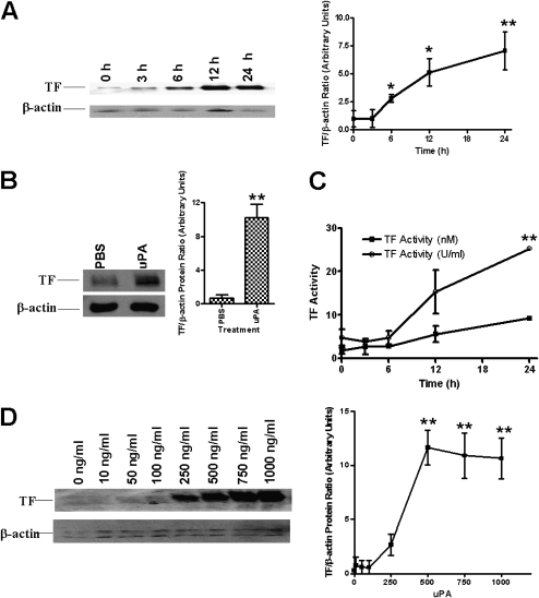
Objectives: This study was designed to investigate whether uPA alters the expression of tissue factor (TF), the major initiator of the coagulation cascade, in lung epithelial cells (ECs).

Methods: Bronchial, primary airway ECs and C57B6 wild-type, uPA-deficient (uPA(-/-)) mice were exposed to phosphate-buffered saline, uPA, or LPS. Immunohistochemistry, protein, cellular, and molecular techniques were used to assess TF expression and activity.

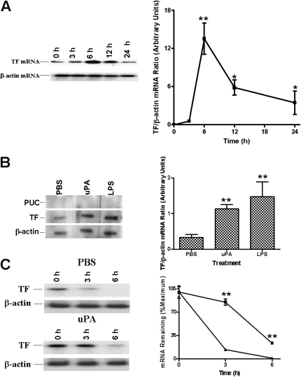
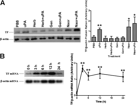
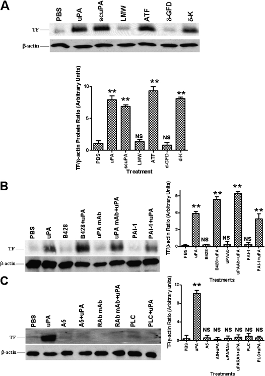
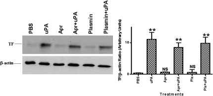
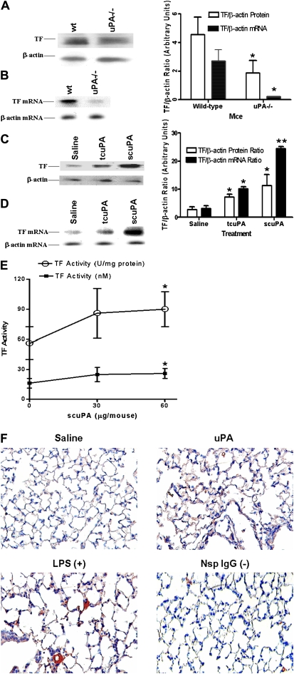
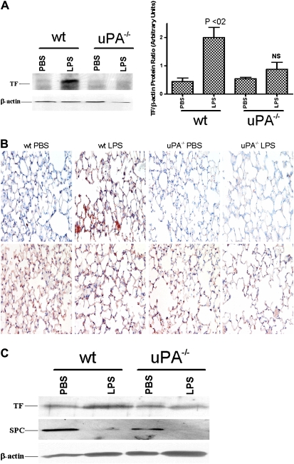
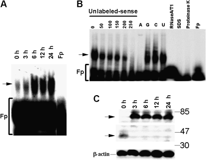
Measurements and main results: uPA enhanced TF mRNA and protein expression, and TF-dependent coagulation in lung ECs. uPA-induced expression of TF involves both increased synthesis and enhanced stabilization of TF mRNA. uPA catalytic activity had little effect on induction of TF. By contrast, deletion of the uPA receptor binding growth factor domain from uPA markedly attenuated the induction of TF, suggesting that uPA receptor binding is sufficient for TF induction. Lung tissues of uPA-deficient mice expressed less TF protein and mRNA compared with wild-type mice. In addition, intratracheal instillation of mouse uPA increased TF mRNA and protein expression and accelerated coagulation in lung tissues. uPA(-/-) mice exposed to LPS failed to induce TF.

Conclusions: uPA increased TF expression and TF-dependent coagulation in the lungs of mice. We hypothesize that uPA-mediated induction of TF occurs in lung ECs to promote increased fibrin deposition in the airways during acute lung injury.


Figures

Figure 1.
Figure 1.
Time- and concentration-dependent induction of tissue factor (TF) expression by two-chain urokinase-type plasminogen activator (tcuPA) in lung epithelial cells (ECs). (A) Human bronchial ECs (Beas2B) were incubated with or without recombinant human tcuPA (1 μg/ml) for 0 to 24 hours. Total protein (3 μg/lane) from the cell lysates were immunoblotted with anti-TF antibody. Same membranes were stripped and later developed with anti–β-actin monoclonal antibody to assess loading. The data illustrated are integrated from at least four independent experiments, and mean densities of the individual bands are presented in the line graph. (B) Analysis of TF protein expression in primary small airway ECs (SAECs) treated with phosphate-buffered saline or tcuPA for 24 hours. Total lysates (3 μg protein/lane) from SAECs were analyzed for TF expression by Western blotting. Total protein (25 μg/lane) from the same samples were blotted with β-actin monoclonal antibody for assessment of loading. The mean and SD of densitometric values of individual bands of three experiments from B are represented as a bar graph. (C) Beas2B cells incubated with tcuPA as described in A were lysed in Hanks' balanced salt solution and analyzed for TF activity using a single-stage clotting assay. The experiments were repeated at least three times and the results are shown as nM TF and U/ml determined as described in Methods. (D) Beas2B cells grown to confluence were incubated with 0 to 1 μg/ml of tcuPA for 24 hours. The total proteins from cell lysates were subjected to immunoblotting as described in A. The blots are representative of three separate experiments. The figure (line graph) shows the mean band densities ± SD of four independent experiments. The differences in TF are significant (*P < 0.005 or **P < 0.001) when compared with its level at 0 hours or 0 ng/ml.
Figure 2.
Figure 2.
Regulation of tissue factor (TF) mRNA expression by two-chain urokinase-type plasminogen activator (tcuPA) in human bronchial epithelial cells (Beas2B). (A) Time-dependent induction of TF mRNA by tcuPA. The cells were treated as described in the legend to Figure 1. Total RNA (20 μg/lane) was subjected to Northern blotting using 32P-labeled TF cDNA. Same blots were stripped and probed for β-actin mRNA. The blot is representative of three separate experiments. The line graph depicts the integrated data (mean ± SD) of four individual experiments. (B) Effect of tcuPA on the rate of transcription of TF mRNA. Nuclei isolated from Beas2B cells, incubated with phosphate-buffered saline (PBS) or tcuPA for 3 hours as described in Figure 1, were subjected to the transcription reaction in the presence of [32P]UTP at 30°C for 30 minutes. 32P-labeled nuclear RNA was hybridized with TF cDNA immobilized on nitrocellulose membrane. β-actin and plasmid from the University of California cDNAs were used as positive and negative loading controls, respectively. (C) Effect of tcuPA on TF mRNA decay stability. Beas2B cells were incubated with PBS or tcuPA for 6 hours, after which 5,6-dichloro-1-β-D-ribofuranosylbenz-imidazole (20 μg/ml) was added for various periods of time. TF mRNA was analyzed by Northern blotting. Same membranes were stripped and developed for β-actin mRNA. In B and C the blots are representative of three separate experiments. The bar and line graphs show the mean and SD of integrated data from three independent experiments. The increased expression of TF mRNA is significant (*P < 0.05 or **P < 0.001) when compared with its level at 0 hours.
Figure 3.
Figure 3.
Effect of tyrosine kinase and phosphatase inhibitors on two-chain urokinase-type plasminogen activator (tcuPA)-mediated tissue factor (TF) expression. (A) Human bronchial epithelial cells (Beas2B) grown to confluence were treated with or without herbimycin A (Herb), genistein (Gen), and sodium orthovanadate (Naor) for 2 hours followed by tcuPA (1 μg/ml) for 24 hours at 37°C and subjected to immunoblotting with anti-TF and β-actin antibodies as described in Figure 1B. (B) Beas2B cells treated with sodium orthovanadate for 0 to 24 hours were analyzed for the expression of TF mRNA by Northern blotting. Same membranes were stripped and developed for β-actin mRNA. In A and B the blots are representative of four and three experiments, respectively, and the bar graphs illustrate the mean band densities ± SD of three independent experiments. The increase in the level of TF expression is significant (*P < 0.05 or **P < 0.001) when compared with its level in cells treated with phosphate-buffered saline or Naor at 0 hours.
Figure 4.
Figure 4.
Effect of urokinase-type plasminogen activator (uPA) enzymatic activity and its receptor interaction on tissue factor (TF) expression in human bronchial epithelial cells (Beas2B). (A) Effect of different fragments of uPA on TF expression. Beas2B cells were incubated with phosphate-buffered saline (PBS) or 1 μg/ml each of active two-chain uPA (tcuPA), single chain uPA (scuPA), the amino-terminal fragments (ATF) or low molecular weight (LMW) fragments of uPA, scuPA growth factor domain deletion (δ-GFD), or kringle domain deletion (δ-K) mutant for 24 hours. Cellular proteins (3 μg and 25 μg/lane) were immunoblotted as described above (Figure 1B) with anti-TF and anti–β-actin antibodies, respectively. The data illustrated are representative of the findings of four independent experiments. Composite densitometric analyses of the effect of deletion fragments of uPA on TF induction in Beas2B cells shown as the mean ± SD of four experiments. (B) Effect of uPA enzymatic activity on TF expression. Beas2B cells were treated with or without B-428 (0.02 mM), anti-uPA monoclonal antibody (uPA mAb) (2 μg/ml), plasminogen activator inhibitor-1 (4 μg/ml) for 2 hours followed by tcuPA (1 μg/ml) for 24 hours at 37°C in a basal media, and the cell lysates were analyzed for TF and β-actin expression as described in A. (C) Effect of uPA interaction with its receptor on TF expression. Beas2B cells were incubated with or without anti-uPAR antibody (RAb, 2 μg/ml), A5 (1 μg/ml) compound, or phosphatidylinositol-phospholipase C (10 units/ml) for 2 hours followed by uPA (1 μg/ml) for 24 hours at 37°C in basal medium. The cell lysates (3 μg and 25 μg total protein/lane) were analyzed for the expression of TF and β-actin, respectively, by Western blotting using anti-TF and β-actin antibodies. In B and C, the blots are representative of three independent experiments and bar graphs show the data from those experiments expressed as the mean ± SD. The increased level of TF expression is significant (**P < 0.001) when compared with phosphate-buffered saline (PBS)-treated cells. NS = not significant compared with PBS-treated controls.
Figure 5.
Figure 5.
Effect of inhibitors of two-chain urokinase-type plasminogen activator (tcuPA) activity on uPA-mediated tissue factor (TF) expression by human bronchial epithelial cells (Beas2B). Beas2B cells were incubated with or without uPA, aprotinin (1 μg/ml), or plasmin (1 μg/ml) alone or in combination with uPA for 24 hours at 37°C. Total proteins from cell lysates were isolated and subjected to immunoblotting as described in Figure 4. The blots are representative of four to five independent experiments and bar graphs show the data from those experiments expressed as the mean ± SD. The increased expression of TF is significant (**P < 0.001) when compared with its level in phosphate-buffered saline (PBS)-treated cells. Apr = aprotinin; NS = not significant compared with PBS treated controls.
Figure 6.
Figure 6.
Effect of urokinase-type plasminogen activator (uPA) on tissue factor (TF) expression by lung tissues in vivo. (A) The lung lysates (20 μg/lane) of wt and uPA-deficient (uPA−/−) mice were analyzed for TF protein expression by Western blotting (three mice per group). The same membranes were stripped and analyzed for β-actin to assess equal loading. (B) Total RNA isolated from lung homogenates of wt and uPA-deficient mice were analyzed for TF mRNA (three mice per group). (C) Saline or uPA (two-chain [tc] uPA or single-chain [sc] uPA) was injected intratracheally into wt mice and the lung lysates (20 μg/ lane) were analyzed for tissue factor expression by Western blotting as described in A (three mice per group). (D) Total RNA isolated from lung homogenates of mice treated with saline or uPA were analyzed for expression of TF and β-actin mRNAs by Northern blotting. The bar represents densitometric values (mean ± SD) of three independent experiments. (E) The lung tissues of saline and scuPA-treated wt mice (n = 3–6) as described in D were lysed in Hanks' balanced salt solution and analyzed for TF activity using a single-stage clotting assay. The results are shown as nM TF and U/ml determined as described in Methods. The asterisks represent differences that are significant (*P < 0.05 and **P < 0.001) versus the saline-treated control. (F) Immunohistochemical staining for TF antigen was performed on lung tissue (5 μm) from mice exposed to saline or murine scuPA using anti-TF antibody. Plates are representative fields from three mice for each condition at 200× magnification. Lung sections from LPS-exposed mice incubated with an anti-TF antibody were used as a positive control (+) and sections incubated with nonimmune rabbit IgG are shown as the negative control (−).
Figure 7.
Figure 7.
Role of urokinase-type plasminogen activator (uPA) in LPS-induced tissue factor (TF) expression in mouse lungs. (A) Total proteins (20 μg/ lane) isolated from the lung homogenates of phosphate-buffered saline– or LPS-challenged wt or uPA−/− mice were separated by sodium dodecylsulfate-polyacrylamide gel electrophoresis (SDS-PAGE) under nonreducing conditions and transferred to nitrocellulose membranes. The membranes were immunoblotted using an anti-TF antibody. The same membranes were later stripped and developed for β-actin to assess loading equality. LPS significantly (P < 0.02) induced TF expression in wt mice when compared with the saline-treated counterparts. In contrast, the difference in TF levels between saline- versus LPS-treated uPA−/− mice did not reach statistical significance (NS). (B) Lung sections (5 μm) from wt and uPA−/− mice exposed to saline or LPS were subjected to IHC staining for TF (upper panel) and SPC (lower panel) antigen as described in Figure 6F. Representative fields for each condition at 200× magnification are presented. (C) Alveolar type (AT)II cells isolated from the lungs of wt and uPA−/− mice exposed to PBS or LPS as described in A were lysed, and the lysates (20 μg/ lane) were separated by SDS-PAGE and transferred to nitrocellulose membranes. The membranes were immunoblotted using an anti-TF antibody. The same membranes were later stripped and developed for surfactant protein C to confirm the isolation of ATII cells and β-actin to assess loading equality.
Figure 8.
Figure 8.
Identification of tissue factor (TF) 3′untranslated region (3′UTR) mRNA binding protein by gel mobility shift assay. (A) Lysates from human bronchial epithelial cells (Beas2B) treated with urokinase-type plasminogen activator (uPA) were incubated with the 32P-labeled TF mRNA 3′UTR. The mRNA-binding protein complexes were analyzed by gel mobility shift assay. Fp = buffer alone. Arrow indicates RNA–protein complex. (B) Determination of specificity of TF mRNABp-TF mRNA 3′UTR interaction by gel mobility shift assay. Beas2B cell lysate was incubated with 32P-labeled probe TF 3′UTR mRNA in the presence of 0 to 250-fold molar excess of unlabeled sense transcript or with a 200-fold molar excess of unlabeled poly (A), poly (C), poly (G), and poly (U) polyribonucleotides. The 32P-labeled TF mRNA probe was added, and the mixtures were digested with RNase T1 and analyzed by gel mobility shift assay. Beas2B cell lysates were treated with proteinase K (2.5 mg/ml) and 0.1% sodium dodecyl sulfate (SDS) for 30 minutes at 37°C and subjected to TF 3′UTR binding by gel mobility shift assay as described above. 32P-labeled TF 3′UTR mRNA predigested with RNase A/T1 mixture before exposure to TF mRNA. Fp = probe alone. (C) Identification of TF mRNA binding protein by Northwestern assay. Lysates from Beas2B cells incubated with 1 μg/ml of uPA for 0 to 24 hours were separated on 8% SDS-polyacrylamide gel electrophoresis, transferred to nitrocellulose membranes. The membranes were later washed and exposed to X-ray film. The same membrane was stripped and analyzed for β-actin by Western blotting.

Similar articles

Cited by

References

    1. Abraham E, Gyetko MR, Kuhn K, Arcaroli J, Strassheim D, Park JS, Shetty S, Idell S. Urokinase-type plasminogen activator potentiates lipopolysaccharide-induced neutrophil activation. J Immunol 2003;170:5644–5651. - PubMed
    1. Julie AB, Ling W, Thomas G, Zhengming W, Kurt HA, Michael AM, Lorraine BW. The alveolar epithelium can initiate the extrinsic coagulation cascade through expression of tissue factor. Thorax 2007;62:608–616. - PMC - PubMed
    1. Altman R, Scazziota AS, Herrera MDL, Gonzalez C. Thrombin generation by activated factor VII on platelet activated by different agonists. Extending the cell-based model of hemostasis. Thromb J 2006;4:5. - PMC - PubMed
    1. Hattori N, Sisson TH, Xu Y, Simon RH. Upregulation of fibrinolysis by adenovirus-mediated transfer of urokinase-type plasminogen activator genes to lung cells in vitro and in vivo. Hum Gene Ther 1999;10:215–222. - PubMed
    1. Chua F, Sly PD, Laurent GJ. Pediatric lung disease: from proteinases to pulmonary fibrosis. Pediatr Pulmonol 2005;39:392–401. - PubMed

Publication types