

Magnetic resonance imaging of hippocampal subfields in posttraumatic stress disorder
- PMID: 20194830
- PMCID: PMC2848481
- DOI: 10.1001/archgenpsychiatry.2009.205
Magnetic resonance imaging of hippocampal subfields in posttraumatic stress disorder
Abstract
Context: Most neuroimaging studies of posttraumatic stress disorder (PTSD) have focused on potential abnormalities in the whole hippocampus, but the subfields of this structure, which have distinctive histological characteristics and specialized functions, have not been investigated. Studies of individual subfields may clarify the role of the hippocampus in PTSD.
Objective: To determine if PTSD is associated with structural alterations in specific subfields of the hippocampus.
Design: Case-control study.
Participants: A total of 17 male veterans with combat trauma and PTSD (mean [SD] age, 41 [12] years) and 19 age-matched male veterans without PTSD who were recruited from the outpatient mental health clinic of the San Francisco Veterans Affairs Medical Center and by advertising in the community.
Interventions: High-resolution magnetic resonance imaging at 4 T.
Main outcome measure: Volumes of hippocampal subfields.
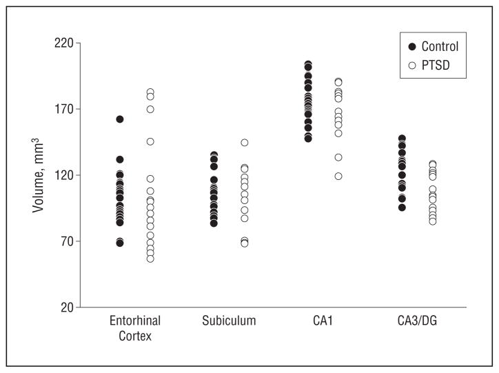
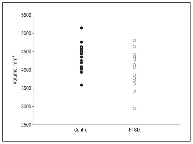
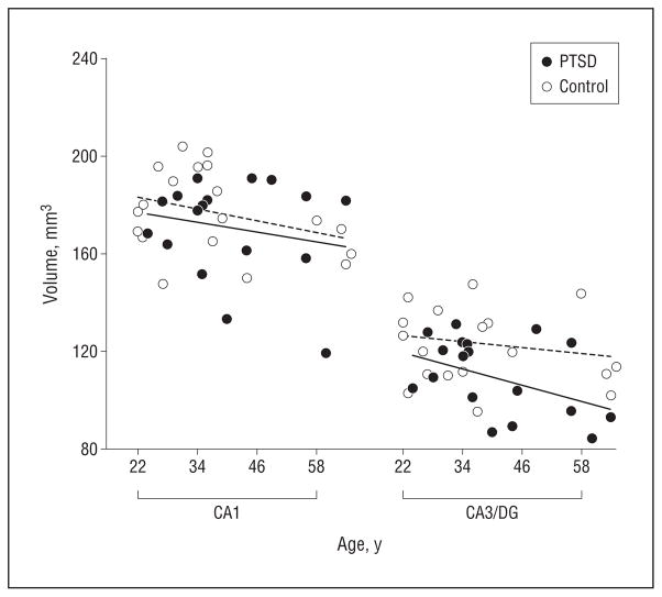
Results: Posttraumatic stress disorder was associated with 11.4% (1.5%) (P = .02) smaller mean (SD) cornu ammonis 3 (CA3)/dentate gyrus subfield volumes, irrespective of age-related alterations, whereas other subfields were spared. Age was associated with reduced volume of the CA1 subfield (P = .03). Total hippocampal volume was also reduced in PTSD by a mean (SD) of 6.5% (0.6%) but, related to both PTSD (P = .05) and age (P = .01), was consistent with the measurements in the subfields.
Conclusions: The findings indicate for the first time in humans that PTSD is associated with selective volume loss of the CA3/dentate gyrus subfields, consistent with animal studies, implying that chronic stress suppresses neurogenesis and dendritic branching in these structures.
Figures

Similar articles
-
Automated measurement of hippocampal subfields in PTSD: Evidence for smaller dentate gyrus volume.J Psychiatr Res. 2017 Dec;95:247-252. doi: 10.1016/j.jpsychires.2017.09.007. Epub 2017 Sep 9. J Psychiatr Res. 2017. PMID: 28923718 Free PMC article.
-
The atrophy and laterality of the hippocampal subfields in parents with or without posttraumatic stress disorder who lost their only child in China.Neurol Sci. 2017 Jul;38(7):1241-1247. doi: 10.1007/s10072-017-2952-3. Epub 2017 Apr 17. Neurol Sci. 2017. PMID: 28417215
-
Smaller hippocampal CA1 subfield volume in posttraumatic stress disorder.Depress Anxiety. 2018 Nov;35(11):1018-1029. doi: 10.1002/da.22833. Epub 2018 Sep 26. Depress Anxiety. 2018. PMID: 30256497 Free PMC article.
-
Bilateral hippocampal volume reduction in adults with post-traumatic stress disorder: a meta-analysis of structural MRI studies.Hippocampus. 2005;15(6):798-807. doi: 10.1002/hipo.20102. Hippocampus. 2005. PMID: 15988763
-
Stress and brain atrophy.CNS Neurol Disord Drug Targets. 2006 Oct;5(5):503-12. doi: 10.2174/187152706778559309. CNS Neurol Disord Drug Targets. 2006. PMID: 17073653 Free PMC article. Review.
Cited by
-
Perceived Stress Is Differentially Related to Hippocampal Subfield Volumes among Older Adults.PLoS One. 2016 May 4;11(5):e0154530. doi: 10.1371/journal.pone.0154530. eCollection 2016. PLoS One. 2016. PMID: 27144832 Free PMC article.
-
Animal models of post-traumatic stress disorder: face validity.Front Neurosci. 2013 May 31;7:89. doi: 10.3389/fnins.2013.00089. eCollection 2013. Front Neurosci. 2013. PMID: 23754973 Free PMC article.
-
Tempering aversive/traumatic memories with cannabinoids: a review of evidence from animal and human studies.Psychopharmacology (Berl). 2019 Jan;236(1):201-226. doi: 10.1007/s00213-018-5127-x. Epub 2019 Jan 2. Psychopharmacology (Berl). 2019. PMID: 30604182 Review.
-
Dysregulation of inflammation, neurobiology, and cognitive function in PTSD: an integrative review.Cogn Affect Behav Neurosci. 2020 Jun;20(3):455-480. doi: 10.3758/s13415-020-00782-9. Cogn Affect Behav Neurosci. 2020. PMID: 32170605 Free PMC article. Review.
-
[Hippocampal subfield volume alteration in post-traumatic stress disorder: a magnetic resonance imaging study].Sheng Wu Yi Xue Gong Cheng Xue Za Zhi. 2018 Apr 25;35(2):252-257. doi: 10.7507/1001-5515.201707056. Sheng Wu Yi Xue Gong Cheng Xue Za Zhi. 2018. PMID: 29745531 Free PMC article. Chinese.
References
-
- Pine DS. Anxiety disorders: clinical features. In: Sadock BJ, Sadock VA, editors. Kaplan and Sadock’s Comprehensive Textbook of Psychiatry. Philadelphia, PA: Lippincott Williams & Wilkins; 2000. pp. 1476–1490.
-
- Kessler RC, Sonnega A, Bromet E, Hughes M, Nelson CB. Posttraumatic stress disorder in the National Comorbidity Survey. Arch Gen Psychiatry. 1995;52 (12):1048–1060. - PubMed
-
- Sapolsky RM. Glucocorticoids and hippocampal atrophy in neuropsychiatric disorders. Arch Gen Psychiatry. 2000;57(10):925–935. - PubMed
Publication types
MeSH terms
Grants and funding
LinkOut - more resources
Full Text Sources
Other Literature Sources
Medical
Miscellaneous
