. 2010 May;185(1):55-64.
doi: 10.1534/genetics.110.115139. Epub 2010 Mar 1.

Condensins promote coorientation of sister chromatids during meiosis I in budding yeast

Affiliations

Condensins promote coorientation of sister chromatids during meiosis I in budding yeast

Ilana L Brito et al. Genetics. 2010 May.

Abstract

The condensin complex is a key determinant of higher-ordered chromosome structure. We show here that the complex is also important for the correct alignment of chromosomes on the meiosis I spindle. Unlike during mitosis and meiosis II, when sister chromatids attach to microtubules emanating from opposite spindle poles (biorientation), accurate meiosis I chromosome segregation requires that sister chromatids attach to microtubules emanating from the same spindle pole (co-orientation). The monopolin complex, consisting of Lrs4, Csm1, and the meiosis-specific component Mam1, brings about meiosis I co-orientation. We find that in the absence of functional condensin complexes, a fraction of sister kinetochores biorient on the meiosis I spindle and association of the monopolin complex subunit Mam1 with kinetochores is decreased. Our studies uncover a new locus-specific effect of the condensin complex.


Figures
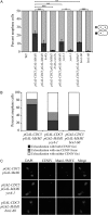
F<sc>igure</sc> 1.—
Figure 1.—
Condensin is required for sister kinetochore coorientation induced by high levels of Mam1 and Cdc5. (A) Wild-type (A5244), pGAL-CDC5 pGAL-MAM1 (A12312), pGAL-CDC5 pGAL-MAM1 lrs4Δ (A15910), pGAL-CDC5 pGAL-MAM1 lrs4Δ csm1Δ (A21128), pGAL-CDC5 pGAL-MAM1 ycs4-1 (A20739), ycs4-1 (A21818), pGAL-CDC5 pGAL-MAM1 brn1-60 (A21712), and brn1-60 cells (A21688), all carrying CENIV GFP dots, were arrested in G1 using 5 μg/ml α-factor in YEP medium containing 2% raffinose. One hour prior to release, galactose (2%) was added to induce MAM1 and CDC5 expression. Cells were released into YEP medium containing 2% raffinose and 2% galactose at 34°. The percentage of anaphase cells in which GFP dots cosegregated (dark gray bars) was determined. Two hundred cells were counted per strain. Bars represent standard deviation. Statistical significance was measured using a single-factor ANOVA test. P-values are represented by the asterisks (*P < 0.05; **P < 0.01; ***P < 0.001). (B and C) pGAL-CDC5 pGAL-MAM1 (A12312), pGAL-CDC5 pGAL-MAM1 ycs4-1 (A20739), and pGAL-CDC5 pGAL-MAM1 brn1-60 (A21712) were grown as described in A to determine the colocalization of Mam1-9MYC with both, one, or no CENIV GFP dots by chromosome spreads of anaphase cells (B). The micrographs in C show Mam1-9MYC (red) and CENIV-GFP (green) localization. At least 50 cells were counted per strain.
F<sc>igure</sc> 2.—
Figure 2.—
Condensin is required for sister kinetochore coorientation during meiosis I. pCLB2-CDC20 (A7118, diamonds), pCLB2-CDC20 mam1Δ (A7316, squares), pCLB2-CDC20 ycg1-2 (A23218, triangles), and pCLB2-CDC20 ycs4-2 (A23220, circles) cells containing heterozygous CENV GFP dots were induced to sporulate at 25°. One hour after transfer into sporulation medium, cells were shifted to 34°. At the indicated times, samples were taken to determine the percentage of cells with metaphase I spindles (left graph) and separated CENV GFP dots (right graph). Two hundred cells were counted per time point.
F<sc>igure</sc> 3.—
Figure 3.—
Condensin is required to maintain pericentromeric structure. (A) 3HA-BRN1 (HY1143) and pCLB2-3HA-BRN1 (3069C) cells were induced to sporulate and 3HA-Brn1 protein levels were examined in wild-type (left) and pCLB2-3HA-BRN1 (right) cells. β-Tubulin was used as a loading control. (B and C) pCLB2-CDC20 (A7118, diamonds), pCLB2-CDC20 mam1Δ (A7316, squares), and pCLB2-CDC20 pCLB2-BRN1 (A22520, triangles) diploid cells containing heterozygous CENV GFP dots were induced to sporulate at 30°. At the indicated times, samples were taken to determine the percentage of cells with metaphase I spindles (B, left graph) and separated CENV GFP dots (B, right graph). (C) Ten hours after transfer into sporulation medium, the percentage of cells displaying stretched CENV GFP signal was determined. Two hundred cells were counted per time point.
F<sc>igure</sc> 4.—
Figure 4.—
Condensins are required for Mam1 localization to kinetochores. (A and B) Wild-type (A7097, circles) and pCLB2-3HA-BRN1 (A22517, squares) cells carrying Mam1-9MYC and Ndc10-6HA fusions were induced to sporulate at 30°. (A) At the indicated times, samples were taken to determine the levels of Mam1-9MYC. Cdc28 was used as a loading control. (B) Samples were also taken to determine the percentage of cells with metaphase I spindles. Two hundred cells were counted per time point. (C and D) Wild-type (A7097) and pCLB2-3HA-BRN1 (A22517) cells carrying Mam1-9MYC and Ndc10-6HA fusions were induced to sporulate at 30°. Chromosome spreads were performed on cells 6 hr after transfer into sporulation medium. (C) The percentage of cells showing >50%, <50%, or no colocalization between Mam1-9MYC (red) and Ndc10-6HA (green) foci was determined. The micrographs in D show examples of Mam1 association with kinetochores (top panels), taken from wild-type cells, and Mam1 association with chromosomes but not kinetochores (bottom panels), taken from Brn1-depleted cells. At least 50 cells were counted per strain.
F<sc>igure</sc> 5.—
Figure 5.—
Lrs4 localizes to kinetochores independently of the condensin complex. Wild-type (A9043) and pCLB2-3HA-BRN1 (A23861) cells carrying Lrs4-13MYC and Ndc10-6HA fusions were induced to sporulate at 30°. (A) At the indicated times, samples were taken to determine the percentage of cells with metaphase I (diamonds) and anaphase I (squares) spindles and the percentage of cells with Lrs4-13MYC released from the nucleolus (open circles). Chromosome spreads were performed on cells 6 hr after transfer into sporulation medium. (B) The percentage of cells showing >50%, <50%, or no colocalization of the Lrs4-13MYC (green) and Ndc10-6HA (red) foci was determined. The micrographs in C show examples of Lrs4 association with kinetochores taken from wild-type cells (top panels) and Brn1-depleted cells (bottom panels). At least 50 cells were counted per strain.
F<sc>igure</sc> 6.—
Figure 6.—
Condensin association with kinetochores is independent of Lrs4-Csm1. (A and B) cdc14-3 (A20336) and cdc14-3 lrs4Δ (A21607) cells carrying Ndc80-GFP and Ycs4-13MYC fusions were released from a pheromone-induced G1 arrest at 37°. (A) Chromosome spreads were performed on samples taken 150 min after release to determine the percentage of cells showing strong, weak, or no colocalization of Ycs4-13MYC with Ndc80-GFP. Strong colocalization refers to Ycs4 staining at Ndc80 foci that is at or above the level of Ycs4 staining at the rDNA. Weak colocalization refers either to Ycs4 signal at only one Ndc80 focus or Ycs4 signal at both Ndc80 foci that is weaker than Ycs4 signal at the rDNA. The micrographs in B show Ycs4-13MYC (red) and Ndc80-GFP (green) localization in cdc14-3 and cdc14-3 lrs4Δ mutants. At least 50 cells were counted per strain. (C and D) cdc14-3 (A21860) and cdc14-3 csm1Δ (A21861) cells carrying Ndc80-GFP and Smc4-13MYC fusions were grown and processed as described in A to determine the percentage of cells showing strong, weak, or no colocalization of Smc4-13MYC with Ndc80-GFP. (C) Categories are described as in A and B. The micrographs in D show Smc4-13MYC (red) and Ndc80-GFP (green) localization in cdc14-3 and cdc14-3 csm1Δ mutants. At least 50 cells were counted per strain.
F<sc>igure</sc> 7.—
Figure 7.—
A speculative model for how condensins and Lrs-Csm1 promote sister kinetochore coorientation and prevent unequal recombination of rDNA repeats. Our data suggest a model where Lrs4 and Csm1 collaborate with condensins to bring about the coorientation of sister chromatids during meiosis I. Lrs4-Csm1 and condensins independently associate with kinetochores and pericentromeric regions, respectively (A). We speculate that once recruited, Lrs4 and Csm1 aggregate condensin complexes, thereby physically constraining the two kinetochores. This permits the association of Mam1, which promotes sister kinetochore attachment to microtubules emanating from the same pole (B). Lrs4-Csm1 function at kinetochores is analogous to their role within the nucleolus. There, the recruitment hierarchy is different, with Fob1 and other RENT complex components recruiting Lrs4-Csm1. These two proteins in turn, bind to condensins and recruit them to the rDNA, where they bring about higher-order chromosome structure. Lrs4-Csm1 “zip up” condensin complexes or restrict their movement with respect to each other, thereby preventing the interaction of rDNA repeats with repeats that are not at the homologous position (C). We suggest that Lrs4-Csm1 collaborate with condensins to create higher-order chromosomal structures.

Similar articles

Cited by

References

    1. Amon, A., 2002. Synchronization procedures. Methods Enzymol. 351 457–467. - PubMed
    1. Bhalla, N., S. Biggins and A. W. Murray, 2002. Mutation of YCS4, a budding yeast condensin subunit, affects mitotic and nonmitotic chromosome behavior. Mol. Biol. Cell 13 632–645. - PMC - PubMed
    1. Chan, R. C., A. F. Severson and B. J. Meyer, 2004. Condensin restructures chromosomes in preparation for meiotic divisions. J. Cell Biol. 167 613–625. - PMC - PubMed
    1. Chelysheva, L., S. Diallo, D. Vezon, G. Gendrot, N. Vrielynck et al., 2005. AtREC8 and AtSCC3 are essential to the monopolar orientation of the kinetochores during meiosis. J. Cell Sci. 118 4621–4632. - PubMed
    1. Clyne, R. K., V. L. Katis, L. Jessop, K. R. Benjamin, I. Herskowitz et al., 2003. Polo-like kinase Cdc5 promotes chiasmata formation and cosegregation of sister centromeres at meiosis I. Nat. Cell. Biol. 5 480–485. - PubMed

Publication types

MeSH terms