. 2010 Feb 24;5(2):e9357.
doi: 10.1371/journal.pone.0009357.

Protective effect of human amniotic fluid stem cells in an immunodeficient mouse model of acute tubular necrosis

Affiliations

Protective effect of human amniotic fluid stem cells in an immunodeficient mouse model of acute tubular necrosis

Laura Perin et al. PLoS One. .

Abstract

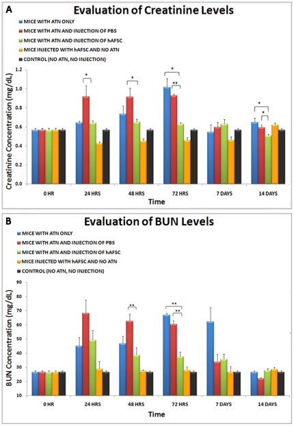
Acute Tubular Necrosis (ATN) causes severe damage to the kidney epithelial tubular cells and is often associated with severe renal dysfunction. Stem-cell based therapies may provide alternative approaches to treating of ATN. We have previously shown that clonal c-kit(pos) stem cells, derived from human amniotic fluid (hAFSC) can be induced to a renal fate in an ex-vivo system. Herein, we show for the first time the successful therapeutic application of hAFSC in a mouse model with glycerol-induced rhabdomyolysis and ATN. When injected into the damaged kidney, luciferase-labeled hAFSC can be tracked using bioluminescence. Moreover, we show that hAFSC provide a protective effect, ameliorating ATN in the acute injury phase as reflected by decreased creatinine and BUN blood levels and by a decrease in the number of damaged tubules and apoptosis therein, as well as by promoting proliferation of tubular epithelial cells. We show significant immunomodulatory effects of hAFSC, over the course of ATN. We therefore speculate that AFSC could represent a novel source of stem cells that may function to modulate the kidney immune milieu in renal failure caused by ATN.


Conflict of interest statement

Competing Interests: The authors have declared that no competing interests exist.

Figures

Figure 1
Figure 1. hAFSC morphology, gene expression and karyotype.
A. Morphology of hAFSC population. After 40 passages in culture under bright field the cells present a fibroblastoid appearance (10×). B. Karyotype of hAFSC after 38 passages. C. RT-PCR of hAFSC before in vivo injection. Neither early nor mature kidney markers are expressed. hACTB is used as a housekeeping gene.
Figure 2
Figure 2. Morphological analysis of glycerol-rhabodomyolysis-induced-ATN model in nu/nu mice.
A. Histological section showing PAS staining of a nu/nu mouse kidney. Proximal and distal tubules and glomeruli, as indicated by the arrows, present normal morphology (40x). B. Histological section showing PAS staining of a nu/nu mouse kidney after 3 days of glycerol-rhabdomyolysis-induced ATN. Destruction of brush borders, intraluminal cast formation as well as general disorganization of the kidney structures is evident (arrow, 40x) C. TUNEL staining of a nu/nu mouse kidney. The level of apoptotic cells is very low, when compared with TUNEL staining of a nu/nu mouse kidney after 3 days of glycerol-induced ATN (D), (10x). E. Graph showing the effect of glycerol ATN on kidney cell apoptosis (number of positive apoptotic nuclei per 300 nuclei) compared with untreated controls. Values are presented as mean ± SEM (*** p< 0.001).
Figure 3
Figure 3. In vivo luciferase detection of hAFSC in ATN damaged kidneys.
hAFSC transduced with luciferase maintain the expression of luciferase under bioluminescence over many population doublings as demonstrated by the presence of optical signal after 20 passages in culture (tube on right) when compared with hAFSC that were not transfected (tube on left) when stimulated with the substrate for luciferase (A). The lowest number of cells that exhibit detectable luciferase signal in vitro is 1×10⁵. Luciferase gain range is shown on right (B). C. In vivo imaging showing bioluminescent detection of hAFSC after injection into a damaged nu/nu mouse kidney over a period of 21 days. In panel 1 is shown a negative control, injected only with luciferin, the luciferse substrate. The optical signal is strongly present during the first 5 hours post injection (panel 2–5), decreases over time (panel 6–8), but is still present after 21 days (panel 9). In panel 10 is shown the bioluminescence gain range. D. RT-PCR demonstrates the presence of the luciferase gene (expressed only by human transfected hAFSC clones) in 5 injected kidneys, compared with the cells before injection (positive control) and in non-injected kidney (negative control). Human ACTB is used as housekeeping gene. E. Immuno-fluorescence staining of injected kidney with hAFSC after 3 weeks. The red fluorescence (arrow) confirms the presence of hAFSC expressing luciferase. The nuclei are stained with DAPI (20×).
Figure 4
Figure 4. Integration and structural differentiation of hAFSC injected into glycerol induced damaged ATN kidneys.
A. Frozen section of a kidney injected with hAFSC after 1 week. The cells are evident as red fluorescence of the surface marker CM-DiI. The nuclei are stained with DAPI (30×). It is noticeable that CM-Dil-labelled-hAFSC locate in the proximity of tubular structures after 3 weeks following injection and are shown to express Peanut Agglutinin (B) as well as Dolichus Biflorus Agglutinin (arrow) (C); hAFSC locate also in close proximity of the glomerular structures and express human Glial Derived Neutrofic Factor (arrow) (D). The nuclei are stained with DAPI (40×). E. Double Immuno-fluorescent staining of injected kidney with luciferase transduced hAFSC (and not CM-Dil labeled), showing the same cells expressing both Aquaporin 2 and luciferase (arrow), 3 weeks after injection. The nuclei are, stained with DAPI. (30×). F. RT-PCR performed on RNA isolated from kidney, 3 weeks after hAFSC injection. The expressed kidney markers such as NPHS1, AQP2, PAX2, OCLN, identified by primers designed with human specific sequences. Human ACTB is used as a housekeeping gene. G RT-PCR showing the specificity, as indicated by human sequences not cross-reacting with mouse.
Figure 5
Figure 5. Protective effect of injected hAFSC as determined by measurements of blood creatinine and BUN levels.
In these graphs the difference in levels of blood creatinine (A) and BUN (B) between the experimental groups are shown (Blue: animals that underwent only ATN; Red: animals that underwent ATN and intrarenal injection of PBS; Green: animals that underwent ATN and intrarenal injection of hAFSC; Orange: animals that underwent intrarenal injection of hAFSC but not ATN; Black: control animals, no ATN and no injections), shown at different time points (24 hours, 48 hours, 72 hours, 1 week and 2 weeks). Values are presented as mean ± SEM (* p<0,05; ** p<0,01).
Figure 6
Figure 6. Protective effect of injected hAFSC, determined by maintenance of the morphological structure in glycerol induced ATN damaged kidneys.
In these figures are shown the most representative PAS-Staining of kidney sections of mice treated only with injection of glycerol over the course time of 24 hours (A), 48 hours (B) and 72 hours, (C) when compared with mice treated with injection of glycerol and hAFSC at 24 hours (D), 48 hours (E) and 72 hours (F) after injection. In the mice treated only with glycerol the level of disruption of brush border (BB), the desegregation of tubular membrane (DTM) and cast formation (CF) increased over time, while the injection of hAFSC preserved the morphology of the tubular structures after they have been damaged following IM glycerol injection. The graph (G) represents the percentage of damaged tubules in the different experimental groups. Blue: animals that underwent only ATN; Red: animals that underwent ATN and intrarenal injection of PBS; Green: animals that underwent ATN and intrarenal injection of hAFSC; Orange: animals that underwent intrarenal injection of hAFSC but not ATN) at different time points (24 hours, 48 hours, 72 hours, 1 weeks and 2 weeks) per total number of tubules in the sections. Values are presented as mean ± SEM (* p<0,05; ** p<0,01).
Figure 7
Figure 7. Protective effect of injected hAFSC as determined by increase of proliferation and decrease of apoptotic tubular cells in glycerol induced ATN kidneys.
In these graphs is shown the proliferation activity in tubular cells (A) and the apoptosis (B) between the experimental groups (Blue: animals that underwent only ATN; Red: animals that underwent ATN and intrarenal injection of PBS; Green: animals that underwent ATN and intrarenal injection of hAFSC; Orange: animals that underwent intrarenal injection of hAFSC but not ATN;) at different time points (24 hours, 48 hours, 72 hours, 1 week and 2 weeks). Values are presented as mean ± SEM (* p<0,05; ** p<0,01; *** p<0,001).
Figure 8
Figure 8. Immunomodulatory effects of hAFSC once injected into glycerol induced ATN-damaged kidneys.
Mouse Cytokines were analyzed among all the experimental groups to evaluate changes in the inflammatory milieu. A. Developed multiplex cytokine assay membranes for mouse with ATN, mouse with ATN and Injection of PBS and Mouse with ATN and injection of hAFSC, showing the markedly different expression patterns of cytokines at 14 days. B. Schematic outline representation of mouse cytokine expression over the 14 days among different groups: 1. Mice with ATN only. As a general trend of cytokine expression, in these animals the pro-Inflammatory cytokines increased or remained highly expressed and anti-inflammatory cytokines showed decreased or low expression; 2. Mice with ATN and injection of hAFSC. As a general trend of cytokine expression, it is evident that the anti inflammatory cytokines increased over the 14 day study period, while pro-Inflammatory cytokines decreased; 3. Mice with ATN and injection of PBS. As a general trend of cytokine expression, in these animals the pro-Inflammatory cytokines increased or remained highly expressed and anti-inflammatory cytokines showed decreased or low expression; 4. Mice without ATN and injection of hAFSC. As a general trend of cytokine expression, in these mice the anti-inflammatory cytokines are mildly increased over the 14 days, while pro-inflammatory cytokines are not expressed or show decreased expression over the 14 days.

Similar articles

Cited by

References

    1. National Kidney and Urologic Diseases Information Clearinghouse. Kidney and Urologic Diseases Statistics for the United States. Available: http://kidney.niddk.nih.gov/kudiseases/pubs/kustats/index.htm#kpClearing....
    1. Thadhani R, Pascual M, Bonventre JV. Acute renal failure. N Engl J Med. 1996;334(22):1448–1460. - PubMed
    1. Yokoo T, Fukui A, Ohashi T, Miyazaki Y, Utsunomiya Y, et al. Xenobiotic kidney organogenesis from human mesenchymal stem cells using a growing rodent embryo. J Am Soc Nephrol. 2006;17(4):1026–1034. - PubMed
    1. Duffield JS, Park KM, Hsiao LL, Kelley VR, Scadden DT, et al. Restoration of tubular epithelial cells during repair of the postischemic kidney occurs independently of bone marrow-derived stem cells. J Clin Invest. 2005;115(7):1743–1755. - PMC - PubMed
    1. Herrera MB, Bussolati B, Bruno S, Fonsato V, Romanazzi GM, et al. Mesenchymal stem cells contribute to the renal repair of acute tubular epithelial injury. Int J Mol Med. 2004;14(6):1035–1041. - PubMed

Publication types

MeSH terms