. 2010 Feb 24;5(2):e9321.
doi: 10.1371/journal.pone.0009321.

Oral biofilm architecture on natural teeth

Affiliations

Oral biofilm architecture on natural teeth

Vincent Zijnge et al. PLoS One. .

Abstract

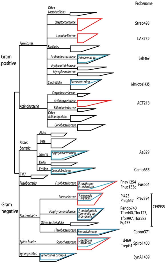
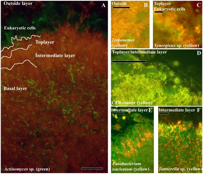
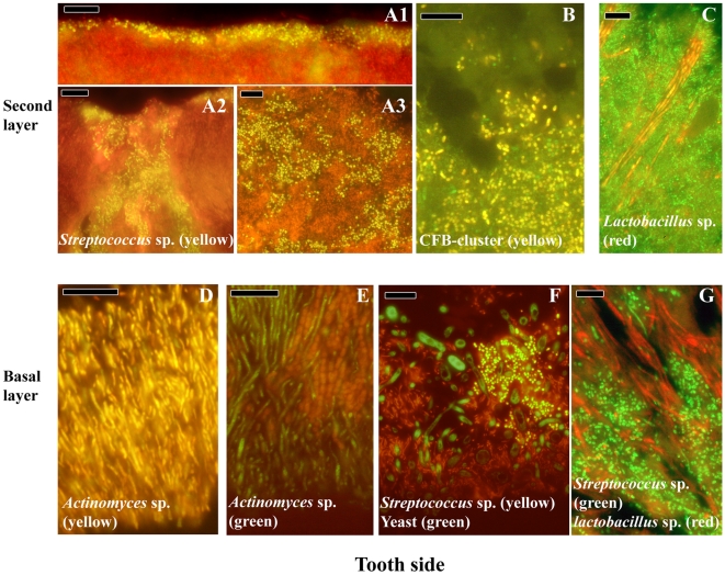
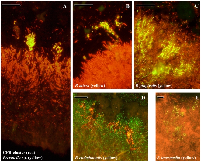
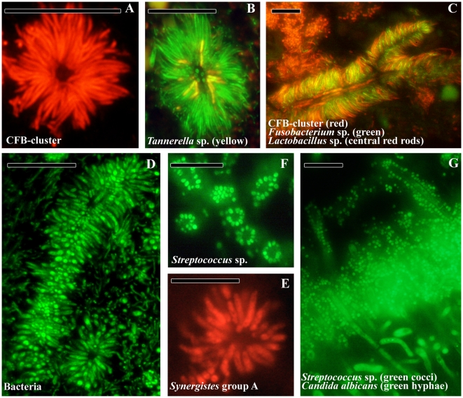
Periodontitis and caries are infectious diseases of the oral cavity in which oral biofilms play a causative role. Moreover, oral biofilms are widely studied as model systems for bacterial adhesion, biofilm development, and biofilm resistance to antibiotics, due to their widespread presence and accessibility. Despite descriptions of initial plaque formation on the tooth surface, studies on mature plaque and plaque structure below the gum are limited to landmark studies from the 1970s, without appreciating the breadth of microbial diversity in the plaque. We used fluorescent in situ hybridization to localize in vivo the most abundant species from different phyla and species associated with periodontitis on seven embedded teeth obtained from four different subjects. The data showed convincingly the dominance of Actinomyces sp., Tannerella forsythia, Fusobacterium nucleatum, Spirochaetes, and Synergistetes in subgingival plaque. The latter proved to be new with a possibly important role in host-pathogen interaction due to its localization in close proximity to immune cells. The present study identified for the first time in vivo that Lactobacillus sp. are the central cells of bacterial aggregates in subgingival plaque, and that Streptococcus sp. and the yeast Candida albicans form corncob structures in supragingival plaque. Finally, periodontal pathogens colonize already formed biofilms and form microcolonies therein. These in vivo observations on oral biofilms provide a clear vision on biofilm architecture and the spatial distribution of predominant species.


Conflict of interest statement

Competing Interests: The authors have declared that no competing interests exist.

Figures

Figure 1
Figure 1. Phylogenetic tree representing oral microbial diversity.
The tree is based on >1500 sequences derived from oral-cavity studies and shows the schematic coverage of the diversity by our probe set. The branching of the tree was simplified for clarity. The boxes represent groups of bacteria. The vertical size of the box reflect the number of sequences and the angular side the genetic diversity. The blue boxes indicate the part that is covered by the species or subgroup-specific probes and the red box indicate genus-specific probes.
Figure 2
Figure 2. Localization of the most abundant species in subgingival biofilms.
(A) Overview of the subgingival biofilm with Actinomyces sp. (green bacteria), bacteria (red) and eukaryotic cells (large green cells on top). (B) Spirochaetes (yellow) outside the biofilm. (C) Detail of Synergistetes (yellow) in the top layer in close proximity to eukaryotic cells (green). (D) CFB-cluster (yellow) in the top and intermediate layer. (E) F. nucleatum in the intermediate layer. (F) Tannerella sp. (yellow) in the intermediate layer. Each panel is double-stained with probe EUB338 labeled with FITC or Cy3. The yellow color results from the simultaneous staning with FITC and Cy3 labeled probes. Bars are 10 µm.
Figure 3
Figure 3. Localization of the most abundant species in supragingival biofilms.
Streptococcus sp. (yellow) form a thin band on top of the biofilm (A1), almost engulfing in the biofilm (A2) or present as small cells scattered through the top layer of the biofilm (A3). (B) Cells from the CFB-cluster of bacteria in the top layer of the biofilm, without defined structure. (C) Lactobacillus sp. (red) forming long strings through the top layer. (D) Actinomyces sp. (yellow) plaque attached to the tooth. (E) Actinomyces sp. (green) and cocci forming initial plaque.(F) Multispecies initial plaque composed of Streptococcus sp. (yellow), yeast cells (green) and bacteria unidentified (red). (G) Streptococcus sp. (green) and Lactobacillus sp. (red) forming initial plaque. Black holes might be channels through the biofilm. Panels A, B, C, E, F are double stained with probe EUB338 labeled with FITC or Cy3. Bars are 10 µm.
Figure 4
Figure 4. Localization of species associated with periodontitis.
(A) Overview of the subgingival biofilm with CFB-cluster species (red) and Prevotella sp. (yellow). Since Prevotella sp. are part of the CFB-cluster of bacteria, cells appear in yellow. (B) Top of the biofilm with a micro-colony of P. micra (yellow). (C) Micro-colonies of P. gingivalis (yellow) in the top layer. (D) Micro-colonies of P. endodontalis (yellow) in the top layer. (E) Micro-colonies of P. intermedia in the top layer. Panels B, C, D and E are double stained with probe EUB338 labeled with FITC or Cy3. Bars are 10 µm.
Figure 5
Figure 5. Bacterial aggregates in oral plaque.
(A) Transversal view of a test-tube brush found in subgingival plaque composed of filamentous cells from the CFB-cluster. (B) Tannerella sp. (yellow) in a test-tube brush. (C) Longitudinal view of a test-tube brush with Lactobacillus sp. (red rods) as central structures. F. nucleatum (green) and CFB-cluster filaments radiating from the central structures. (D) Longitudinal and transversal view of a test-tube brush stained with the eubacterial probe. (E) Transversal view of the test-tube brush in panel D, composed of Synergistetes group A species. (F) Transversal view of Streptococcus sp. (green) aggregation around a central cell (not stained) in supragingival plaque. (G) Transversal view of supragingival plaque with Streptococcus sp. (green cocci) and Candida albicans (green hyphae) in the top layer of the biofilm and forming corn cob structures growing outwards. Bars are 10 µm.

References

    1. Wood SR, Kirkham J, Marsh PD, Shore RC, Nattress B, et al. Architecture Of Intact Natural Human Plaque Biofilms Studied By Confocal Laser Scanning Microscopy. J Dent Res. 2000;79:21–27. - PubMed
    1. Socransky SS, Haffajee AD. Dental Biofilms: Difficult Therapeutic Targets. Periodontol 2000. 2002;28:12–55. - PubMed
    1. Reese S, Guggenheim B. A Novel TEM Contrasting Technique For Extracellular Polysaccharides In In Vitro Biofilms. Microsc Res Tech. 2007;70:816–822. - PubMed
    1. Bos R, Van Der Mei HC, Busscher HJ. Physico-Chemistry Of Initial Microbial Adhesive Interactions–Its Mechanisms And Methods For Study. FEMS Microbiol Rev. 1999;23:179–230. - PubMed
    1. Busscher HJ, Van Der Mei HC. Physico-Chemical Interactions In Initial Microbial Adhesion And Relevance For Biofilm Formation. Adv Dent Res. 1997;11:24–32. - PubMed

Publication types

MeSH terms