Review
. 2009 Feb 13;1(2):157-81.
doi: 10.18632/aging.100024.

Immune physiology in tissue regeneration and aging, tumor growth, and regenerative medicine

Affiliations
Review

Immune physiology in tissue regeneration and aging, tumor growth, and regenerative medicine

Antonin Bukovsky et al. Aging (Albany NY). .

Abstract

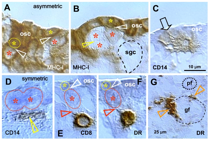
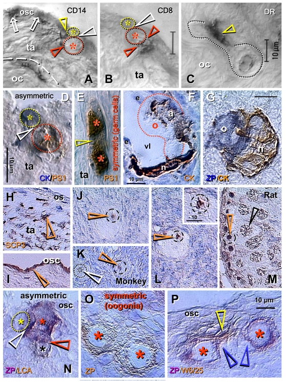
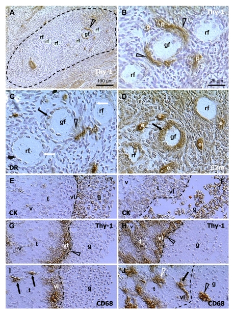
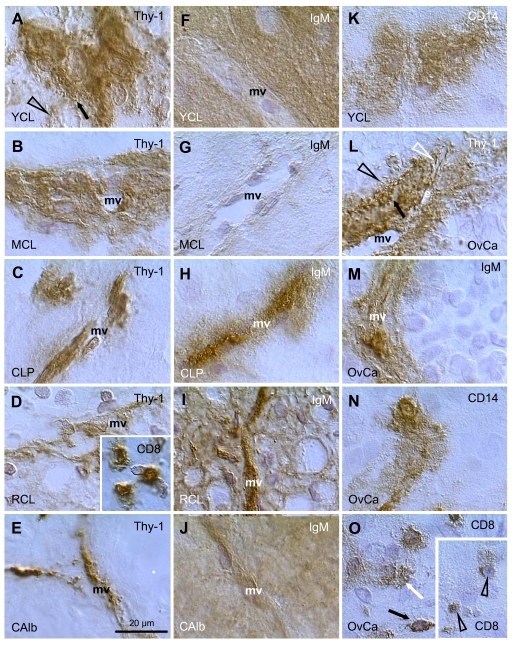
The immune system plays an important role in immunity (immune surveillance), but also in the regulation of tissue homeostasis (immune physiology). Lessons from the female reproductive tract indicate that immune system related cells, such as intraepithelial T cells and monocyte-derived cells (MDC) in stratified epithelium, interact amongst themselves and degenerate whereas epithelial cells proliferate and differentiate. In adult ovaries, MDC and T cells are present during oocyte renewal from ovarian stem cells. Activated MDC are also associated with follicular development and atresia, and corpus luteum differentiation. Corpus luteum demise resembles rejection of a graft since it is attended by a massive influx of MDC and T cells resulting in parenchymal and vascular regression. Vascular pericytes play important roles in immune physiology, and their activities (including secretion of the Thy-1 differentiation protein) can be regulated by vascular autonomic innervation. In tumors, MDC regulate proliferation of neoplastic cells and angiogenesis. Tumor infiltrating T cells die among malignant cells. Alterations of immune physiology can result in pathology, such as autoimmune, metabolic, and degenerative diseases, but also in infertility and intrauterine growth retardation, fetal morbidity and mortality. Animal experiments indicate that modification of tissue differentiation (retardation or acceleration) during immune adaptation can cause malfunction (persistent immaturity or premature aging) of such tissue during adulthood. Thus successful stem cell therapy will depend on immune physiology in targeted tissues. From this point of view, regenerative medicine is more likely to be successful in acute rather than chronic tissue disorders.

Keywords: aging; animal models; follicular renewal; immune physiology; mesenchymal-epithelial interactions; proliferation; regeneration; regenerative medicine; tissue homeostasis; tumor growth.


Conflict of interest statement

The authors declare that they have no competing financial interests.

Figures

Figure 1.
Figure 1.. Schematic drawing of the basic "tissue control unit," which consists of monocyte-derived cells (marked M in the figure), vascular pericytes (P), and autonomic innervation (AI, dashed arrow), and the involvement of other components of the tissue control system (solid arrows).
Monocyte-derived cells physically interact with adjacent epithelial (Ep) and endothelial cells (En) through the basement membranes (dotted lines), and influence pericytes, which secrete intercellular vesicles (ICV). These vesicles collapse into the so-called empty spikes (ES) releasing their content (growth factor/cytokine) after reaching target cells. The activity of pericytes is stimulated or inhibited by autonomic innervation (+ or -) which controls quantitative aspects of tissues. Interaction of MDC with endothelial cells may stimulate homing of T lymphocytes (T) and monocyte-derived dendritic cell precursors (DCP; also known as veiled cells) differentiating into mature dendritic cells (DC). The dendritic cell precursors and T cells interact themselves and stimulate advanced differrentiation of epithelial cells. IgMs regulate early (IgM1), mid (IgM2), and late differentiation (apoptosis) of epithelial cells (IgM3), and IgG associates with aged cells (see Figure 2 and 3). The monocyte-derived cell system (including intraepithelial DCP and mature DC) is postulated to play a dominant role in the regulation of qualitative aspects of tissue-specific cells, including expression of ligands for intraepithelial T cells and regulating autoantibody action. Monocyte-derived cells also carry "stop effect" information (Figure 10B), presumptively encoded at the termination of immune adaptation (Figure 10A), which determines the highest state of epithelial cell differentiation allowed for a particular tissue. For details see Ref. [3,4,33]. Reprinted from Ref. [4], © Antonin Bukovsky.
Figure 2.
Figure 2.. Peroxidase immunohistochemistry (brown color) of stratified epithelium of uterine ectocervix as indicated above columns and in the inset.
(A) CD14 primitive MDC in lamina propria (lp) associate with the epithelium basement membrane (dotted line). Dashed box indicates detail shown in (B). b, basal layer; pb, parabasal layer; im, intermediate layer; s, superficial layer. Arrowheads, basal/parabasal interface; dashed line, parabasal/intermediate interface; dashed/dotted line, intermediate/superficial interface. Ki67 staining (inset) of epithelial cells in lower parabasal layer (arrowheads). (B) CD14 MDC (arrows) exhibit extensions among basal cells (arrowhead). (C) Pericytes of microvasculature (arrows) associate with the basement membrane. (D) Detail from (C) shows intercellular Thy-1 vesicles (arrow) secreted by pericytes and migrating among basal cells (short black arrowhead) to basal/parabasal interface (long arrowhead). Yellow arrowhead indicates residual empty structures ("spikes"). (E) Strong MHC class I expression (W6/32 antibody specific for heavy chain) is characteristic of para-basal cells, and diminishes in lower intermediate layers. Dashed box indicates detail shown in (F). (F) Basal cells show no MHC class I expression. Reprinted from Ref. [4], © Antonin Bukovsky.
Figure 3.
Figure 3.. Uterine ectocervix immunohistochemistry as indicated above columns.
(A) Dendritic cell (DC) precursors secrete HLA-DR among parabasal cells (arrows) and differentiate into mature DC (arrowheads). (B) T cells migrate through parabasal layer (arrow) to parabasal/intermediate interface (dashed line) and show fragmentation after entering the intermediate layer (arrowheads). (C) Transformation of DC precursors into mature DC at the top of parabasal layer is associated with CD68 expression (arrow). Mature DC (black arrowheads) secrete CD68 material in intermediate layer accompanying mature (intermediate) and aged (superficial) epithelial cells (white arrowheads). (D) CD1a is expressed by DC precursors (arrows) and mature DC (black arrowheads). Mature DC (Langerhans' cells) undergo fragmentation in the mid intermediate layer (white arrowheads). (E) Strong IgM binding (arrowheads) in upper parabasal [1], upper intermediate [2] and upper superficial layers [3]. (F) IgG binds to the entire superficial layer. For abbreviations see Figure 2. Reprinted from Ref. [4], © Antonin Bukovsky.
Figure 4.
Figure 4.
Uterine cervix dual color immunohistochemistry (HLA-DR peroxidase/CD8 FITC) viewed in dark field visible light (A), incident fluorescence (B) and dark field fluorescence (C). (A) Interface (dashed line) between parabasal and intermediate layers. White arrowhead shows differentiating DC, yellow arrowhead shows mature DC. Arrow indicates activated T cell with HLA-DR expression (see below). (B) White arrow indicates T cell exhibiting unusual elongated shape at the interface. Yellow arrows indicate residual CD8 expression in fragmented T cell among adjacent im epithelial cells. (C) Activated T cell with HLA-DR expression (white arrow) interacts with differentiating DC (white arrowhead). Mature DC (yellow arrowhead) accompany T cell fragmentation (yellow arrows). Reprinted from Ref. [4], © Antonin Bukovsky.
Figure 5.
Figure 5.. Expression of MHC class I heavy chain (MHC-I), CD14 of primitive MDC, CD8 of T cells, and HLA-DR (DR) of activated MDC and T cells, as indicated in the panels, in human fetal ovary obtained at midpregnancy (24 weeks).
Asymmetric division (white arrowheads, panels A and B) of OSC (osc) gives rise to the OSC (yellow asterisks) and the germ cell daughters (red asterisks). Symmetric division of germ cells follows (yellow arrowhead, panel B), which is required for crossing over, and the secondary germ cells (sgc) attain the ameboid shape (dashed line, no hematoxylin counterstain) to leave the OSC layer and enter cortex. CD14+ primitive MDC interact with the OSC (arrow, panel C) and accompany (arrowhead, panel D) symmetric division of secondary germ cells. CD8 T cells (panel E) and DR+ cells of lymphocyte type (panel F) accompany (red arrowheads) asymmetric division of OSC (white arrowheads) resulting in emergence of secondary germ cells. DR+ MDC (arrowheads, panel G) associate with growing (gf) but not resting primordial follicles (pf). Bar in C for A-F. Adapted from Ref. [36], © Humana Press.
Figure 6.
Figure 6.
Origin of new oocytes (neo-oogenesis), primordial follicles, and SCP3 expression in adult human and monkey ovaries (A-M), and oogenesis in adult rat ovaries (N-P). (A) During asymmetric division (white arrowhead), the CD14 MDC interact with both the OSC daughter (yellow arrowhead) and germ cell daughter (red arrowhead). (B) T lymphocytes, however, interact with the germ cell daughter only (red arrowheads). (C) Ameboid germ cells (dotted line) migrating through the dense ovarian cortex (oc) are accompanied by activated MDC (arrowhead). (D) Asymmetrically dividing OSC produce a new PS1+ germ cell (red asterisk) and CK+ progenitor cell (yellow asterisk). (E) In the tunica albuginea (ta) germ cells (asterisks) symmetrically divide (arrowhead). (F) Capture of oocyte (o) from the blood circulation by an arm (a) of granulosa cell nest (n) lining the venule lumen (vl); e, endothelial cells. (G) Oocyte nest assembly. (H) Segments of tunica albuginea (ta) in ovaries with follicular renewal (early luteal phase) showed strong SCP3 expression of mesenchymal (arrowheads) OSC precursors under ovarian surface (os). (I) Staining of OSC (osc and arrowhead) was apparent in other segments - note lack of staining of tunica albuginea under developed OSC. (J) Postovulatory human ovaries showed staining of oocyte nucleoli (arrowhead) in some primordial follicles. (K) In monkey ovaries, similar staining of oocyte nucleoli in some primordial follicles was observed (red vs. white arrowhead). (L) Staining of paired chromosomes oocyte was observed in human ovaries (inset shows higher magnification). (M) Adult rat testis (positive control) showed staining of condensed chromosomes in spermatogonia (red arrowhead) and progression of meiotic division in primary spermatocytes (black arrowhead). Oogenesis in adult rat ovaries is initiated by asymmetric division of OSC (white arrowhead, N) showing unstained OSC daughter (yellow asterisk) and ZP+ (magenta color) germ cell daughter (red asterisk) accompanied like in human ovaries by a lymphocyte (black asterisk and brown color). Symmetric division of ZP+ oogonia (asterisks, O) follows, and is accompanied (P) by MDC (yellow arrowhead). Blue arrowheads in (P) indicate association of primitive granulosa cells with this process. ZP, zona pellucida; LCA, leukocyte common antigen; W6/25, marker of rat MDC. Details in text. Adapted A-C from Ref. [57], © Blackwell Munksgaard, D-G from Ref. [35], © Antonin Bukovsky, H-M from Ref. [71], © Landes Bioscience, N-P from Ref. [72], © Landes Bioscience.
Figure 7.
Figure 7.
Selection of secondary (A-D) and preovulatory (dominant) follicles (E-F) in the adult human ovary. Staining for Thy-1, HLA-DR (DR), MHC class I light chain (β2m), cytokeratin 18 (CK) and CD68 of mature MDC, as indicated in panels. Dashed line in (A) indicates an area exhibiting diminution of Thy-1 expression by stromal cells. (B), detail from (A). (C) and (D) are semi-parallel sections to (B). Dashed line in (E-J), follicular basement membrane. rf, resting follicles; gf, growing follicle; p, pericytes; e, endothelial cells; v, microvasculature in theca interna (t); vl, vascular layer adjacent to the follicular basement membrane; g, granulosa layer. Details in text. Adapted from Ref. [70], © Wiley-Blackwell.
Figure 8.
Figure 8.
Staining for Thy-1, IgM, CD8, and CD14, as indicated in panels, in human corpora lutea and ovarian adenocarcinomas (OvCa). YCL, young CL; MCL, mature CL; CLP, CL of pregnancy; RCL, regressing CL (subsequent follicular phase); CAlb, corpus albicans. mv, microvasculature. Scale bar in E applies to panels A-O, including insets. Details in text. Adapted from Ref. [109], © Elsevier.
Figure 9.
Figure 9.. Macrophages, cytokines and ovarian cancer.
Epithelial inclusion cysts (EIC), showing infiltration of the cyst wall (A) and lumen (B) by CD68 positive MDC (arrows), ciliated cells (arrowheads), and Ki67 positive (arrow in C) proliferating cells. Immunohistochemical staining of cancerous ovarian tissues for IL-6. (D) and TNF-alpha (E) (x400). A-C adapted from Ref. [111], © Elsevier, and D and E from Ref. [113], © John Libbey Eurotext Ltd.
Figure 10.
Figure 10.
Immune adaptation and TCS "stop effect." (A) Immune adaptation (IA) and tissue longevity. The heart differentiates from early stages of ontogeny (LONG IA) and functions throughout life. The ovary differentiates later (MODERATE IA), and its normal function is limited by follicular renewal (until 35-40 years of age). Aging primordial follicles (apf) persist until exhausted (physiologic menopause). SHORTER period of ovarian development during IA causes earlier termination of follicular renewal during adulthood and results in POF. SHORT period of ovarian development during IA causes no follicular renewal and results in primary amenorrhea. Absence of corpora lutea (CL) during immune adaptation causes their cyclic degeneration, except during pregnancy, which is accompanied by immune suppression. fpf, fetal primordial follicles; fr, follicular renewal; POF, premature ovarian failure; CL, corpus luteum. Adapted from Ref. [31]. (B) Stages of cell differentiation during immune adaptation (left) sets TCS "stop effect" (StE) for tissue physiology and pathology during adulthood. Arrowheads indicate a tendency to StE "shifts" with age. Adapted from Ref. [3,30,33,108].
Figure 11.
Figure 11.
Oocyte and parthenote development in vitro. (A) The oocyte development in OSC culture is accompanied by a satellite (black arrow) and neuronal (white arrow) cells. White arrowhead indicates neuronal extension. (B) DAPI staining of (A). (C) The parthenote shows a blastocoele (white arrow) and inner cell mass (black arrow). (D) DAPI staining of (C). Four cell embryo. (E - G) and morula (H and I). J and K panels show a blastocyst consisting of blastocoele (bc), trophectoderm (te), and inner cell mass (icm) releasing ESC (esc). Left insert in panel K shows enhanced DAPI staining of dividing ESC vs. low DAPI staining of other cells in the culture (right insert). Details in text. Adapted in part from Ref. [137], © Cambridge Journals.
Figure 12.
Figure 12.
Human ovarian epithelial stem cell cultures (representative images from four experiments): untreated (A and B); pre-treated for 1 day with E2 and 1h after TP+PG treatment (C-G); pre-treated for 1 day with E2 and 3h after TS+PG treatment (H-O). Lack of SSEA-1 expression (A) and moderate Thy-1 expression by some epithelial cells (B). SSEA-1 is strongly expressed in some small cells resembling stem cells (black vs. white arrowheads, C and D) and one of the cells originating by asymmetric division (E). Similar cells show strong expression of Thy-1 (F). The NCAM expression was also detected in some cells (G). Two hours later, the cells reached neuronal morphology and exhibited SSEA-1 expression in the cell bodies but not extending processes (H, black vs. white arrowheads), Thy-1 and NCAM expression in both (I and J, black arrowheads), and SSEA-4 expression slightly exceeding that of SSEA-1 (K vs. H). No staining was observed in the immunohistochemistry control (L). Panels M-O show phase contrast microscopy with neuronal and epithelial cells (M), floating numerous putative NSC (N), and putative NSC exhibiting bubble type anchors (arrowheads, O). Numbers above bars indicate microns. For details see text. Adapted from Ref. [138], © Landes Bioscience.

Similar articles

Cited by

References

    1. Carrel A. Growth-promoting function of leukocytes. J Exp Med. 1922;36:385–391. - PMC - PubMed
    1. Fidler IJ. Lymphocytes are not only immunocytes. Biomedicine. 1980;32:1–3. - PubMed
    1. Bukovsky A, Caudle MR, Keenan JA. Dominant role of monocytes in control of tissue function and aging. Med Hypotheses. 2000;55:337–347. - PubMed
    1. Bukovsky A, Caudle MR, Keenan JA, Upadhyaya NB, Van Meter S, Wimalasena J, Elder RF. Association of mesenchymal cells and immunoglobulins with differentiating epithelial cells. BMC Dev Biol. 2001;1:11. - PMC - PubMed
    1. Havran WL, Jameson JM, Witherden DA. Epithelial cells and their neighbors. III. Interactions between intraepithelial lymphocytes and neighboring epithelial cells. Am J Physiol Gastrointest Liver Physiol. 2005;289:G627–G630. - PubMed

Publication types