. 2009 Jul 23;1(8):723-32.
doi: 10.18632/aging.100072.

Modeling of replicative senescence in hematopoietic development

Affiliations

Modeling of replicative senescence in hematopoietic development

Anna Marciniak-Czochra et al. Aging (Albany NY). .

Abstract

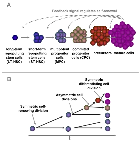
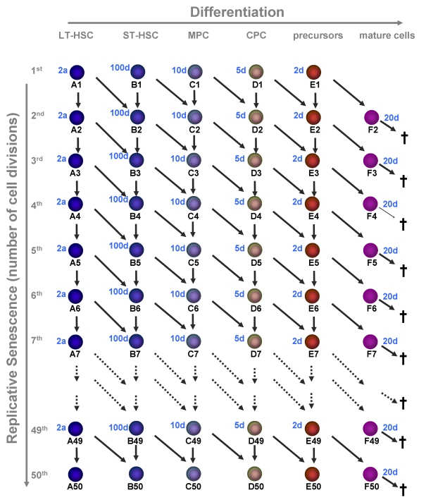
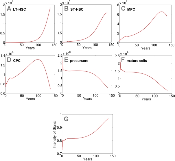
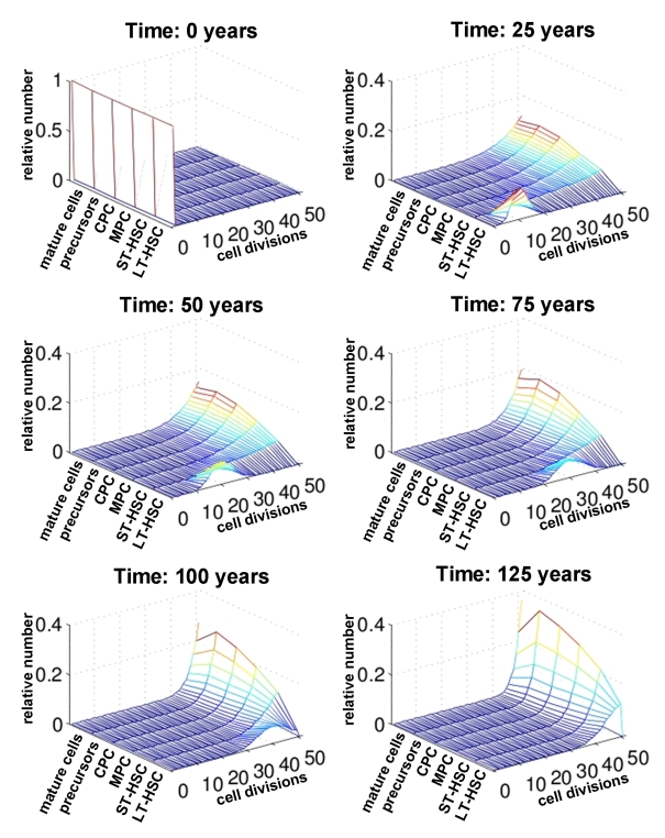
Hematopoietic stem cells (HSC) give rise to an enormous number of blood cells throughout our life. In contrast their number of cell divisions preceding senescence is limited underin vitro culture conditions. Here we consider the question whether HSC can rejuvenate indefinitely or if the number of cell divisions is restricted. We have developed a multi-compartmental model for hematopoietic differentiation based on ordinary differential equations. The model is based on the hypothesis that in each step of maturation, the percentage of self-renewal versus differentiation is regulated by a single external feedback mechanism. We simulate the model under the assumption that hematopoietic differentiation precedes the six steps of maturation and the cells ultimately cease to proliferate after 50 divisions. Our results demonstrate that it is conceivable to maintain hematopoiesis over a life-time if HSC have a slow division rate and a high self-renewal rate. With age, the feedback signal increases and this enhances self-renewal, which results in the increase of the number of stem and progenitor cells. This study demonstrates that replicative senescence is compatible with life-long hematopoiesis and that model predictions are in line with experimental observations. Thus, HSC might not divide indefinitely with potentially important clinical implications.

Keywords: aging; hematopoietic stem cells; mathematical model; replicative senescence; self-renewal.


Conflict of interest statement

The authors in this manuscript have no conflict of interests to declare.

Figures

Figure 1.
Figure 1.. Self-renewal and differentiation in hematopoiesis.
Hematopoietic differentiation is a multi-step process. A small group of long term repopulating hematopoietic stem cells (LT-HSC) replicates very slowly. The down-stream compartments are more and more committed to a specific linage and replicate at faster rates. Some of the progeny have to self-renew to keep the pool of hematopoietic stem and progenitor cells. Our model is based on the hypothesis this percentage of self-renewal versus differentiation is regulated by a feedback mechanism that is related to the number of mature cells in the blood (A). There is evidence, that the dual function of self-renewal and differentiation is regulated by asymmetric cell divisions where one daughter cell retains the stem cell function whereas the other differentiating cell becomes a faster proliferating precursor cell. Alternatively, cells can undergo symmetric cell divisions to produce either two identical, self-renewing cells or two differentiated daughter cells (B).
Figure 2.
Figure 2.. Replicative senescence in hematopoietic development.
In this model we have addressed the question if hematopoiesis is compatible with a restriction in cell divisions (e.g. 50 cell divisions). Upon each division the daughter cells may either remain on the same maturation level or proceed to the next step of differentiation. Proliferation rates increase upon differentiation and the estimated times are indicated for each maturation step. Mature cells are post-mitotic and die after 20 days.
Figure 3.
Figure 3.. Modeling of replicative senescence in hematopoiesis.
Cell numbers of the different compartments are plotted over a time course of 140 years (A: LT-HSC; B: ST-HSC; C: MPC; D: CPC; E: precursors; F: mature cells). The plots A-F depict the dynamics of the cells. In addition the progression of the signal is demonstrated (G). Input cell numbers were chosen close to the local equilibrium. Our model demonstrates that, under the assumptions on model parameters, hematopoiesis can be maintained for more than 100 years with a restriction to 50 cell divisions. However, the number of mature cells declines over time and the feedback signal increases correspondingly. Therefore the percentage of self-renewal increases resulting in a higher number of stem cells and progenitor cells.
Figure 4.
Figure 4.. Number of cell divisions over time. For each compartment of differentiation the relative number of cells is plotted against the number of cell divisions (0 to 50). The distribution is compared at different time points (0, 25, 50, 75 and 100 years). This indicates that changes upon aging are more prominent in the stem cell compartment than in mature cells.

Similar articles

Cited by

References

    1. Ho AD, Wagner W, Mahlknecht U. Stem cells and ageing. The potential of stem cells to overcome age-related deteriorations of the body in regenerative medicine. EMBO Rep. 2005;6 Spec No:S35–S38. - PMC - PubMed
    1. Wagner W, Horn P, Bork S, Ho AD. Aging of hematopoietic stem cells is regulated by the stem cell niche. Experimental Gerontology. 2008;43:974–980. - PubMed
    1. Globerson A. Hematopoietic stem cells and aging. Exp Gerontol. 1999;34:137–146. - PubMed
    1. Linton PJ, Dorshkind K. Age-related changes in lymphocyte development and function. Nat Immunol. 2004;5:133–139. - PubMed
    1. Kim M, Moon HB, Spangrude GJ. Major age-related changes of mouse hematopoietic stem/progenitor cells. Ann N Y Acad Sci. 2003;996:195–208. - PubMed

Publication types

LinkOut - more resources