

The lectin concanavalin-A signals MT1-MMP catalytic independent induction of COX-2 through an IKKgamma/NF-kappaB-dependent pathway
- PMID: 20195390
- PMCID: PMC2821472
- DOI: 10.1007/s12079-009-0084-0
The lectin concanavalin-A signals MT1-MMP catalytic independent induction of COX-2 through an IKKgamma/NF-kappaB-dependent pathway
Abstract
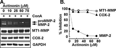
The lectin from Canavalia ensiformis (Concanavalin-A, ConA), one of the most abundant lectins known, enables one to mimic biological lectin/carbohydrate interactions that regulate extracellular matrix protein recognition. As such, ConA is known to induce membrane type-1 matrix metalloproteinase (MT1-MMP) which expression is increased in brain cancer. Given that MT1-MMP correlated to high expression of cyclooxygenase (COX)-2 in gliomas with increasing histological grade, we specifically assessed the early proinflammatory cellular signaling processes triggered by ConA in the regulation of COX-2. We found that treatment with ConA or direct overexpression of a recombinant MT1-MMP resulted in the induction of COX-2 expression. This increase in COX-2 was correlated with a concomitant decrease in phosphorylated AKT suggestive of cell death induction, and was independent of MT1-MMP's catalytic function. ConA- and MT1-MMP-mediated intracellular signaling of COX-2 was also confirmed in wild-type and in Nuclear Factor-kappaB (NF-kappaB) p65(-/-) mutant mouse embryonic fibroblasts (MEF), but was abrogated in NF-kappaB1 (p50)(-/-) and in I kappaB kinase (IKK) gamma(-/-) mutant MEF cells. Collectively, our results highlight an IKK/NF-kappaB-dependent pathway linking MT1-MMP-mediated intracellular signaling to the induction of COX-2. That signaling pathway could account for the inflammatory balance responsible for the therapy resistance phenotype of glioblastoma cells, and prompts for the design of new therapeutic strategies that target cell surface carbohydrate structures and MT1-MMP-mediated signaling. Concise summary Concanavalin-A (ConA) mimics biological lectin/carbohydrate interactions that regulate the proinflammatory phenotype of cancer cells through yet undefined signaling. Here we highlight an IKK/NF-kappaB-dependent pathway linking MT1-MMP-mediated intracellular signaling to the induction of cyclooxygenase-2, and that could be responsible for the therapy resistance phenotype of glioblastoma cells.
Keywords: Cancer; Concanavalin-A; Cyclooxygenase; Inflammation; MT1-MMP.
Figures


Similar articles
-
A MT1-MMP/NF-kappaB signaling axis as a checkpoint controller of COX-2 expression in CD133+ U87 glioblastoma cells.J Neuroinflammation. 2009 Mar 9;6:8. doi: 10.1186/1742-2094-6-8. J Neuroinflammation. 2009. PMID: 19272160 Free PMC article.
-
Concanavalin-A-induced autophagy biomarkers requires membrane type-1 matrix metalloproteinase intracellular signaling in glioblastoma cells.Glycobiology. 2012 Sep;22(9):1245-55. doi: 10.1093/glycob/cws093. Epub 2012 Jun 12. Glycobiology. 2012. PMID: 22692046
-
Concanavalin-A triggers inflammatory response through JAK/STAT3 signalling and modulates MT1-MMP regulation of COX-2 in mesenchymal stromal cells.Exp Cell Res. 2012 Nov 15;318(19):2498-506. doi: 10.1016/j.yexcr.2012.08.003. Epub 2012 Aug 27. Exp Cell Res. 2012. PMID: 22971618
-
MEMBRANE TYPE 1-MATRIX METALLOPROTEINASE (MT1-MMP) IDENTIFIED AS A MULTIFUNCTIONAL REGULATOR OF VASCULAR RESPONSES.Fukushima J Med Sci. 2015;61(2):91-100. doi: 10.5387/fms.2015-15. Epub 2015 Sep 11. Fukushima J Med Sci. 2015. PMID: 26370683 Free PMC article. Review.
-
Concanavalin A as a promising lectin-based anti-cancer agent: the molecular mechanisms and therapeutic potential.Cell Commun Signal. 2022 Oct 26;20(1):167. doi: 10.1186/s12964-022-00972-7. Cell Commun Signal. 2022. PMID: 36289525 Free PMC article. Review.
Cited by
-
TIMP-2 Interaction with MT1-MMP Activates the AKT Pathway and Protects Tumor Cells from Apoptosis.PLoS One. 2015 Sep 2;10(9):e0136797. doi: 10.1371/journal.pone.0136797. eCollection 2015. PLoS One. 2015. PMID: 26331622 Free PMC article.
-
Abrus agglutinin suppresses human hepatocellular carcinoma in vitro and in vivo by inducing caspase-mediated cell death.Acta Pharmacol Sin. 2014 Jun;35(6):814-24. doi: 10.1038/aps.2014.15. Epub 2014 May 5. Acta Pharmacol Sin. 2014. PMID: 24793310 Free PMC article.
-
Impact of Concanavalin-A-Mediated Cytoskeleton Disruption on Low-Density Lipoprotein Receptor-Related Protein-1 Internalization and Cell Surface Expression in Glioblastomas.Biomark Cancer. 2016 May 19;8:77-87. doi: 10.4137/BIC.S38894. eCollection 2016. Biomark Cancer. 2016. PMID: 27226736 Free PMC article.
-
Tetracycline derivative minocycline inhibits autophagy and inflammation in concanavalin-a-activated human hepatoma cells.Gene Regul Syst Bio. 2014 Mar 4;8:63-73. doi: 10.4137/GRSB.S13946. eCollection 2014. Gene Regul Syst Bio. 2014. PMID: 24634581 Free PMC article.
-
Probing into the chemopreventive properties of synthetic 1,3,6-tri-O-galloyl-α-D-glucose (α-TGG) against glioblastoma and triple-negative breast cancer-derived cell models.Curr Res Pharmacol Drug Discov. 2025 Apr 5;8:100219. doi: 10.1016/j.crphar.2025.100219. eCollection 2025. Curr Res Pharmacol Drug Discov. 2025. PMID: 40248812 Free PMC article.
References
-
- Anilkumar N, Uekita T, Couchman JR, Nagase H, Seiki M, Itoh Y. Palmitoylation at Cys574 is essential for MT1-MMP to promote cell migration. FASEB J. 2005;19:1326–1328. - PubMed
-
- Annabi B, Lee YT, Martel C, Pilorget A, Bahary JP, Béliveau R. Radiation induced-tubulogenesis in endothelial cells is antagonized by the antiangiogenic properties of green tea polyphenol (–) epigallocatechin-3-gallate. Cancer Biol Ther. 2003;2:642–649. - PubMed
-
- Annabi B, Thibeault S, Lee YT, Bousquet-Gagnon N, Eliopoulos N, Barrette S, Galipeau J, Beliveau R. Matrix metalloproteinase regulation of sphingosine-1-phosphate-induced angiogenic properties of bone marrow stromal cells. Exp Hematol. 2003;31:640–649. doi: 10.1016/S0301-472X(03)00090-0. - DOI - PubMed
Grants and funding
LinkOut - more resources
Full Text Sources
Research Materials
