. 2010 Mar;4(1):39-50.
doi: 10.1007/s12079-010-0085-z. Epub 2010 Feb 9.

CCN3/CCN2 regulation and the fibrosis of diabetic renal disease

CCN3/CCN2 regulation and the fibrosis of diabetic renal disease

Bruce L Riser et al. J Cell Commun Signal. 2010 Mar.

Abstract

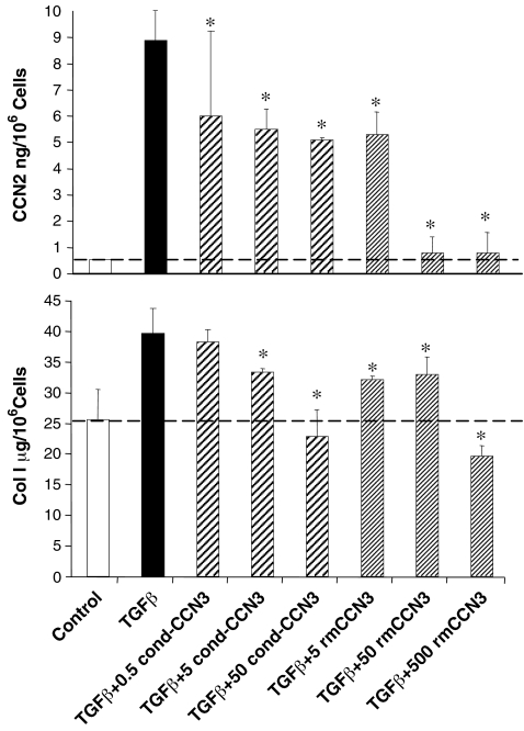
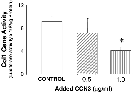
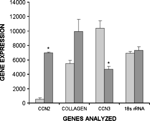
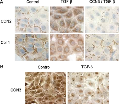
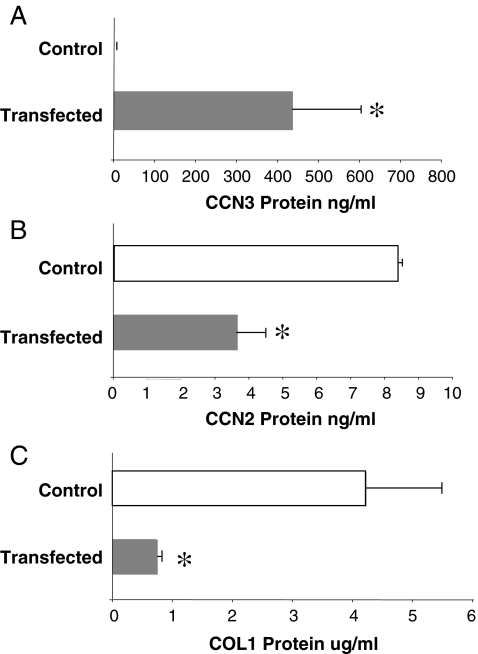
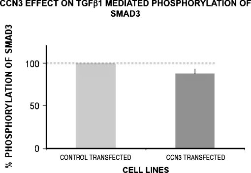
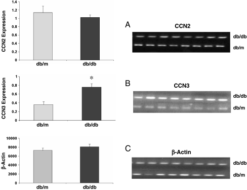
Prior work in the CCN field, including our own, suggested to us that there might be co-regulatory activity and function as part of the actions of this family of cysteine rich cytokines. CCN2 is now regarded as a major pro-fibrotic molecule acting both down-stream and independent of TGF-beta1, and appears causal in the disease afflicting multiple organs. Since diabetic renal fibrosis is a common complication of diabetes, and a major cause of end stage renal disease (ESRD), we examined the possibility that CCN3 (NOV), might act as an endogenous negative regulator of CCN2 with the capacity to limit the overproduction of extracellular matrix (ECM), and thus prevent, or ameliorate fibrosis. We demonstrate, using an in vitro model of diabetic renal fibrosis, that both exogenous treatment with CCN3 and transfection with the over-expression of the CCN3 gene in mesangial cells markedly down-regulates CCN2 activity and blocks ECM over-accumulation stimulated by TGF-beta1. Conversely, TGF-beta1 treatment reduces endogenous CCN3 expression and increases CCN2 activity and matrix accumulation, indicating an important, novel yin/yang effect. Using the db/db mouse model of diabetic nephropathy, we confirm the expression of CCN3 in the kidney, with temporal localization that supports these in vitro findings. In summary, the results corroborate our hypothesis that one function of CCN3 is to regulate CCN2 activity and at the concentrations and conditions used down-regulates the effects of TGF-beta1, acting to limit ECM turnover and fibrosis in vivo. The findings suggest opportunities for novel endogenous-based therapy either by the administration, or the upregulation of CCN3.

Keywords: Anti-fibrotic therapy; CCN regulation; Diabetic nephropathy; Fibrosis.


Figures

Fig. 1
Fig. 1
CCN3 reduces TGFβ1-stimulated CCN2 and COL1 production in cultured rat renal mesangial cells (MC). CCN2 secretion is significantly increased by addition of TGFβ1 (2 ng/ml) alone to the culture medium for 96 h, and this stimulated production is reduced in a dose-dependent fashion by adding either a conditioned medium (0.5–50 ng/ml, cond-CCN3) from NCI-H295R cells enriched in CCN3, or direct addition of recombinant mouse CCN3 (5–500 ng/ml, rmCCN3) for 1 h prior to treatment with TGFβ1. A similar pattern is observed with TGFβ1-stimulated COL1 production in MC. The error bars represent the mean ± the standard deviation for three measurements. * indicates a significant difference (P < 0.05) from TGF-β1 stimulated conditions due to CCN3 treatment
Fig. 2
Fig. 2
CCN3 inhibits COL1 promoter activity in cultured MC. Human col1 promoter was linked to luciferase in MC, and the COL1 promoter activity was measured as luciferase activation. Renilla-luciferase pRL-SV40 was used as a control to normalize for transfection efficiency (data not shown). CCN3 (0.5 or 1.0 μg/ml) was administered 24 h after transfection of the promoter construct, and significantly inhibited COL1 promoter activity in a dose dependent manner. The error bars represent the mean ± the standard deviation for three separate transfections. * indicates a significant difference of P = 0.001
Fig. 3
Fig. 3
TGFβ1 exposure induces opposite effects on mRNA levels for CCN2 and COL1 versus CCN3 in cultured MC. TGFβ1 (2 ng/ml) significantly increased expression of CCN2 (* is P = 0.00001) and COL1 (P = 0.06) when added to the culture medium, but reduced CCN3 expression (* is P = 0.01) as determined by semiquantitative RT-PCR measured in the linear range. This semi-quantitative data were obtained by scanning PCR DNA fragments using densitometry (arbitrary units). The values were normalized utilizing 18s rRNA as an internal control. TGFβ1 did not affect expression of 18s RNA. The error bars represent the mean ± the standard deviation among three replicates. The experiement was repeated with similar results
Fig. 4
Fig. 4
TGFβ1 causes converse effects on CCN2 and COL1 localization in cultured MC (48 h) compared to CCN3, and pre-treatment with CCN3 for 1 h reverses the localization pattern for CCN2 and COL1 in response to TGFβ1. Using immunocytochemistry, CCN2 and COL1 localize as dense staining in the peripheral regions of the cells under control conditions (a), but this localization is lost when TGFβ1 (5 ng/ml) is added to the incubation medium, and a new pattern of diffuse but intense, staining throughout the cytoplasm is observed (a). In each case, addition of CCN3 (500 ng/ml) to the incubation medium reverses the effects of TGFβ1 (a). Conversely, CCN3 is homogenously distributed throughout the cytoplasm in the absence of TGFβ1 (b), but becomes more diffusely distributed and accumulates as dense bodies at the cell periphery with addition of TGFβ1 (b)
Fig. 5
Fig. 5
Transfection of cultured MC with human recombinant CCN3 (hCCN3) causes a reduction in CCN2 and COL1 production. Panel A confirms that transfection of MC with hrCCN3 produces a significant increase in CCN3 protein, as determined by indirect ELISA (anti-human antibody). Specificity is indicated by a zero control value (a). This increased production of endogenous CCN3 causes a resultant reduction in CCN2 (b) and COL1 (c) protein in the cells. The error bars represent the mean ± the standard deviation for three measurements. * indicates a significant difference at P < 0.05
Fig. 6
Fig. 6
Transfection of cultured MC with CCN3 does not significantly reduce TGF-β1 stimulated pSmad3 activity. CCN3 over-expressing cells were stimulated with TGF-β1 (3 ng/ml). The transfected cells had 88.1 ± 5.7 (mean±SD) percent of control pSmad3 activity (P = 0.1) for two separate experiments
Fig. 7
Fig. 7
CCN3 expression is increased at a late stage in a model of type two diabetic renal fibrosis, and CCN2 expression is not significantly different from control. Graphs show CCN2 (a) and CCN3 (b) expression in kidney cortex of diabetic (db/db) and control (db/m) mice, using semi-quantitative RT-PCR, after 5 months of disease. Data is expressed as the intensity of RT-PCR bands (arbritrary densitometry units). Values are normalized by using β-actin as a housekeeping gene (c). There is a significant large increase (* is P < 0.01) in CCN3 mRNA, and no significant change in CCN2 mRNA. Error bars represent the mean ± the standard error for eight measurements
Fig. 8
Fig. 8
CCN3 is excreted in the urine of diabetic mice with late stage fibrosis, but not in non-diabetic control animals. Western blot analyses of the urines from diabetic (db/db,) and control (db/m) mice show multiple immunoreactive forms of CCN3 in the diabetic animals, but virtually none in healthy control mice. This includes a strong band in the diabetic mice at approximately 55 kDa (likely representing full-length CCN3), and a band at approximately 27 kDa (likely representing a half-fragment of CCN3)
Fig. 9
Fig. 9
A working hypothesis for the regulation of CCN2 and COL1 formation in wound healing and fibrosis. In response to stimuli favoring wound healing, TGFβ causes an increase in CCN2 production, which in turn stimulates COL1 formation and synthesis of the extracellular matrix (ECM). CCN3, an endogenous inhibitor of CCN2 production and COL1 formation, is transiently inhibited by TGFβ, thus allowing remodeling of the ECM. During the later stages of wound healing, however, inhibition of CCN3 is reversed by a mechanism not yet defined (?), thereby suppressing COL1 production and terminating the process of wound healing. During fibrosis, the normal up-regulation of CCN3 following the initial phase of COL1 production is prevented, resulting in an uncontrolled production of extracellular matrix leading to fibrotic changes

References

    1. Abdel-Wahab N, Weston BS, Roberts T, Mason RM. Connective tissue growth factor and regulation of the mesangial cell cycle: role in cellular hypertrophy. J Am Soc Nephrol. 2002;13(10):2437–2445. doi: 10.1097/01.ASN.0000031828.58276.02. - DOI - PubMed
    1. Bollineni JS, Reddi AS. Transforming growth factor-beta 1 enhances glomerular collagen synthesis in diabetic rats. Diabetes. 1993;42(11):1673–1677. doi: 10.2337/diabetes.42.11.1673. - DOI - PubMed
    1. Breyer MD, Bottinger E, Brosius FC, 3rd, Coffman TM, Harris RC, Heilig CW, et al. Mouse models of diabetic nephropathy. J Am Soc Nephrol. 2005;16(1):27–45. doi: 10.1681/ASN.2004080648. - DOI - PubMed
    1. Brigstock DR, Steffen CL, Kim GY, Vegunta RK, Diehl JR, Harding PA. Purification and characterization of novel heparin-binding growth factors in uterine secretory fluids. Identification as heparin-regulated Mr 10,000 forms of connective tissue growth factor. J Biol Chem. 1997;272(32):20275–20282. doi: 10.1074/jbc.272.32.20275. - DOI - PubMed
    1. Brigstock DR, Goldschmeding R, Katsube KI, Lam SC, Lau LF, Lyons K, et al. Proposal for a unified CCN nomenclature. Mol Pathol. 2003;56(2):127–128. doi: 10.1136/mp.56.2.127. - DOI - PMC - PubMed