Comparative Study
. 2010 Feb 26;6(2):e1000788.
doi: 10.1371/journal.ppat.1000788.

Quantitative comparison of HTLV-1 and HIV-1 cell-to-cell infection with new replication dependent vectors

Affiliations
Comparative Study

Quantitative comparison of HTLV-1 and HIV-1 cell-to-cell infection with new replication dependent vectors

Dmitriy Mazurov et al. PLoS Pathog. .

Abstract

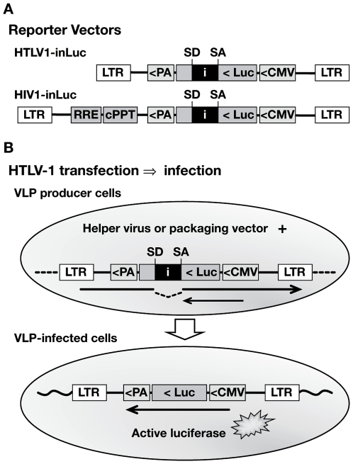
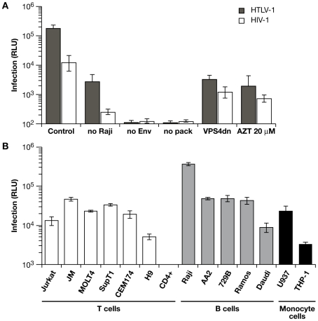
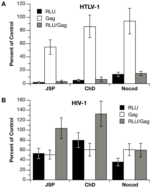
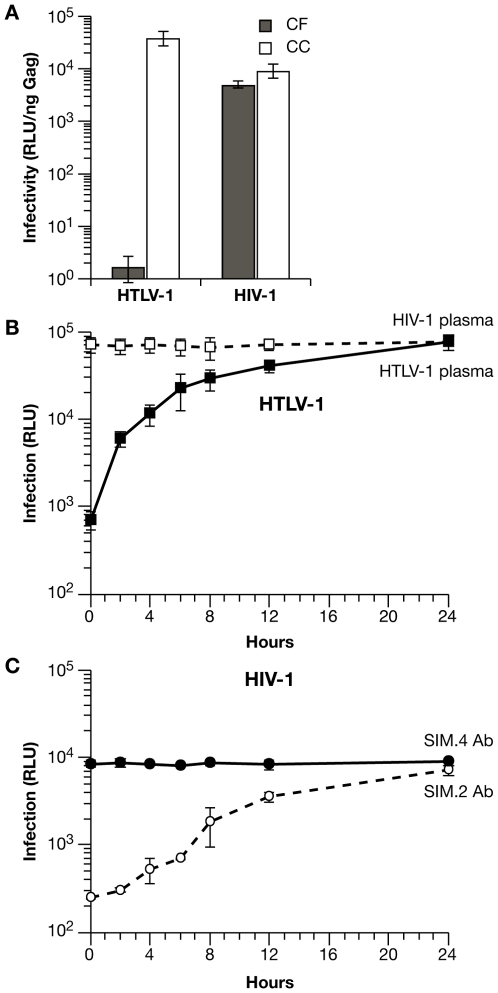
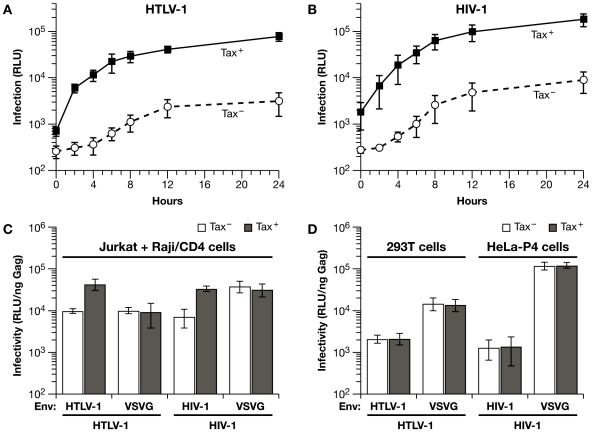
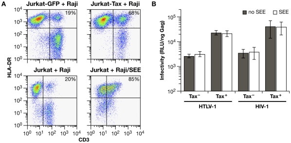
We have developed an efficient method to quantify cell-to-cell infection with single-cycle, replication dependent reporter vectors. This system was used to examine the mechanisms of infection with HTLV-1 and HIV-1 vectors in lymphocyte cell lines. Effector cells transfected with reporter vector, packaging vector, and Env expression plasmid produced virus-like particles that transduced reporter gene activity into cocultured target cells with zero background. Reporter gene expression was detected exclusively in target cells and required an Env-expression plasmid and a viral packaging vector, which provided essential structural and enzymatic proteins for virus replication. Cell-cell fusion did not contribute to infection, as reporter protein was rarely detected in syncytia. Coculture of transfected Jurkat T cells and target Raji/CD4 B cells enhanced HIV-1 infection two fold and HTLV-1 infection ten thousand fold in comparison with cell-free infection of Raji/CD4 cells. Agents that interfere with actin and tubulin polymerization strongly inhibited HTLV-1 and modestly decreased HIV-1 cell-to-cell infection, an indication that cytoskeletal remodeling was more important for HTLV-1 transmission. Time course studies showed that HTLV-1 transmission occurred very rapidly after cell mixing, whereas slower kinetics of HIV-1 coculture infection implies a different mechanism of infectious transmission. HTLV-1 Tax was demonstrated to play an important role in altering cell-cell interactions that enhance virus infection and replication. Interestingly, superantigen-induced synapses between Jurkat cells and Raji/CD4 cells did not enhance infection for either HTLV-1 or HIV-1. In general, the dependence on cell-to-cell infection was determined by the virus, the effector and target cell types, and by the nature of the cell-cell interaction.


Conflict of interest statement

The authors have declared that no competing interests exist.

Figures

Figure 1
Figure 1. Specialized HTLV-1 and HIV-1 reporter vectors to measure single-cycle replication in coculture infection.
(A) Replication-dependent HTLV-1 and HIV-1 transfer vectors contain an antisense-oriented expression cassette, which consists of a CMV promoter, firefly luciferase (luc) gene, and TK polyA signal (PA). The luc gene is interrupted by an intron (i), which is oriented in the sense direction, indicated by splice donor (SD) and splice acceptor (SA) sites. The HIV1-inLuc vector contains a Rev responsive element (RRE) and central polypurine tract (cPPT). Analogous vectors were constructed that contain a yellow fluorescent protein gene with intron (inYFP). (B) Translation of the reporter protein mRNA (small arrow) in transfected cells is blocked by the intron. The vector mRNA (large arrow) is spliced and packaged into VLPs; after infection and replication, a provirus is formed that lacks the intron and is capable of reporter gene expression.
Figure 2
Figure 2. Characteristics of the replication dependent vectors in coculture infections.
(A) HTLV1-inLuc and HIV1-inLuc vectors were transfected into Jurkat cells with respective packaging vectors and Env expression plasmids; transfected Jurkat cells were combined with an equal number of Raji/CD4 cells 24 h later. Infection is expressed as relative light units (RLU) of luciferase activity, measured 48 h after cell mixing. Coculture of Raji/CD4 cells with Jurkat cells transfected with wild type vectors is designated as control. The level of infection between Jurkat cells was determined in cultures without Raji/CD4 cells (no Raji). In other cocultures, Jurkat cells transfected with reporter vectors but without Env-expression plasmid (no Env) or without packaging plasmid (no pack) produced no Luc activity above background. Cotransfection of the viral vectors with a dominant-negative VPS4A plasmid (VPSdn), which inhibits virus budding, or addition of the reverse transcriptase inhibitor azidotimidine (AZT 20 µm) inhibited Luc transduction. (B) Jurkat cells were transfected with HTLV-1 packaging plasmid and HTLV1-inLuc reporter vector and then incubated with an equal number of the indicated cells. Luc activity in the target cells was assayed 48 h after cell mixing. Luc activity was not detected in activated human CD4+ T cells from different donors in repeated experiments. Graphs represent the mean of at least three independent experiments with error bars indicating standard deviations.
Figure 3
Figure 3. Effects of cytoskeleton disrupting agents on coculture infection with HTLV-1 and HIV-1 vectors.
(A) Jurkat cells were transfected with HTLV-1 vectors or (B) HIV-1 vectors (plus HTLV-1 Tax expression plasmid) and then mixed with an equal number of Raji/CD4 cells. Cocultures were left untreated or treated for the duration of the coculture with 100 nM jasplakinolide (Jsp), 5 µM cytochalasin D (ChD), or 5 µM nocodozole (nocod). Values for infection (RLU of Luc activity) and Gag concentration in culture supernatants (Gag) are expressed as percent relative to untreated controls. For HTLV-1 vectors, Luc activity was 3.54×10⁵ RLU and Gag (p19) concentration was 1.18 ng per ml in the untreated control. For HIV-1 vectors, Luc activity was 2.83×10⁵ RLU and Gag (p24) concentration was 1.81 ng per ml in the untreated control. Graphs represent the mean of at least three independent experiments with error bars indicating standard deviations.
Figure 4
Figure 4. Differences in the mechanisms of transmission for HTLV-1 and HIV-1 in Jurkat-Raji/CD4 cocultures.
(A) Comparison of cell-free (CF) and coculture (CC) infection. Raji/CD4 cells were infected with cell-free VLPs produced from transfected 293T cells or by coculture with transfected Jurkat cells. Luc activity is normalized relative to the amount of viral Gag protein in the supernatants. (B) The time course of HTLV-1 coculture infection was examined by blocking virus entry and infection with HTLV-1-infected human plasma (filled squares) at the indicated times after mixing transfected Jurkat cells and Raji/CD4 cells. Luc activity was measured 48 h after the start of coculture (0 h). For control infections, human plasma from HIV-1-infected patients was used (open squares). (C) Time course of HIV-1 coculture infection was examined by blocking virus entry and infection as in (B) except that anti-CD4 mAb SIM.2 (open circles) was used to block entry and non-blocking anti-CD4 mAb SIM.4 was used as control (filled circles). Luc activity was measured 48 h after the start of coculture (0 hr). These experiments were performed at least 3 times; error bars depict standard deviations.
Figure 5
Figure 5. HTLV-1 Tax expression in Jurkat cells enhances coculture infection of Raji/CD4 cells with HIV-1 and HTLV-1 vectors.
(A) Time course coculture infection experiments were performed as in figure 4B except that Jurkat cells were transfected with either wt (Tax+) or mutated (Tax) HTLV-1 packaging vectors. (B) A coculture infection time course experiment was performed as in figure 4C except that Jurkat cells were transfected with HIV-1 vectors with either a Tax-expression vector (Tax+) or empty vector (Tax). (C) HIV-1 and HTLV-1 VLPs were pseudotyped with VSV-G or their own Env and examined in Jurkat-Raji/CD4 coculture infection system with or without Tax expression. Infectivity is expressed as Luc activity (RLU) relative to the amount of Gag protein in the culture medium at 48 h. (D) Human 293T cells or HeLa-P4 cells were transfected with the same plasmids as in panel C. The medium was replaced the next day and the infection was allowed to develop until 48 h after transfection. Infectivity is expressed relative to the concentration of Gag in the supernatant. All results represent the mean of at least three independent experiments with standard deviations represented by error bars.
Figure 6
Figure 6. A superantigen-induced immunological synapse does not enhance infection with HIV-1 or HTLV-1 VLPs.
(A) Conjugate formation between Jurkat cells and Raji/CD4 cells was quantified by flow cytometry. One hour after mixing, cells were fixed and stained with anti-CD3-FITC (Jurkat cells) and anti-HLA-DR-PE antibodies (Raji/CD4 cells); cell-cell conjugates are identified as double-positive signals. The basal level of conjugate formation was approximately 20% (left hand panels). In the upper panels, Jurkat cells were transduced with lentivirus vectors encoding GFP only (left) or Tax-IRES-GFP (right) 48 hrs before mixing with Raji/CD4 cells. In the lower panels, Raji/CD4 cells were untreated (left) or were pulsed with SEE (right) prior to mixing with Jurkat cells. Typical Dot Plots from one of three experiments are presented. (B) Effects of SEE-induced synapse formation on HIV-1 and HTLV-1 infection in Jurkat-Raji/CD4 cocultures. Luc activity is normalized relative to the amount of Gag in culture supernatants at the end of infection. The histograms represent the mean of four independent experiments.

Similar articles

Cited by

References

    1. Johnson DC, Huber MT. Directed egress of animal viruses promotes cell-to-cell spread. J Virol. 2002;76:1–8. - PMC - PubMed
    1. Jolly C, Kashefi K, Hollinshead M, Sattentau QJ. HIV-1 cell to cell transfer across an Env-induced, actin-dependent synapse. J Exp Med. 2004;199:283–293. - PMC - PubMed
    1. Marsh M, Helenius A. Virus entry: open sesame. Cell. 2006;124:729–740. - PMC - PubMed
    1. Phillips DM. The role of cell-to-cell transmission in HIV infection. Aids. 1994;8:719–731. - PubMed
    1. Piguet V, Sattentau Q. Dangerous liaisons at the virological synapse. J Clin Invest. 2004;114:605–610. - PMC - PubMed

Publication types