. 2010 Feb 26;6(2):e1000863.
doi: 10.1371/journal.pgen.1000863.

Cdk2 is required for p53-independent G2/M checkpoint control

Affiliations

Cdk2 is required for p53-independent G2/M checkpoint control

Jon H Chung et al. PLoS Genet. .

Abstract

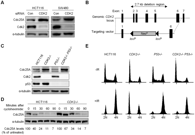
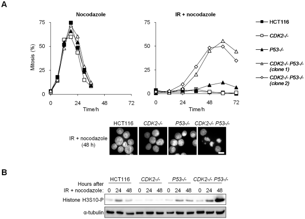
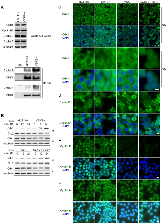
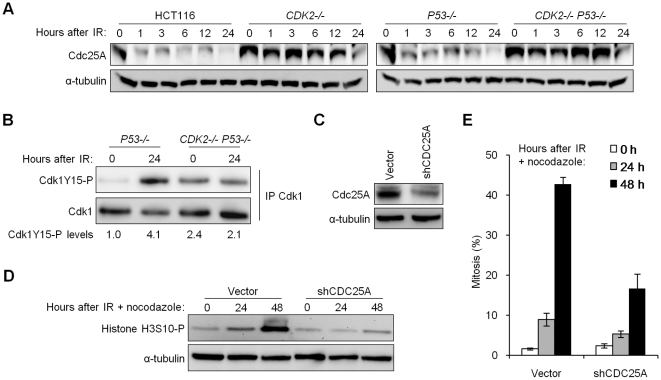
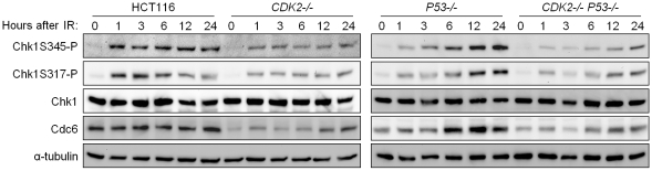
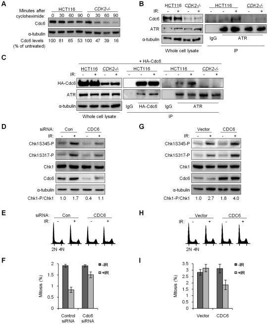
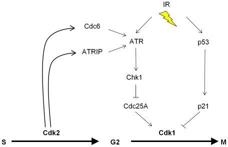
The activation of phase-specific cyclin-dependent kinases (Cdks) is associated with ordered cell cycle transitions. Among the mammalian Cdks, only Cdk1 is essential for somatic cell proliferation. Cdk1 can apparently substitute for Cdk2, Cdk4, and Cdk6, which are individually dispensable in mice. It is unclear if all functions of non-essential Cdks are fully redundant with Cdk1. Using a genetic approach, we show that Cdk2, the S-phase Cdk, uniquely controls the G(2)/M checkpoint that prevents cells with damaged DNA from initiating mitosis. CDK2-nullizygous human cells exposed to ionizing radiation failed to exclude Cdk1 from the nucleus and exhibited a marked defect in G(2)/M arrest that was unmasked by the disruption of P53. The DNA replication licensing protein Cdc6, which is normally stabilized by Cdk2, was physically associated with the checkpoint regulator ATR and was required for efficient ATR-Chk1-Cdc25A signaling. These findings demonstrate that Cdk2 maintains a balance of S-phase regulatory proteins and thereby coordinates subsequent p53-independent G(2)/M checkpoint activation.


Conflict of interest statement

The authors have declared that no competing interests exist.

Figures

Figure 1
Figure 1. Altered cell cycle regulation and increased Cdc25A protein in Cdk2-deficient human cancer cells.
(A) Human colorectal cancer cells HCT116 and SW480 transfected with control (Con) or CDK2 (CDK2) siRNA. Levels of Cdc25A and Cdk2 were analyzed by immunoblot. α-tubulin was assessed as a loading control. (B) CDK2 targeting strategy. Exons 2-5 at the CDK2 locus were replaced upon integration of the knockout vector. (C) Levels of Cdc25A, Cdk2 and p53 were determined by immunoblot in parental cells and single and double knockout cells. (D) Isogenic HCT116 cells were harvested at the indicated times after cycloheximide treatment. Cdc25A protein levels were analyzed by immunoblot, quantitated, normalized to α-tubulin and represented as a percentage relative to untreated control cells. (E) Isogenic HCT116 cells were fixed and stained with Hoechst 33258 and subjected to flow cytometry, before and 24 h after treatment with 12 Gy IR. Positions of cell populations with 2N and 4N DNA content are indicated.
Figure 2
Figure 2. Cdk2 is required for G2/M arrest in p53-deficient cells.
(A,B) Entry into mitosis was assessed by incubation of untreated or irradiated (12 Gy) isogenic HCT116 cells in 0.2 µg/ml nocodazole. Two independent CDK2−/− P53−/− clones are shown. Cells were collected at the indicated times. (A) Cells were fixed and stained with Hoechst 33258. Mitotic chromosome condensation was determined by fluorescence microscopy, images were taken at 63× magnification (scale bar, 10 µm). (B) Levels of histone H3S10-P were determined by immunoblot.
Figure 3
Figure 3. Aberrant localization of Cdk1 in Cdk2-deficient cells after IR treatment.
(A) Association between Cdk1 and cyclins E and A was determined by immunoprecipitation/immunoblot. Non-denatured cell lysates were subjected to immunoprecipitation (IP) with anti-Cdk1 or control (IgG) antibodies. Samples not subjected to immunoprecipitation were analyzed as whole cell lysate. Levels of Cdk1 and cyclins B1, E, and A were determined by immunoblot. (B) Cdk1 levels in nuclear and cytoplasmic fractions were determined by immunoblot at the indicated times after treatment with 12 Gy IR. Loading of nuclear and cytoplasmic proteins was assessed by probing levels of Orc2 and α-tubulin, respectively. (C) Localization of Cdk1 by indirect immunofluorescence. Isogenic HCT116 cells were fixed 24 h after treatment with 12 Gy IR, stained with Cdk1 antibody (green) and counterstained with DAPI (blue). Representative fields are shown under low (20×) and high (63×) magnification (scale bar, 50 µm under 20× magnification and 10 µm under 63× magnification). (D–F) Localization of cyclins by indirect immunofluorescence. Isogenic HCT116 cells were fixed 24 h after treatment with 12 Gy IR, stained with (D) cyclin B1, (E) cyclin E or (F) cyclin A antibodies (green) and counterstained with DAPI (blue). Representative fields are shown under 40× magnification (scale bar, 10 µm).
Figure 4
Figure 4. Impaired degradation of Cdc25A in Cdk2-deficient cells contributes to G2/M checkpoint defect.
(A) Isogenic HCT116 cells were harvested at the indicated times after treatment with 12 Gy IR. Cdc25A levels were determined by immunoblot. (B) Non-denatured cell lysates were collected before and 24 h after 12 Gy IR. Cdk1 was immunoprecipitated (IP) and analyzed for phosphorylation on Y15 by immunoblot. Cdk1Y15-P was quantitated and normalized to total Cdk1. (C) Stable knockdown of Cdc25A in CDK2−/− P53−/− cells by retrovirus-mediated delivery of empty vector control (vector) or CDC25A shRNA (shCDC25A), assessed by immunoblot. (D) Cdc25A knockdown or control CDK2−/− P53−/− cells were treated with 12 Gy IR and 0.2 µg/ml nocodazole. Histone H3S10-P levels were determined by immunoblot. (E) Mitotic indices of Cdc25A-knockdown and control CDK2−/− P53−/− cells at the indicated times after treatment with 12 Gy IR and 0.2 µg/ml nocodazole. Error bars represent s.e.m. from six knockdown clones.
Figure 5
Figure 5. Impaired phosphorylation of Chk1 in Cdk2-deficient cells.
Isogenic cells were treated with 12 Gy IR and harvested at the indicated time points. Levels of the Chk1 phosphoproteins, Chk1S345-P and Chk1S317-P, Chk1 and Cdc6 were determined by immunoblot. Levels of α-tubulin were assessed as a loading control.
Figure 6
Figure 6. Stabilized Cdc6 facilitates Chk1 phosphorylation, associates with ATR, and controls mitotic entry.
(A) Cdc6 protein stability was analyzed by immunoblot in wild-type and CDK2−/− cells at the indicated times after cycloheximide treatment. Cdc6 levels were quantitated, normalized to α-tubulin, and represented as a percentage relative to untreated control cells. (B) Physical interaction between endogenous Cdc6 and ATR. Non-denatured cell lysates from the indicated cell lines were collected before or 4 h after 12 Gy IR. Lysates were subjected to immunoprecipitation (IP) with anti–ATR or isotypic control (IgG) antibodies. Samples not subjected to immunoprecipitation were analyzed as whole cell lysate. Levels of ATR and Cdc6 were determined by immunoblot. (C) Isogenic HCT116 cells were transfected with a plasmid expressing HA-tagged Cdc6 (HA-Cdc6). Transfected cells were untreated or treated with 12 Gy IR and lysed after 4 h. Non-denatured cell lysates were subjected to immunoprecipitation (IP) with anti–HA, anti–ATR, or isotypic control (IgG) antibodies. Samples not subjected to immunoprecipitation were analyzed as whole cell lysate. Levels of ATR and HA-Cdc6 were determined by immunoblot. (D–F) U2OS cells were transfected with control (Con) or CDC6 (CDC6) siRNA. (D) Lysates were taken before and 4 h after treatment with 12 Gy IR. Chk1 and Cdc6 levels were analyzed by immunoblot. Relative Chk1-phosphorylation (Chk1-P/Chk1) was determined by quantitation of Chk1S317-P plus Chk1S345-P followed by normalization to total Chk1. (E) Cells were fixed and stained with Hoechst 33258 and subjected to flow cytometry before and 4 h after treatment with 12 Gy IR. (F) Cells fixed and stained for histone H3S10-P before and 24 h after treatment with 4 Gy IR and 1 µg/ml nocodazole, mitotic index was determined by indirect immunofluorescence. Error bars represent s.e.m. from three independent experiments. (G–I) CDK2−/− P53−/− cells were transfected with an empty vector or Cdc6 expression vector. (G) Lysates were taken before and 4 h after treatment with 12 Gy IR. Chk1 and Cdc6 levels were analyzed by immunoblot. (H) Cells were fixed and stained with Hoechst 33258 and subjected to flow cytometry before and 4 h after treatment with 12 Gy IR. (I) Cells fixed and stained with Hoechst 33258 before and 24 h after treatment with 12 Gy IR and 0.2 µg/ml nocodazole, mitotic index was determined by fluorescence microscopy. Error bars represent s.e.m. from six independent experiments.
Figure 7
Figure 7. The proposed role of Cdk2 in the p53-independent regulation of Cdk1.
Cdk2 directly phosphorylates at least two proteins, ATRIP and Cdc6, that directly regulate the G2-M transition via the ATR-Chk1-Cdc25A pathway. The activity of Cdk1 is independently inhibited upon activation of the p53-p21 pathway, which is defective in many cancer cells. See text for additional details.

References

    1. Malumbres M, Barbacid M. Mammalian cyclin-dependent kinases. Trends Biochem Sci. 2005;30(11):630–641. - PubMed
    1. Sherr CJ, Roberts JM. Living with or without cyclins and cyclin-dependent kinases. Genes Dev. 2004;18(22):2699–2711. - PubMed
    1. Hochegger H, Takeda S, Hunt T. Cyclin-dependent kinases and cell-cycle transitions: Does one fit all? Nat Rev Mol Cell Biol. 2008;9(11):910–916. - PubMed
    1. Malumbres M, Barbacid M. Cell cycle, CDKs and cancer: A changing paradigm. Nat Rev Cancer. 2009;9(3):153–166. - PubMed
    1. Berthet C, Kaldis P. Cell-specific responses to loss of cyclin-dependent kinases. Oncogene. 2007;26(31):4469–4477. - PubMed

Publication types

MeSH terms