. 2010 Feb 26;6(2):e1000784.
doi: 10.1371/journal.ppat.1000784.

A major role for the Plasmodium falciparum ApiAP2 protein PfSIP2 in chromosome end biology

Affiliations

A major role for the Plasmodium falciparum ApiAP2 protein PfSIP2 in chromosome end biology

Christian Flueck et al. PLoS Pathog. .

Abstract

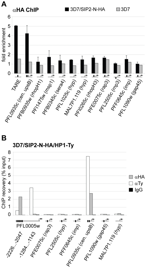
The heterochromatic environment and physical clustering of chromosome ends at the nuclear periphery provide a functional and structural framework for antigenic variation and evolution of subtelomeric virulence gene families in the malaria parasite Plasmodium falciparum. While recent studies assigned important roles for reversible histone modifications, silent information regulator 2 and heterochromatin protein 1 (PfHP1) in epigenetic control of variegated expression, factors involved in the recruitment and organization of subtelomeric heterochromatin remain unknown. Here, we describe the purification and characterization of PfSIP2, a member of the ApiAP2 family of putative transcription factors, as the unknown nuclear factor interacting specifically with cis-acting SPE2 motif arrays in subtelomeric domains. Interestingly, SPE2 is not bound by the full-length protein but rather by a 60kDa N-terminal domain, PfSIP2-N, which is released during schizogony. Our experimental re-definition of the SPE2/PfSIP2-N interaction highlights the strict requirement of both adjacent AP2 domains and a conserved bipartite SPE2 consensus motif for high-affinity binding. Genome-wide in silico mapping identified 777 putative binding sites, 94% of which cluster in heterochromatic domains upstream of subtelomeric var genes and in telomere-associated repeat elements. Immunofluorescence and chromatin immunoprecipitation (ChIP) assays revealed co-localization of PfSIP2-N with PfHP1 at chromosome ends. Genome-wide ChIP demonstrated the exclusive binding of PfSIP2-N to subtelomeric SPE2 landmarks in vivo but not to single chromosome-internal sites. Consistent with this specialized distribution pattern, PfSIP2-N over-expression has no effect on global gene transcription. Hence, contrary to the previously proposed role for this factor in gene activation, our results provide strong evidence for the first time for the involvement of an ApiAP2 factor in heterochromatin formation and genome integrity. These findings are highly relevant for our understanding of chromosome end biology and variegated expression in P. falciparum and other eukaryotes, and for the future analysis of the role of ApiAP2-DNA interactions in parasite biology.


Conflict of interest statement

The authors have declared that no competing interests exist.

Figures

Figure 1
Figure 1. Identification of the ApiAP2 protein PfSIP2 as the SPE2-interacting protein.
(A) Gel shift assay to test the course of affinity purification. Lane 1: probe only; lane 2: nuclear extract (input); lanes 3–5: supernatants after incubation of nuclear extract with beads carrying single-stranded DNA (3), SPE2 (4) or mutated SPE2M (5); lanes 7–10: proteins eluted from SPE2-loaded beads (SPE2 E). Lanes 11 and 12: A second eluate (SPE2 E2) and proteins bound to mutated SPE2M (SPE2mut E) contain no SPE2-binding activity. Faster migrating bands are probably due to degradation of PfSIP2-N during affinity purification. (B) Schematic representation of the ApiAP2 protein PfSIP2 encoded by PFF0200c and recombinant epitope-tagged PfSIP2 proteins expressed in either P. falciparum (SIP2-Ty; SIP2-N-HA; SIP2-N-Ty) or E. coli (SIP2-N-HIS_A; SIP2-N-HIS_B). Dark and light blue boxes indicated AP2 domains 1 and 2. (C) Size estimation of the endogenous SPE2-binding activity in parasite nuclear extracts by UV-crosslinking. The arrow highlights the size of the crosslinked DNA-protein complex (lanes 2 and 4). Complex formation is competed by a 25-fold molar excess of SPE2 (lane 3) but not by mutated SPE2M (lane 4). The DNA-protein complex is not observed in absence of radiolabeled SPE2 (lane 1) or without prior UV-crosslinking (lane 5). (D) Gel shift assay to confirm the identity of PFF0200c as the SPE2-binding protein. Lane 1: probe only; lanes 2–5: 3D7 nuclear extract; lanes 6–9: nuclear extract of 3D7/SIP2-N-HA over-expressing SIP2-N-HA (aa 1–387); lane 10: untransformed E. coli control lysate; lanes 11–14: lysate of E. coli expressing recombinant SIP2-N-HIS_A (aa 1–387); lanes 15–18: lysate of E. coli expressing recombinant SIP2-N-HIS_B (aa 171–387). Lanes 3, 7, 12 and 16: 100-fold molar excess of unlabeled SPE2 competitor. Lanes 4, 8, 13 and 17: 100-fold molar excess of mutated competitor SPE2M. Lanes 5, 9, 14 and 18: EMSA supershift in presence of anti-HA or anti-6×HIS antibodies. (E) Anti-Ty Western blot of 3D7/SIP2-Ty nuclear extracts prepared at five consecutive timepoints during the intra-erythrocytic developmental cycle (IDC). Expression of full-length PfSIP2-Ty and subsequent processing occur during schizogony. The bottom panel shows the presence of PfSIP2-N by EMSA in schizont extracts. Protein extracted from equal numbers of nuclei were used in each lane. hpi: hours post-invasion. (F) Pull-down of full-length PfSIP2-Ty and PfSIP2-N-Ty based on affinity to SPE2. PfSIP2-N-Ty binds efficiently to immobilized SPE2 whereas neither full-length PfSIP2-Ty nor the C-terminal processed fragment PfSIP2-C-Ty interact with SPE2. None of the proteins bound to mutated SPE2M DNA. SN: supernatant.
Figure 2
Figure 2. PfSIP2-N localizes to P. falciparum chromosome end clusters.
(A) IFA detects discrete perinuclear PfSIP2-N-HA signals in late trophozoites (LT) and early (ES) and late (LS) schizont stage 3D7/SIP2-N-HA parasites. The expression cassette is schematically depicted on the left. (B) Co-localisation of PfSIP2-N with PfHP1-containing subtelomeric heterochromatin in late trophozoites (LT) and early (ES) and late (LS) schizonts in the double transgenic parasite line 3D7/SIP2-N-HA/HP1-Ty (over-expressing both proteins as epitope-tagged versions simultaneously). Expression cassettes are schematically depicted on the left. (C) Targeted ChIP-qPCR analysis demonstrates the specific binding of PfSIP2-N-HA to SPE2 upstream of upsB var gene PFL0005w in 3D7/SIP2-N-HA. Fold enrichment of cross-linked PfSIP2-N-HA-associated chromatin was determined for five regions across the PFL0005w locus. Values represent the mean of three independent experiments (error bars indicate the standard deviation). The location of the SPE2 repeat array and the positions of qPCR primers are indicated by nucleotide positions with respect to the ATG start codon. 3D7 wild-type parasites were used as negative control.
Figure 3
Figure 3. Binding of PfSIP2-N to SPE2 is highly sequence-specific and requires a bipartite motif and both adjacent AP2 domains.
(A) SIP2-N in 3D7 nuclear extracts binds only to the bipartite SPE2 motif but not to a probe of identical length containing the SPE2 half site GTGCA only (GATACATGTGCAAACATGAA). C: SPE2/PfSIP2-N complex. (B) Both AP2 domains are required for binding of PfSIP2-N to SPE2. Left panel: Coomassie-stained SDS-PAGE gel showing recombinantly expressed PfSIP2-GST fusions in E. coli lysates. Lane 1: non-induced control lysate; lane 2: SIP2-AP2_1 consisting of the first AP2 domain only (aa 174–252); lane 3: SIP2-AP2_2 carries the second AP2 domain only (aa 231–311); lane 4: SIP2-AP2_12 contains both adjacent AP2 domains (aa 174–311). Right panel: EMSA showing that single isolated AP2 domains are unable to bind to SPE2 and that both AP2 domains are required for binding. C: SPE2/PfSIP2-GST complex. (C) Competition EMSA using recombinant SIP2-N-HIS_B to determine the minimal sequence requirements for binding of PfSIP2-N to a SPE2 consensus site. The gel was rotated by 90° clockwise for simpler display (the arrow on top indicates the direction of electrophoretic separation). Lane 1: radiolabeled 28bp SPE2 probe only; lane 2: SPE2/PfSIP2-N-HIS_B interaction in absence of competitor; lanes 3–19: SPE2/PfSIP2-N-HIS_B interaction in presence of a 50-fold molar excess of specific competitors. The names and sequences of all competitors (all 28bp) are indicated to the left. The dashed lines group competitors into artificially mutated SPE2 motifs or into naturally occurring SPE2-like elements upstream of P. falciparum invasion genes or upsB promoters (M1.C1, M1.A1C2). Altered nucleotides in the left or right half site of the original SPE2 sequence are highlighted in red. Additional nucleotides in the 4bp spacer are indicated in green. Red squares identify competitors unable to interact with PfSIP2-N-HIS_B, green squares highlight competitors that were able to compete with the SPE2/PfSIP2-N-HIS_B interaction. The experimentally determined SPE2 consensus site is shown at the bottom. (D) SPE2 consensus motifs define subtelomeric landmarks at P. falciparum chromosome ends. The position of all SPE2 motifs (arrowheads) at the right end of chromosome 3 is shown. Blue and purple arrowheads represent SPE2 motifs with four and five bp spacing between the half sites, respectively. Arrowhead orientation indicates the presence of SPE2 motifs on the sense or antisense strand. T1-T6: TARE 1 to 6. The telomere is depicted as a black box. The upsB var gene PFC1120c represents the first coding sequence downstream of TARE6. See also Tables S3 and S4 for more information. Chromosomal coordinates are according to PlasmoDB version5.5 annotation (www.plasmodb.org).
Figure 4
Figure 4. PfSIP2-N binds to SPE2 landmarks in subtelomeric regions of P. falciparum chromosomes in vivo.
(A) Genome-wide PfSIP2-N-HA occupancy as determined by high-density ChIP-on-chip. Schematic display (SignalMap) and localization of genomic regions bound by PfSIP2-N-HA in 3D7/SIP2-N-HA schizont stage parasites (blue lines) and the position of predicted SPE2 consensus sites (red lines) on all 14 parasite chromosomes. Genomic regions were considered occupied by PfSIP2-N-HA if the average of log2 ratios (ChIP over input) for all probes in a 1000bp window was higher than 1.4. Chromosome numbers are indicated on the left, chromosomal positions on top. Solid black lines indicate regions not represented on the microarray (see also Table S3). (B) Regional zoom-in of the PfSIP2-N-HA ChIP-on-chip profile and predicted SPE2 motifs at the left arm of chromosome 10. PfSIP2-N-HA-occupied regions (peaks) have been identified by the built-in algorithm of SignalMap as explained above. The locations of the upsB var gene and downstream rif genes are indicated below. Chromosomal coordinates are according to PlasmoDB version5.5 annotation (www.plasmodb.org).
Figure 5
Figure 5. PfSIP2-N-HA binds exclusively to SPE2 elements located in heterochromatic TARE2/3 and upsB var gene domains.
(A) PfSIP2-N-HA binds exclusively to SPE2 sites in TARE2/3 and upstream of upsB var genes. A region located between TARE2 and TARE3, the regions upstream of the only internal upsB var gene PFL0935c, promoters of five loci carrying predicted SPE2 consensus sites, and promoters of five genes lacking a SPE2 consensus site were tested by ChIP-qPCR for in vivo binding of PfSIP2-N-HA. qPCR primers were directed against upstream regions (represented by horizontal lines with arrows). Fold enrichment values represent the mean of three independent experiments on 3D7/SIP2-N-HA schizont stage samples (error bars indicate the standard deviation). 3D7 wild-type parasites were used as negative control. Gene names and accession numbers are according to PlasmoDB version5.5 (www.plasmodb.org). SPE2 elements are indicated by thick vertical black lines. (B) PfSIP2-N/SPE2 is embedded in PfHP1-containing heterochromatin. Parallel ChIP-qPCR was performed on 3D7/SIP2-N-HA/HP1-Ty schizont stage parasites using anti-HA and anti-Ty antibodies to test for occupancy by PfSIP2-N-HA and PfHP1-Ty, respectively. Normal rabbit IgG was used as negative control for ChIP. qPCR primers were directed against upstream regions (represented by horizontal lines with arrows). Vertical thick lines indicate the presence of SPE2 consensus sites. PfSIP2-N-HA and PfHP1-Ty co-occupancy at these loci was confirmed by ChIP-re-ChIP in independent experiments (Figure S4).
Figure 6
Figure 6. Overexpression of PfSIP2-N-HA has no effect on global gene transcription.
Global transcript profiles in 3D7/SIP2-N-HA parasites were compared to the control line 3D7/camHG at four timepoints across the IDC. TP1: ring stages 4–14 hours post-invasion (hpi); TP2: late ring stages 14–24 hpi; TP3: trophozoites 24–34 hpi; TP4: schizonts 32–42 hpi. All genes transcribed at greater 3-fold difference in 3D7/SIP2-N-HA compared to the control in at least one timepoint are listed. Over-expression of pfsip2-n is shown on top. The heat map indicates fold differences in transcript abundance on a gradual scale (green: down-regulated; red: up-regulated). Gene annotations are listed to the right and are according to PlasmoDB version5.5 (www.plasmodb.org).

Similar articles

Cited by

References

    1. Pryde FE, Gorham HC, Louis EJ. Chromosome ends: all the same under their caps. Curr Opin Genet Dev. 1997;7:822–828. - PubMed
    1. Taddei A, Hediger F, Neumann FR, Gasser SM. The function of nuclear architecture: a genetic approach. Annu Rev Genet. 2004;38:305–345. - PubMed
    1. Louis EJ, Vershinin AV. Chromosome ends: different sequences may provide conserved functions. Bioessays. 2005;27:685–697. - PubMed
    1. Tham WH, Zakian VA. Transcriptional silencing at Saccharomyces telomeres: implications for other organisms. Oncogene. 2002;21:512–521. - PubMed
    1. Moazed D. Common themes in mechanisms of gene silencing. Mol Cell. 2001;8:489–498. - PubMed

Publication types

MeSH terms