. 2010 Feb 25;5(2):e9415.
doi: 10.1371/journal.pone.0009415.

SPARC deficiency results in improved surgical survival in a novel mouse model of glaucoma filtration surgery

Affiliations

SPARC deficiency results in improved surgical survival in a novel mouse model of glaucoma filtration surgery

Li-Fong Seet et al. PLoS One. .

Abstract

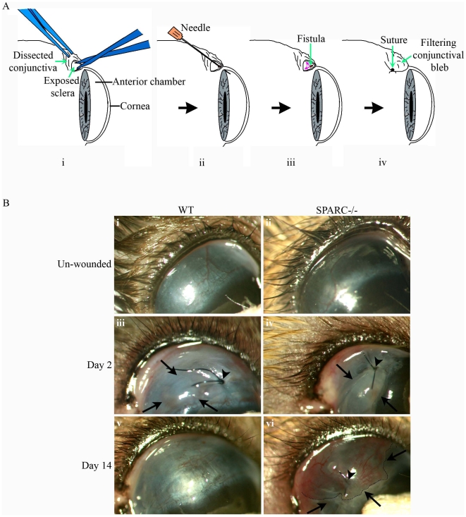
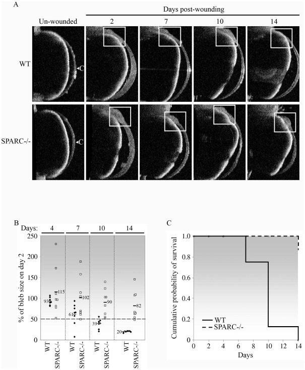
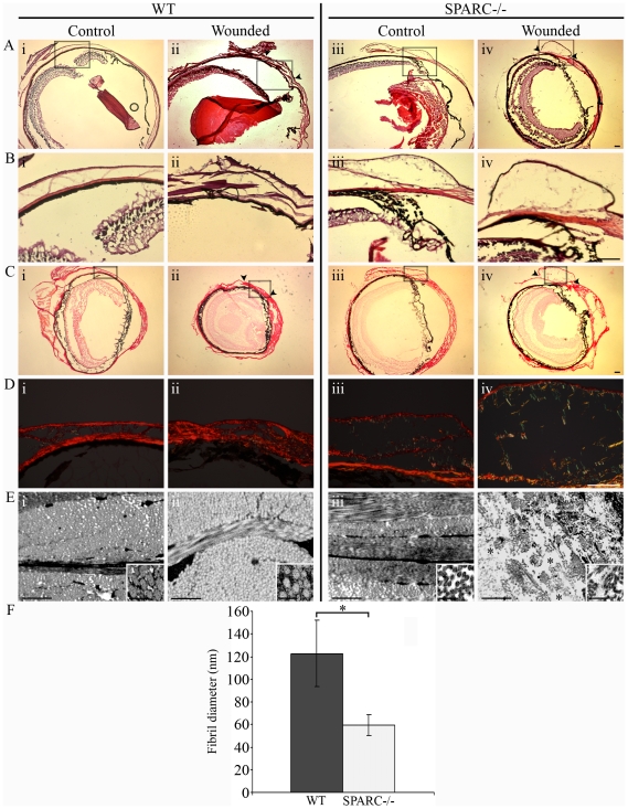
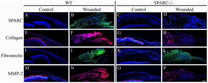
Glaucoma is a disease frequently associated with elevated intraocular pressure that can be alleviated by filtration surgery. However, the post-operative subconjunctival scarring response which blocks filtration efficiency is a major hurdle to the achievement of long-term surgical success. Current application of anti-proliferatives to modulate the scarring response is not ideal as these often give rise to sight-threatening complications. SPARC (secreted protein, acidic and rich in cysteine) is a matricellular protein involved in extracellular matrix (ECM) production and organization. In this study, we investigated post-operative surgical wound survival in an experimental glaucoma filtration model in SPARC-null mice. Loss of SPARC resulted in a marked (87.5%) surgical wound survival rate compared to 0% in wild-type (WT) counterparts. The larger SPARC-null wounds implied that aqueous filtration through the subconjunctival space was more efficient in comparison to WT wounds. The pronounced increase in both surgical survival and filtration efficiency was associated with a less collagenous ECM, smaller collagen fibril diameter, and a loosely-organized subconjunctival matrix in the SPARC-null wounds. In contrast, WT wounds exhibited a densely packed collagenous ECM with no evidence of filtration capacity. Immunolocalization assays confirmed the accumulation of ECM proteins in the WT but not in the SPARC-null wounds. The observations in vivo were corroborated by complementary data performed on WT and SPARC-null conjunctival fibroblasts in vitro. These findings indicate that depletion of SPARC bestows an inherent change in post-operative ECM remodeling to favor wound maintenance. The evidence presented in this report is strongly supportive for the targeting of SPARC to increase the success of glaucoma filtration surgery.


Conflict of interest statement

Competing Interests: The authors have declared that no competing interests exist.

Figures

Figure 1
Figure 1. Effect of TGF-β2 on collagen I, fibronectin, and α-SMA mRNA expression.
Cells treated with TGF-β2 for 72 h were measured for collagen I, fibronectin or α-SMA mRNA abundance by real-time quantitative PCR. (A) In the presence of TGF-β2, collagen I mRNA was increased in WT (*, P = 1.0×10⁻⁵) but not in SPARC−/− conjunctival fibroblasts. The unstimulated level of collagen I mRNA in SPARC−/− cells was significantly lower than that in WT cells (P = 4.3×10⁻⁹). (B) In the presence of TGF-β2, fibronectin mRNA was enhanced in WT (*, P = 0.002) but not in SPARC−/− conjunctival fibroblasts. (C) In the presence of TGF-β2, α-SMA mRNA was increased in both WT and SPARC−/− conjunctival fibroblasts (*, P<0.001). The β-actin transcript was used for normalization. Data are shown as fold induction compared with unstimulated WT cells. Data shown are representative of three independent experiments.
Figure 2
Figure 2. Reduction in the rate of collagen gel contraction by SPARC−/− fibroblasts is associated with a reduction in the expression of MT1-MMP and activity of MMP-2.
(A) SPARC−/− conjunctival fibroblasts contracted free-floating collagen gels to a lesser extent, in comparison to that of WT cells. WT or SPARC−/− fibroblasts were seeded in triplicate in collagen solutions which were allowed to polymerize. Gels were immediately detached and gel contraction was digitally photodocumented on day-1, -3 and −5. Gel contraction was measured as a reduction of gel surface area, expressed as a % of the initial gel size measured immediately after detachment. Values are the means of triplicates; bars indicate SD. The data shown are representative of three independent experiments. The rates of contraction from day-3 to day-5 are shown in Table 1. (B) MMP-2 activity was reduced in medium conditioned by SPARC−/− fibroblasts seeded in collagen gels. Left panel, MMP-2 activity in medium conditioned by WT or SPARC−/− fibroblasts seeded in collagen gels for 5 days was analysed by gelatin zymography. Proteolytic activities corresponding to the molecular weights of MMP-9 (92 kDa) and pro- and active MMP-2 (72 and 66 kDa respectively) are indicated by arrowheads. Right panel, densitometric analysis of total MMP-2 (sum of pro- and active MMP-2) showed that the total MMP-2 secreted by SPARC−/− cells was significantly lower that of WT cells (P = 2.8×10⁻⁵). Values shown are the mean of three independent experiments; bars indicate SD. (C) MT1-MMP expression was reduced in SPARC−/− fibroblasts. Left panel, WT or SPARC/− fibroblasts seeded in collagen gels for 5 days were lysed, and 40 µg of each protein preparation was subjected to immunoblotting with antibodies specific for MT1-MMP and GAPDH (control). Right panel, upon normalization with the housekeeping protein GAPDH, densitometry analysis revealed that SPARC−/− fibroblasts produced significantly less MT1-MMP protein compared to WT cells (P = 0.04). Values shown are the mean of three independent experiments and a representative Western blot is shown; bars indicate SD. (D) The reduced rate of collagen gel contraction observed in SPARC−/− cells did not involve α-SMA. WT or SPARC/− fibroblasts seeded in collagen gels for 5 days were lysed, and the corresponding SPARC or α-SMA mRNAs were quantified by real-time quantitative PCR. The data confirmed that SPARC expression was sustainably reduced at day-5 in the collagen gels. Values shown are the means of triplicates; bars indicate SD.
Figure 3
Figure 3. Creation of a filtering bleb in a mouse model of human glaucoma filtration surgery.
(A) Schematic diagram of the surgical process involved in the creation of a functioning wound in the mouse conjunctiva. The conjunctiva was first surgically dissected to expose the underlying sclera (i) which was then cannulated with a 30-gauge needle through the sclera into the anterior chamber of the eye (ii) to allow aqueous humor to escape (small red arrows, iii). Finally, the conjunctiva was closed by suturing over the newly-created fistula (iv). The conjunctiva overlying the wound site can be observed as a filtering bleb (iv). (B) Slit lamp examination of wound sites revealed the bleb in SPARC−/− conjunctiva 14 days post-surgery. The conjunctiva in the unwounded eye is flat and smooth (i, ii). On day-2 post-wounding, the wound site close to the suture (arrowhead) assumed the appearance of a bleb (demarcated by arrows), indicative of functionality due to the collection of aqueous fluid in the conjunctiva (iii, iv). On day-14 post-surgery, the WT bleb belonging to the same eye as (iii) has completely flattened down to resemble superficially the unwounded conjunctiva (v), whereas the SPARC−/− bleb belonging to the same eye in (iv) remained visibly elevated (vi, outlined by dotted line and demarcated by arrows).
Figure 4
Figure 4. Bleb survival is prolonged in the absence of SPARC.
(A) Anterior segment optical coherence tomography imaging of the mouse conjunctiva confirmed bleb survival in the SPARC−/− conjunctiva. The unwounded and wounded eyes on day-2, -7, -10 and −14 were examined. The location of the bleb is indicated by the white box. The bleb was seen to diminish progressively in the WT wounded eye until its virtual disappearance on day-14, whereas the bleb remained visibly present in the SPARC−/− eye. Images shown belong to the same WT or SPARC−/− eye at each of the days examined and are representative of eight eyes of each genotype. C, cornea. (B) Bleb sizes of SPARC−/− mice were larger than those of WT animals. Each bleb at the indicated day post-wounding was measured centrally in at least five OCT imaging fields, and the mean size was expressed as a percentage of the initial size on day-2 post-surgery. Each symbol represents a single eye (♦, WT, n = 8; □, SPARC−/−, n = 8). The horizontal bars and numbers indicate the mean % bleb sizes. (C) Kaplan-Meier curve showing that targeted inactivation of SPARC resulted in increased bleb survival in comparison to that observed in WT eyes. Individual eyes were scored as positive for bleb survival when the bleb size was ≥50% of the initial day-2 bleb as shown in (B). Bleb failure in WT eyes progressed rapidly from 25% (2/8) on day-7 to 87.5% (7/8) and 100% (8/8) on days-10 and -14 post-surgery, respectively, whereas SPARC−/− blebs experienced only 12.5% failure (1/8) on day-14 post-surgery.
Figure 5
Figure 5. Differential organization of the SPARC−/− versus WT subconjunctival tissue matrix is associated with a distinct wounding response.
In vivo confocal imaging of the unoperated WT (A) and SPARC−/− (B) conjunctiva revealed an optical difference in the composition and organization of the subconjunctival matrix. On day-14 post-surgery, the WT subconjunctival matrix appeared optically dense and filled with a tightly packed fibrous material (C), whereas the SPARC−/− matrix was expanded and optically diffuse, with a loosely-organized fibrous network (D). Images shown are representative of 3 eyes of each genotype. CE, conjunctival epithelium; *, subconjunctival space; S, sclera.
Figure 6
Figure 6. Collagen deposition is altered in the SPARC−/− conjunctiva.
(A) Hematoxylin and eosin staining revealed a flattened conjunctiva at the 14 day-old WT wound site (ii, arrowheads), whereas the SPARC−/− wound site was observed as a spongy protuberance in the conjunctiva (iv, arrowheads). (B) Higher magnification of insets (boxed areas in A) illustrate differing thickness in the SPARC−/− (iii and iv) versus WT (i and ii) conjunctival epithelium, independent of wounding. (C) Picrosirius red-stained sections of the same eyes at low magnification and visualized by brightfield microscopy. (D) Higher magnification of insets (boxed areas in C) and polarization microscopy revealed a meshwork organization of collagen fibers in unoperated WT conjunctiva (i) compared to the well-aligned and tightly packed fibers in WT wound (ii), as well as a delicate meshwork in both unoperated SPARC−/− conjunctiva (iii) and operated SPARC−/− bleb (iv). (E) Transmission electron microscopy confirmed a tightly-packed matrix in the WT conjunctival wound (ii), in contrast to the presence of areas of loosely-organized matrix with open spaces in the SPARC−/− wound (*, iv). Ultrastructural analyses also illustrate the intrinsic deposition of smaller collagen fibrils in the unoperated SPARC−/− eye (iii, inset) compared to WT (i, inset). (F) Graphical representation of the mean diameter of collagen fibrils (± SD) from about 10 fields of view of unoperated WT (filled bar, n = 400) and SPARC−/− conjunctiva (open bar, n = 405). *, P = 5.1×10−159. Bars: (A to D) 100 µm; (E) 1 µm; (E, insets) 200 nm.
Figure 7
Figure 7. SPARC−/− conjunctival wounds exhibit reduced levels of ECM proteins.
Immunofluorescence analyses of normal conjunctiva and wound sites in the WT and SPARC−/− eye revealed that wounding in the WT conjunctiva was associated with increased production of SPARC (B), collagen I (F), fibronectin (J) and MMP-2 (N) in comparison to normal WT conjunctiva (A, E, I, M). The SPARC−/− conjunctiva exhibited no SPARC expression (C, D) and apparently low levels of collagen I (G), and MMP-2 (O) which were not enhanced upon surgical wounding (H, P). There was an apparent increase in fibronectin in the SPARC−/− bleb (L) relative to that in the unoperated SPARC−/− conjunctiva (K). Mouse anti-SPARC and anti-fibronectin antibodies were visualized with anti-mouse secondary antibody conjugated to AlexaFluro-488 (green); rabbit anti-collagen and anti-MMP-2 antibodies were visualized with anti-rabbit secondary antibody conjugated to AlexaFluro-594 (red). Bar, 100 µm.

Similar articles

Cited by

References

    1. Resnikoff S, Pascolini D, Etya'ale D, Kocur I, Pararajasegaram R, et al. Global data on visual impairment in the year 2002. Bull World Health Organ. 2004;82:844–851. - PMC - PubMed
    1. Quigley HA, Broman A. The number of persons with glaucoma worldwide in 2010 and 2020. Br J Ophthalmol. 2006;90:151–156. - PMC - PubMed
    1. The AGIS Investigators. The advanced glaucoma intervention study (AGIS): 7. The relationship between control of intraocular pressure and visual field deterioration. Am J Ophthalmol. 2000;130:429–440. - PubMed
    1. Reddick R, Merritt JC, Ross G, Avery A, Peiffer RL. Myofibroblasts in filtration operations. Ann Ophthalmol. 1985;17:200–203. - PubMed
    1. The Fluorouracil Filtering Surgery Study Group. Fluorouracil filtering surgery study one-year follow-up. Am J Ophthalmol. 1989;108:625–635. - PubMed

Publication types

MeSH terms