. 2010 Feb 25;5(2):e9432.
doi: 10.1371/journal.pone.0009432.

Helper T cell epitope-mapping reveals MHC-peptide binding affinities that correlate with T helper cell responses to pneumococcal surface protein A

Affiliations

Helper T cell epitope-mapping reveals MHC-peptide binding affinities that correlate with T helper cell responses to pneumococcal surface protein A

Rajesh Singh et al. PLoS One. .

Abstract

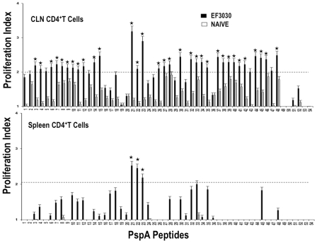
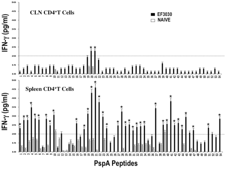
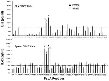
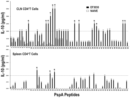
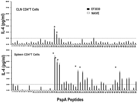
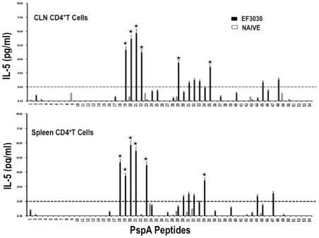
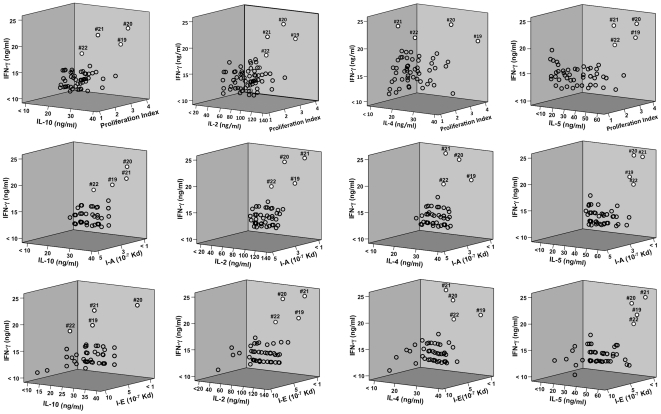
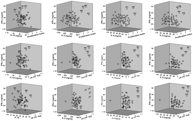
Understanding the requirements for protection against pneumococcal carriage and pneumonia will greatly benefit efforts in controlling these diseases. Several proteins and polysaccharide capsule have recently been implicated in the virulence of and protective immunity against Streptococcus pneumonia. Pneumococcal surface protein A (PspA) is highly conserved among S. pneumonia strains, inhibits complement activation, binds lactoferrin, elicits protective systemic immunity against pneumococcal infection, and is necessary for full pneumococcal virulence. Identification of PspA peptides that optimally bind human leukocyte antigen (HLA) would greatly contribute to global vaccine efforts, but this is hindered by the multitude of HLA polymorphisms. Here, we have used an experimental data set of 54 PspA peptides and in silico methods to predict peptide binding to HLA and murine major histocompatibility complex (MHC) class II. We also characterized spleen- and cervical lymph node (CLN)-derived helper T lymphocyte (HTL) cytokine responses to these peptides after S. pneumonia strain EF3030-challenge in mice. Individual, yet overlapping peptides, 15 amino acids in length revealed residues 199 to 246 of PspA (PspA(199-246)) consistently caused the greatest IFN-gamma, IL-2, IL-5 and proliferation as well as moderate IL-10 and IL-4 responses by ex vivo stimulated splenic and CLN CD4(+) T cells isolated from S. pneumonia strain EF3030-challeged F(1) (B6xBALB/c) mice. IEDB, RANKPEP, SVMHC, MHCPred, and SYFPEITHI in silico analysis tools revealed peptides in PspA(199-246) also interact with a broad range of HLA-DR, -DQ, and -DP allelles. These data suggest that predicted MHC class II-peptide binding affinities do not always correlate with T helper (Th) cytokine or proliferative responses to PspA peptides, but when used together with in vivo validation can be a useful tool to choose candidate pneumococcal HTL epitopes.


Conflict of interest statement

Competing Interests: The authors have declared that no competing interests exist.

Figures

Figure 1
Figure 1. Modular PspA amino acid sequence showing regions of predicted immunogenicity and secondary structure.
Major domains of PspA are indicated. The aligned amino acid sequence shows the previously defined PspA windows A, A*, B and C. The PspA amino acid (AA) sequence was used to predict helical (H), coiled (C), α strand (E), β turns (t), and asparagine endopeptidase sites (N).
Figure 2
Figure 2. Proliferation responses of PspA peptide-specific systemic and mucosal CD4+ T cells during pneumococcal carriage.
Spleen and cervical lymph node (CLN) lymphocytes were isolated from F1 (B6×Balb/c) mice, 28 days after intranasal challenge with Streptococcus pneumonia strain EF3030 (▪) and naïve (□). CD4+ T cells were incubated with 1 µM of PspA peptide (15 amino acid peptides that overlapped every 11 residues) plus mitomycin C-treated naïve syngeneic feeder cells, for 3 days, at a ratio of 5∶1×10⁶ cells. Proliferation was measured by BrdU incorporation, which was measured by ELISA. The data presented are the mean OD450. Experimental groups consisted of 10 mice. The results were expressed as the mean ± the standard error mean (SEM) of the response from 3 replicate determinations of three independent experiments.
Figure 3
Figure 3. PspA peptide-specific IFN-γ secretion by CD4+ T cell following pneumococcal challenge.
Groups of 10 F1 (B6×Balb/c) mice were intranasally challenged with 107 CFUs of S. pneumonia strain EF3030 in a 15 µl volume of Ringer's solution. Spleen and cervical lymph node (CLN) lymphocytes were isolated from mice, 28 days after intranasal challenge with Streptococcus pneumonia strain EF3030 (▪) and naïve (□). CD4+ T cells were incubated with 1 µM of PspA peptide (15 amino acid peptides that overlapped every 11 residues) plus mitomycin C-treated naïve syngeneic feeder cells, for 3 days, at a ratio of 5∶1×10⁶ cells. The results were expressed as the mean ± the standard error mean (SEM) of IFN-γ supernatant levels from 3 replicate determinations of three independent experiments. IFN-γ production of cultured supernatants was determined by Luminex capable of detecting >2 pg/ml of IFN-γ.
Figure 4
Figure 4. PspA peptide-specific IL-2 secretion by CD4+ T cell following pneumococcal challenge.
Groups of 10 F1 (B6×Balb/c) mice were intranasally challenged with 107CFUs of S. pneumonia strain EF3030 in a 15 µl volume of Ringer's solution. Spleen and Cervical lymph node (CLN) lymphocytes were isolated from mice, 28 days after intranasal challenge with Streptococcus pneumoniae strain EF3030 (▪) and naïve (□). CD4+ T cells were incubated with 1 µM of PspA peptide (15 amino acid peptides that overlapped every 11 residues) plus mitomycin C-treated naïve syngeneic feeder cells, for 3 days, at a ratio of 5∶1×10⁶ cells. The results were expressed as the mean ± the standard error mean (SEM) of IL-2 supernatant levels from 3 replicate determinations of three independent experiments. IL-2 production of cultured supernatants was determined by Luminex capable of detecting >2 pg/ml of IL-2.
Figure 5
Figure 5. PspA peptide-specific IL-10 secretion by CD4+ T cell following pneumococcal challenge.
Groups of 10 F1 (B6×Balb/c) mice were intranasally challenged with 107CFUs of S. pneumonia strain EF3030 in a 15 µl volume of Ringer's solution. Spleen and Cervical lymph node (CLN) lymphocytes were isolated from mice, 28 days after intranasal challenge with Streptococcus pneumonia strain EF3030 (▪) and naïve (□). CD4+ T cells were incubated with 1 µM of PspA peptide (15 amino acid peptides that overlapped every 11 residues) plus mitomycin C-treated naïve syngeneic feeder cells, for 3 days, at a ratio of 5∶1×10⁶ cells. The results were expressed as the mean ± the standard error mean (SEM) of IL-10 supernatant levels from 3 replicate determinations of three independent experiments. IL-10 production of cultured supernatants was determined by Luminex capable of detecting >2 pg/ml of IL-10.
Figure 6
Figure 6. PspA peptide-specific IL-4 secretion by CD4+ T cell following pneumococcal challenge.
Groups of 10 F1 (B6×Balb/c) mice were intranasally challenged with 107CFUs of S. pneumonia strain EF3030 in a 15 µl volume of Ringer's solution. Spleen and Cervical lymph node (CLN) lymphocytes were isolated from mice, 28 days after intranasal challenge with Streptococcus pneumonia strain EF3030 (▪) and naïve (□). CD4+ T cells were incubated with 1 µM of PspA peptide (15 amino acid peptides that overlapped every 11 residues) plus mitomycin C-treated naïve syngeneic feeder cells, for 3 days, at a ratio of 5∶1×10⁶ cells. The results were expressed as the mean ± the standard error mean (SEM) of IL-4 supernatant levels from 3 replicate determinations of three independent experiments. IL-4 production of cultured supernatants was determined by Luminex capable of detecting >2 pg/ml of IL-4.
Figure 7
Figure 7. PspA peptide-specific IL-5 secretion by CD4+ T cell following pneumococcal challenge.
Groups of 10 F1 (B6×Balb/c) mice were intranasally challenged with 107CFUs of S. pneumonia strain EF3030 in a 15 µl volume of Ringer's solution. Spleen and Cervical lymph node (CLN) lymphocytes were isolated from mice, 28 days after intranasal challenge with Streptococcus pneumonia strain EF3030 (▪) and naïve (□). CD4+ T cells were incubated with 1 µM of PspA peptide (15 amino acid peptides that overlapped every 11 residues) plus mitomycin C-treated naïve syngeneic feeder cells, for 3 days, at a ratio of 5∶1×10⁶ cells. The results were expressed as the mean ± the standard error mean (SEM) of IL-5 supernatant levels from 3 replicate determinations of three independent experiments. IL-5 production of cultured supernatants was determined by Luminex capable of detecting >2 pg/ml of IL-5.
Figure 8
Figure 8. 3D plot of Th1/Th2 cytokine secretion relative to proliferation or I-A/I-E predicted peptide-binding by cervical lymph node-derived CD4+ T cells.
The panels summarize IFN-γ, IL-10, IL-2, IL-4, IL-5 and proliferation responses of PspA peptide-specific CD4+ T cells isolated from cervical lymph nodes of F1 (B6×Balb/c) mice, 28 days after S. pneumonia strain EF3030- challenge and predicted I-A or I-E binding affinities. Y-axis and X-axis indicate the concentration (ng/ml) of IFN-γ and IL-10, IL-2, IL-4, IL-5 respectively, secreted by PspA peptide-stimulated CD4+ T cells. The Z- axis represents the predicted I-A or I-E binding affinities (Kd). PspA peptides 19, 20, 21 and 22 appear as white circles, while remaining peptides are open circles.
Figure 9
Figure 9. 3D plot of Th1/Th2 cytokine secretion relative to proliferation or I-A/I-E predicted peptide-binding by spleen- derived CD4+ T cells.
The panels summarize IFN-γ, IL-10, IL-2, IL-4, IL-5 and proliferation responses of PspA peptide-specific CD4+ T cells isolated from spleen of F1 (B6×Balb/c) mice, 28 days after S. pneumonia strain EF3030-challenge and predicted I-A or I-E binding affinities. Y-axis and X-axis indicate the concentration (ng/ml) of IFN-γ and IL-10, IL-2, IL-4,IL-5 respectively, secreted by PspA peptide-stimulated CD4+ T cells. The Z-axis represents the predicted I-A or I-E binding affinities (Kd). PspA peptides 19, 20, 21 and 22 appear as white circles, while remaining peptides are open circles.

Similar articles

Cited by

References

    1. Nasrin D, Collignon PJ, Wilson EJ, Pilotto LS, Douglas RM. Antibiotic resistance in Streptococcus pneumoniae isolated from children. Journal of Paediatrics & Child Health. 1999;35:558–561. - PubMed
    1. Hooper DC. Emerging mechanisms of fluoroquinolone resistance. Emerging Infectious Diseases. 2001;7:337–341. - PMC - PubMed
    1. Morita JY, Zell ER, Danila R, Farley MM, Hadler J, et al. Association between antimicrobial resistance among pneumococcal isolates and burden of invasive pneumococcal disease in the community. Clinical Infectious Diseases. 2002;35:420–427. - PubMed
    1. Richter SS, Heilmann KP, Coffman SL, Huynh HK, Brueggemann AB, et al. The molecular epidemiology of penicillin-resistant Streptococcus pneumoniae in the United States, 1994–2000. Clinical Infectious Diseases. 2002;34:330–339. - PubMed
    1. Bignell GR, Huang J, Greshock J, Watt S, Butler A, et al. High-resolution analysis of DNA copy number using oligonucleotide microarrays. Genome Research. 2004;14:287–295. - PMC - PubMed

Publication types

MeSH terms