. 2010 Feb 25;5(2):e9429.
doi: 10.1371/journal.pone.0009429.

miR-24 regulates apoptosis by targeting the open reading frame (ORF) region of FAF1 in cancer cells

Affiliations

miR-24 regulates apoptosis by targeting the open reading frame (ORF) region of FAF1 in cancer cells

Wenming Qin et al. PLoS One. .

Abstract

Background: microRNAs (miRNAs) are small noncoding RNAs that regulate cognate mRNAs at the post-transcriptional stage. Several studies have shown that miRNAs modulate gene expression in mammalian cells by base pairing to complementary sites in the 3'-untranslated region (3'-UTR) of the target mRNAs.

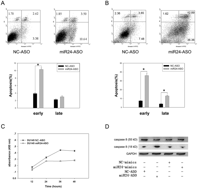
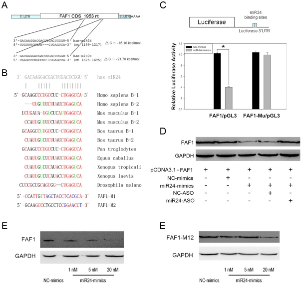
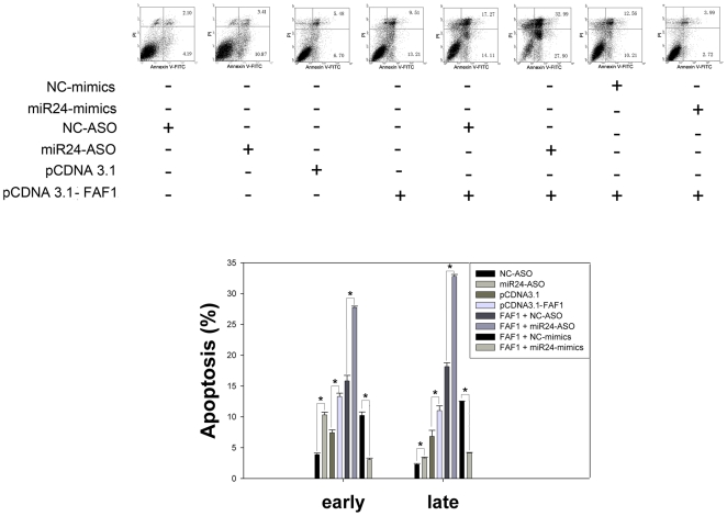
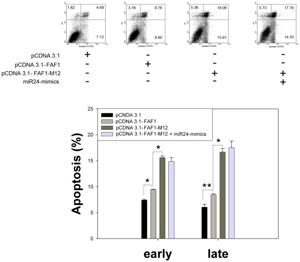
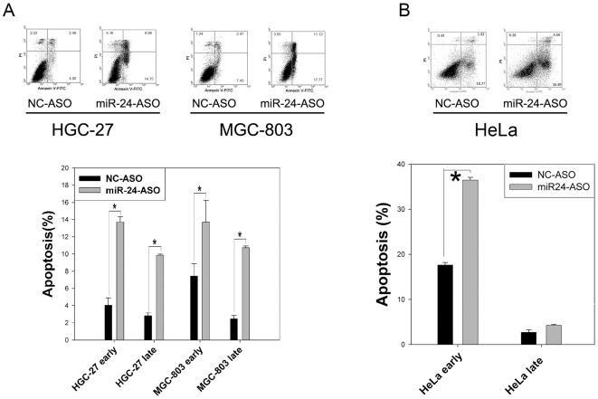
Methodology/principal findings: In the present study, miR-24 was found to target fas associated factor 1(FAF1) by binding to its amino acid coding sequence (CDS) region, thereby regulating apoptosis in DU-145 cells. This result supports an augmented model whereby animal miRNAs can exercise their effects through binding to the CDS region of the target mRNA. Transfection of miR-24 antisense oligonucleotide (miR-24-ASO) also induced apoptosis in HGC-27, MGC-803 and HeLa cells.

Conclusions/significance: We found that miR-24 regulates apoptosis by targeting FAF1 in cancer cells. These findings suggest that miR-24 could be an effective drug target for treatment of hormone-insensitive prostate cancer or other types of cancers. Future work may further develop miR-24 for therapeutic applications in cancer biology.


Conflict of interest statement

Competing Interests: The authors have declared that no competing interests exist.

Figures

Figure 1
Figure 1. Down-regulation of miR-24 induces apoptosis and affects proliferation in DU-145 cells.
(A) DU-145 cells were treated with 20 nM miR-24-ASO or NC-ASO for 48 h. Apoptosis was measured by FACS with Annexin V and propidium iodide staining (n = 3±SE; *p<0.01). Down-regulation of miR-24 induced apoptosis in DU-145 cells. (B) Apoptosis was induced for 2 h with 1 µM staurosporine, 48 h after transfection 20 nM miR-24-ASO or NC-ASO (n = 3±SE; *p<0.01). Apoptosis sensitivity increased significantly after transfection with miR-24-ASO. (C) Cell proliferation was measured by CCK-8 kit at the indicated time points after transfection with 20 nM miR-24-ASO or NC-ASO (n = 3±SE). Down-regulation of miR-24 affected the proliferation of DU-145 cells. (D) Caspase-8 activation was detected by western blot analysis of pro-caspase 8 and the mature form of caspase-8 after 24 h of treatment (as above). Caspase-8 was activated after transfection with miR-24-ASO.
Figure 2
Figure 2. miR-24 regulates FAF1 by targeting its ORF region.
(A) RNA22 software was used to predict that the CDS region of FAF1 mRNA harbors two putative miR-24 binding sites. The sequence of miR-24, its binding sites, and energies are shown. (B) The predicted binding sites of FAF1 were conserved in eight different species. The bases altered in the mutant FAF1 constructs are also shown. Red: paired bases; green: G:U pair; blue: mutant bases. (C) 293T cells were transfected with a reporter vector consisting of a luciferase gene containing the wild type or mutated miR-24 binding sites) in its 3′-UTR region (FAF1/pGL3 or FAF1-M12/pGL3, 20 ng/well in each 24-well plate). The cells were also transfected with a CMV-Renilla luciferase vector (20 ng/well in each 24-well plate) as an internal standard. Expression of the luciferase containing predicted miR-24 binding sequences was decreased by treatment with 20 nM miR-24-mimic compared to treatment with NC-mimic, while the luciferase containing mutated sequences could not be down-regulated (n = 3±SE; *p<0.01). (D) miR-24 regulates the FAF1 gene. 293T cells were treated as indicated and transfected with 100 ng pcDNA3.1-FAF1. The expression of FAF1 was down-regulated after transfection with 20 nM miR-24-mimic and rescued after 100 nM miR-24-ASO being transfected. (E) Dose-dependent suppression of FAF1 by miR-24 in 293T cells. 293T cells were co-transfected with 100 ng pcDNA3.1-FAF1 and 1, 5, or 20 nM miR-24-mimic after 24 h and immunoblotted as above. (F) Mutant FAF1 could not be down-regulated upon transfection with 1 nM or 5 nM of miR-24-mimic for 24 h. FAF1 was down-regulated slightly after transfection with 20 nM miR-24-mimic; this might because the mutation to FAF1 was somewhat small.
Figure 3
Figure 3. miR-24 regulates apoptosis by targeting FAF.
DU-145 cells were treated with the indicated reagents and apoptosis was measured by FACS 48 h after transfection (n = 3±SE, *p<0.01). Not only did down-regulation of miR-24 induce apoptosis, but overexpression of FAF1 also induced apoptosis in DU-145 cells. The percentage of apoptotic cells became much higher when pcDNA3.1-FAF1 and miR-24-ASO were co-transfected. Apoptosis induced by the over-expression of FAF1 could be rescued by the miR-24-mimic. These data show that over-expression of miR-24 can protect DU-145 cells from FAF1-induced apoptosis, and that down-regulation of miR-24 can increase the apoptosis induced by FAF1 in DU-145 cells. Vectors were transfected at the concentration of 100 ng per well; mimics and ASO were transfected at the concentration of 20 nM per well in a 12-well plate.
Figure 4
Figure 4. miR-24 regulates apoptosis by binding to the ORF region of the FAF1 gene.
DU-145 cells were treated as indicated and apoptosis was measured by FACS 36 h after transfection (n = 3±SE, *p<0.01, **p<0.05). Synonymous mutations at the predicted miR-24 binding sites within FAF1 induced greater levels of apoptosis. Over-expression of miR-24 did not protect DU-145 cells from apoptosis induced by mutant FAF1. Vectors were transfected at a concentration of 100 ng per well; mimics and ASO were transfected at a concentration of 20 nM per well in a 12-well plate.
Figure 5
Figure 5. Down-regulation of miR-24 induced apoptosis in HGC-27 and MGC-803 cells, and increased staurosporine sensitivity in HeLa cells.
(A) HGC-27 and MGC-803 cells were treated with 20 nM miR-24-ASO or NC-ASO and apoptosis was measured by FACS with Annexin V and propidium iodine staining after 48 h (n = 3±SE; *p<0.01). Down-regulation of miR-24 induced apoptosis in HGC-27 and MGC-803 cells. (B) Apoptosis was induced with 1 µM staurosporine for 2 h after 48 h of transfection using 20 nM miR-24-ASO or NC-ASO (n = 3±SE; *p<0.01). The apoptotic response to staurosporine was increased after transfection with miR-24-ASO.

Similar articles

Cited by

References

    1. Olena AF, Patton JG. Genomic organization of microRNAs. J Cell Physiol. 222:540–545. - PMC - PubMed
    1. Axtell MJ. Evolution of microRNAs and their targets: are all microRNAs biologically relevant? Biochim Biophys Acta. 2008;1779:725–734. - PubMed
    1. Yekta S, Shih IH, Bartel DP. MicroRNA-directed cleavage of HOXB8 mRNA. Science. 2004;304:594–596. - PubMed
    1. Bartel DP. MicroRNAs: genomics, biogenesis, mechanism, and function. Cell. 2004;116:281–297. - PubMed
    1. Alvarez-Garcia IM, EA. MicroRNA functions in animal development and human disease. Development. 2005;132:10. - PubMed

Publication types

MeSH terms