. 2010 May;62(5):263-72.
doi: 10.1007/s00251-010-0428-1. Epub 2010 Mar 2.

Domain architecture evolution of pattern-recognition receptors

Affiliations

Domain architecture evolution of pattern-recognition receptors

Qing Zhang et al. Immunogenetics. 2010 May.

Abstract

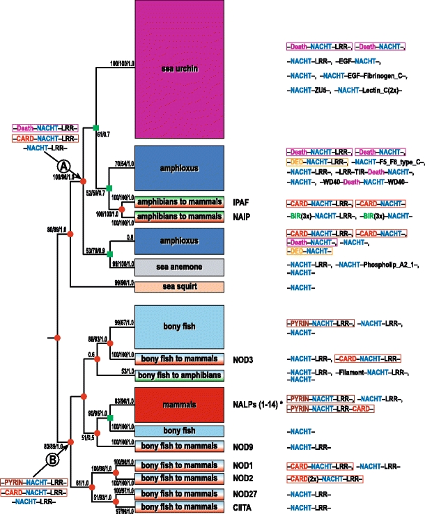
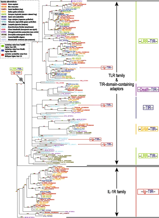
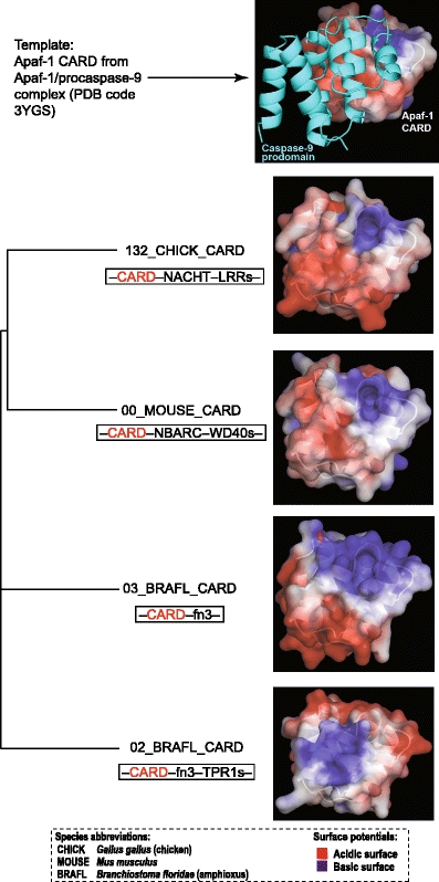
In animals, the innate immune system is the first line of defense against invading microorganisms, and the pattern-recognition receptors (PRRs) are the key components of this system, detecting microbial invasion and initiating innate immune defenses. Two families of PRRs, the intracellular NOD-like receptors (NLRs) and the transmembrane Toll-like receptors (TLRs), are of particular interest because of their roles in a number of diseases. Understanding the evolutionary history of these families and their pattern of evolutionary changes may lead to new insights into the functioning of this critical system. We found that the evolution of both NLR and TLR families included massive species-specific expansions and domain shuffling in various lineages, which resulted in the same domain architectures evolving independently within different lineages in a process that fits the definition of parallel evolution. This observation illustrates both the dynamics of the innate immune system and the effects of "combinatorially constrained" evolution, where existence of the limited numbers of functionally relevant domains constrains the choices of domain architectures for new members in the family, resulting in the emergence of independently evolved proteins with identical domain architectures, often mistaken for orthologs.


Figures

Fig. 1
Fig. 1
Overview of the phylogeny and domain organization of the NACHT protein family. On the phylogenetic tree, branches are assigned with different background colors according to species. The numbers associated with each clade indicate bootstrap values for minimum evolution and maximum likelihood and the posterior probability from Bayesian inference, respectively (values lower than 50% are not shown). The size of each branch is proportional to the number of proteins inside that branch with the domain architectures shown on the right side. Duplication event is indicated by a red circle and speciation by a green square. Domain architectures at the internal nodes A and B are speculated. The detailed sequence information for each terminal node is shown in Supplementary Fig. 1. Some unusual NACHT gene models in sea urchin and amphioxus require further experimental verification. *The NALPs branch appears to be mammalian-specific, with the exception of one protein from chicken with the canonical PYRIN–NACHT–LRR architecture
Fig. 2
Fig. 2
Phylogeny of the TIR protein family. Protein names are assigned to different colors according to species origin. Lineage-specific subtrees are collapsed for clarity with the number of sequences shown in brackets. Support values for each clade are indicated by differently colored boxes to increase their visibility. Bootstrap values higher than 50% from minimum evolution and maximum likelihood approaches are colored in green and blue, respectively. Posterior probability values from a Bayesian approach higher than 0.5 are colored in red. The simplified domain architectures of each branch are presented on the right side of the phylogenetic tree. Domain architectures at the internal node A are speculated. Ig–TIR domain-containing sequences from amphioxus and sea anemone are separate from the vertebrate IL-1R branch, which has the same domain architecture. The detailed sequence information can be found in Supplementary Table 2
Fig. 3
Fig. 3
Structure comparison between different CARD domains. A phylogeny based on the CARD domains is shown on the left, and the related structure models are displayed on the right. The electrostatic potential was mapped on the protein surface of each 3D model. The acidic surface (negatively charged) is colored in red, and the basic surface (positively charged) is in blue. All protein surfaces are displayed in the same view with the putative binding surface (the surface used by the Apaf-1 CARD that binds the Caspase-9 prodomain (Qin et al. 1999) shown on the top panel) facing the viewer. The potential binding surface of the last structure (02_BRAFL_CARD) is dominated by a positive charge while the other three structures are mainly negatively charged

References

    1. Altschul SF, Madden TL, Schaffer AA, Zhang J, Zhang Z, Miller W, Lipman DJ. Gapped BLAST and PSI-BLAST: a new generation of protein database search programs. Nucleic Acids Res. 1997;25:3389–3402. doi: 10.1093/nar/25.17.3389. - DOI - PMC - PubMed
    1. Apic G, Gough J, Teichmann SA. Domain combinations in archaeal, eubacterial and eukaryotic proteomes. J Mol Biol. 2001;310:311–325. doi: 10.1006/jmbi.2001.4776. - DOI - PubMed
    1. Baker NA, Sept D, Joseph S, Holst MJ, McCammon JA. Electrostatics of nanosystems: application to microtubules and the ribosome. Proc Natl Acad Sci U S A. 2001;98:10037–10041. doi: 10.1073/pnas.181342398. - DOI - PMC - PubMed
    1. Basu MK, Carmel L, Rogozin IB, Koonin EV. Evolution of protein domain promiscuity in eukaryotes. Genome Res. 2008;18:449–461. doi: 10.1101/gr.6943508. - DOI - PMC - PubMed
    1. Bateman A, Coin L, Durbin R, et al. The Pfam protein families database. Nucleic Acids Res. 2004;32:D138–D141. doi: 10.1093/nar/gkh121. - DOI - PMC - PubMed

Publication types

MeSH terms

Substances