. 2010 Jun;15(5):667-75.
doi: 10.1007/s00775-010-0633-2. Epub 2010 Mar 2.

Metal ion determinants of conantokin dimerization as revealed in the X-ray crystallographic structure of the Cd(2+)/Mg (2+)-con-T[K7gamma] complex

Affiliations

Metal ion determinants of conantokin dimerization as revealed in the X-ray crystallographic structure of the Cd(2+)/Mg (2+)-con-T[K7gamma] complex

Sara E Cnudde et al. J Biol Inorg Chem. 2010 Jun.

Abstract

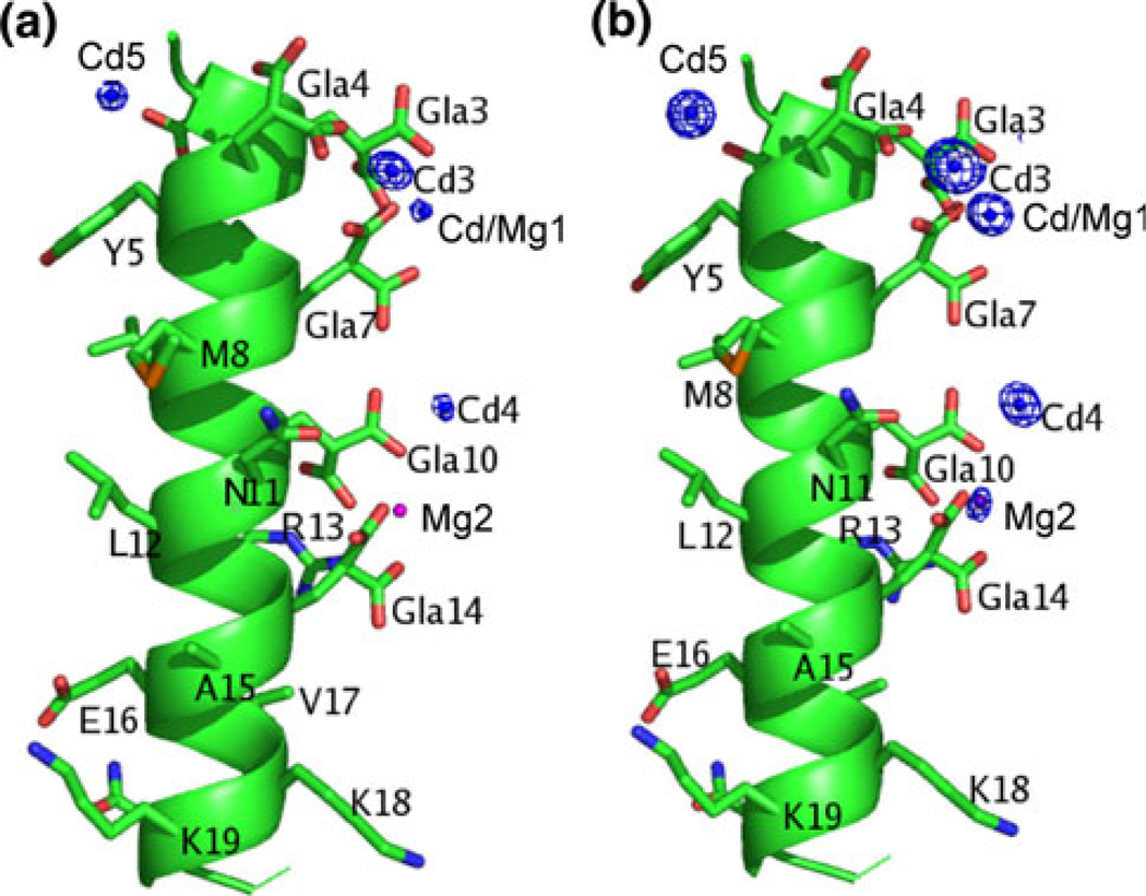
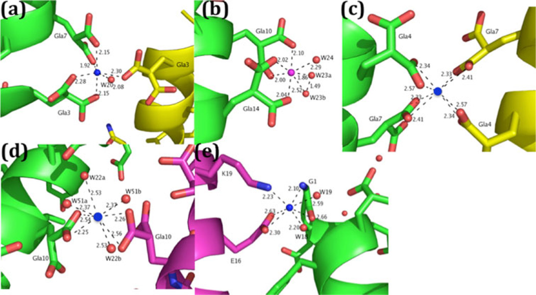
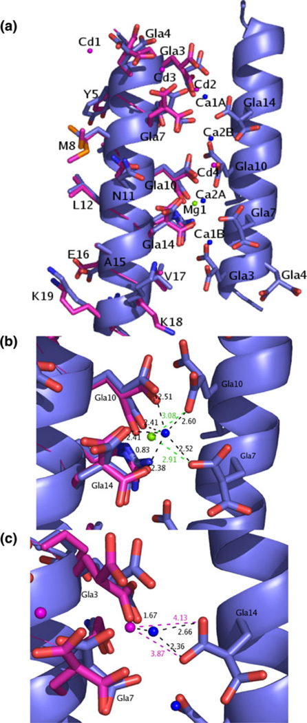
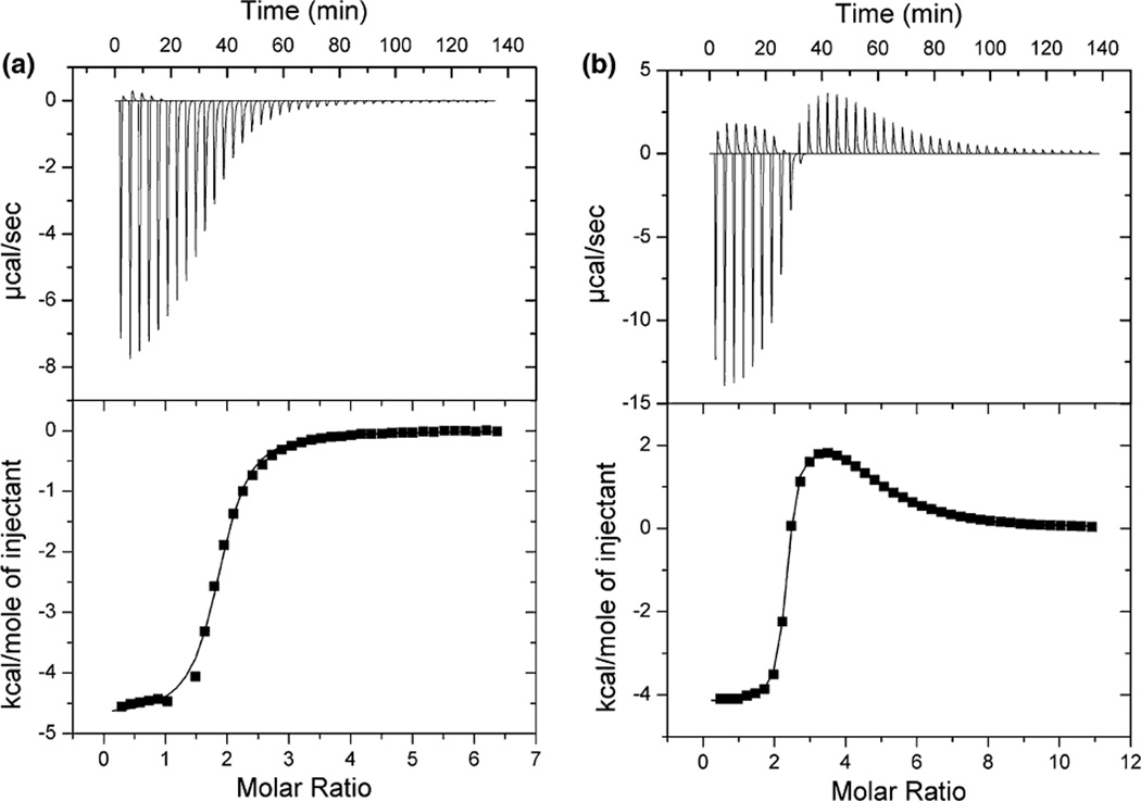
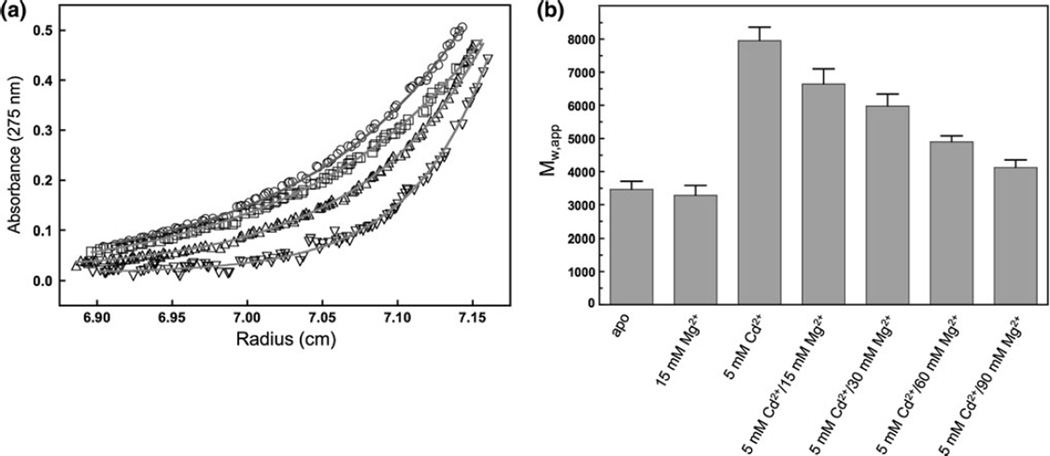
Predatory sea snails from the Conus family produce a variety of venomous small helical peptides called conantokins that are rich in gamma-carboxyglutamic acid (Gla) residues. As potent and selective antagonists of the N-methyl-D: -aspartate receptor, these peptides are potential therapeutic agents for a variety of neurological conditions. The two most studied members of this family of peptides are con-G and con-T. Con-G has Gla residues at sequence positions 3, 4, 7, 10, and 14, and requires divalent cation binding to adopt a helical conformation. Although both Ca(2+) and Mg(2+) can fulfill this role, Ca(2+) induces dimerization of con-G, whereas the Mg(2+)-complexed peptide remains monomeric. A variant of con-T, con-T[K7gamma] (gamma is Gla), contains Gla residues at the same five positions as in con-G and behaves very similarly with respect to metal ion binding and dimerization; each peptide binds two Ca(2+) ions and two Mg(2+) ions per helix. To understand the difference in metal ion selectivity, affinity, and the dependence on Ca(2+) for dimer formation, we report here the structure of the monomeric Cd(2+)/Mg(2+)-con-T[K7gamma] complex, and, by comparison with the previously published con-T[K7gamma]/Ca(2+) dimer structure, we suggest explanations for both metal ion binding site specificity and metal-ion-dependent dimerization.


Figures

Fig. 1
Fig. 1
The structure contains a total of three Cd ions, one mixed-occupancy Mg/Cd site, and one Mg2+. a The overall structure of Cd2+/Mg2+–con-T[K7γ] with an anomalous difference map contoured at 8σ. The four Cd2+ are shown as blue spheres and the Mg2+ is shown as a magenta sphere. Cd3 had the highest peak in the anomalous map at 24σ and is 100% occupied. Cd1 showed a peak at 14σ and is 80% occupied. Cd2 and Cd4 both had peaks in the anomalous map at 11σ and both are 50% occupied. b The overall structure of Cd2+/Mg2+–con-T[K7γ] with a FoFc map contoured at 5σ. The four Cd2+ are shown as blue spheres and the Mg2+ is shown as a magenta sphere. Mg1 is 100% occupied. The side chains are colored by atom
Fig. 2
Fig. 2
Metal coordination in Cd2+/Mg2+–con-T[K7γ] (shown in green). a Mg/Cd1 is six-coordinate. A crystallographically related α-helix is colored yellow. Note the shorter coordination lengths. b Mg2 is octahedrally coordinated. Mg2 makes bidentate coordination with both γ10 and γ14 and also coordinates with waters W24 and W23. W23 has two different conformations, W23a and W23b. c Cd3 is tetrahedrally coordinated. Cd3 sits directly on a crystallographic twofold axis and coordinates with both γ4 and γ7 of the structure and its symmetry-related molecule (yellow). d Cd4 is octahedrally coordinated. Cd4 coordinates with γ10 of the structure and a crystallographically related molecule (magenta) along with waters W22a and W51a and the symmetry-related waters W22b and W51b. c Cd5 is six-coordinate. Cd5 coordinates with Glu16 and Lys19 of a symmetry-related molecule (magenta) and also coordinates with G1 and two waters, W18 and W19. All distances are shown in angstroms. Cd ions are blue spheres, the waters are red spheres, and Mg1 is a magenta sphere. The side chains are colored by atom
Fig. 3
Fig. 3
Comparison of Ca2+- and Cd2+/Mg2+-bound con-T[K7γ]. Overlay of the Cd2+/Mg2+–con-T[K7γ] structure onto the Ca2+–con-T[K7γ] dimer structure. The Cd2+/Mg2+–con-T[K7γ] structure is shown in magenta and the Ca2+–con-T[K7γ] dimer structure is shown in blue. The four Cd2+ are shown as magenta spheres and the Mg2+ is shown as a green sphere. The Ca2+ are shown as blue spheres. The side chains are colored by atom. b A close-up of a in the region around the Mg2 site of Cd2+/Mg2+–con-T[K7γ] and the Ca2 site of the Ca2+–con-T[K7γ] dimer. Mg2 is shown as a green sphere, whereas Ca2 is shown as a blue sphere. Distances involving Mg2 are colored green. c A close-up of a in the region around the Ca1 site of the Ca2+–con-T[K7γ] dimer showing that cross-helix contacts are too long for chelation from the metal ion position seen in the Cd2+/ Mg2+–con-T[K7γ] structure. c A close-up of a at the Ca1 binding site. Mg/Cd1 is shown as a magenta sphere and Ca1 is shown as a blue sphere. The distance between Mg/Cd1 and Ca1 is 1.67 Å. Distances involving Mg/Cd1 are colored magenta
Fig. 4
Fig. 4
Representative calorimetric titrations of con-T[K7γ] with MgCl2 and CdCl2. Increments (5–10 µl) of the ligand solutions were added to the peptide at 200-s intervals. The upper panels depict the heat changes accompanying ligand injection and the lower panels represent the binding isotherms corresponding to the integrated heat changes. Titrations were conducted at 20 °C and in buffer consisting of 20 mM 2-morpholinoethanesulfonic acid, 100 mM NaCl, pH 6.5. a Con-T[K7γ] (0.2 mM) titrated with10 mMMgCl2. Thefitted data for this experiment yielded n = 1.9, Kd = 8.3 µM, and ΔH = −5.2 kcal/mol. b Con-T[K7γ] (0.2 mM) titrated with 10 mM CdCl2. The fitted data for this experiment yielded n1 = 2.2, Kd1 = 205 nM, ΔH1 = −4.2 kcal/mol, n2 = 2.9, Kd2 = 58 µM, and ΔH2 = 2.3 kcal/mol
Fig. 5
Fig. 5
Sedimentation equilibrium data corresponding to con-T[K7γ] in the apo form or in the presence of Mg2+ and/or Cd2+. a Overlay of sedimentation equilibrium scans obtained and the calculated fits for apo con-T[K7γ] (circles), con-T[K7γ] + 15 mM Mg2+ (squares), con-T[K7γ] + 5 mM Cd2+ (inverted triangles), and con-T[K7γ] + 5 mM Cd2+/60 mM Mg2+ (upright triangles). The data were collected at 20 °C at a rotor speed of 52,000 rpm in 20 mM 2-morpholinoethanesulfonic acid, 100 mM NaCl, pH 6.5. The peptide concentration was 0.2 mM. The chloride salts of the metal ions were used. The Mw,app values corresponding to the fits for the individual scans depicted were 3,320 (apo), 3,290 (15 mM Mg2+), 7,800 (5 mM Cd2+), and 4,800 (5 mM Mg2+/5 mM Cd2+). b Comparison of the Mw,app values obtained for con-T[K7γ] in the absence and presence of Mg2+ and/or Cd2+. Mw,app values were obtained through global fitting of multiple equilibrium scans acquired at rotor speeds of 48,000 and 52,000 rpm. The error bars represent the standard deviations

Similar articles

Cited by

References

    1. McIntosh J, Olivera BM, Cruz L, Gray W. J Biol Chem. 1984;259:14343–14346. - PubMed
    1. Haack JA, Rivier J, Parks TN, Mena EE, Cruz LJ, Olivera BM. J Biol Chem. 1990;265:6025–6029. - PubMed
    1. Prorok M, Warder SE, Blandl T, Castellino FJ. Biochemistry. 1996;35:16528–16534. - PubMed
    1. Skjaerbaek N, Nielsen KJ, Lewis RJ, Alewood P, Craik DJ. J Biol Chem. 1997;272:2291–2299. - PubMed
    1. Dai Q, Prorok M, Castellino FJ. J Mol Biol. 2004;336:731–744. - PubMed

Publication types