Review
. 2010 Jul;67(14):2387-404.
doi: 10.1007/s00018-010-0307-9. Epub 2010 Mar 2.

Metabolism of the neuromodulator D-serine

Affiliations
Review

Metabolism of the neuromodulator D-serine

Loredano Pollegioni et al. Cell Mol Life Sci. 2010 Jul.

Abstract

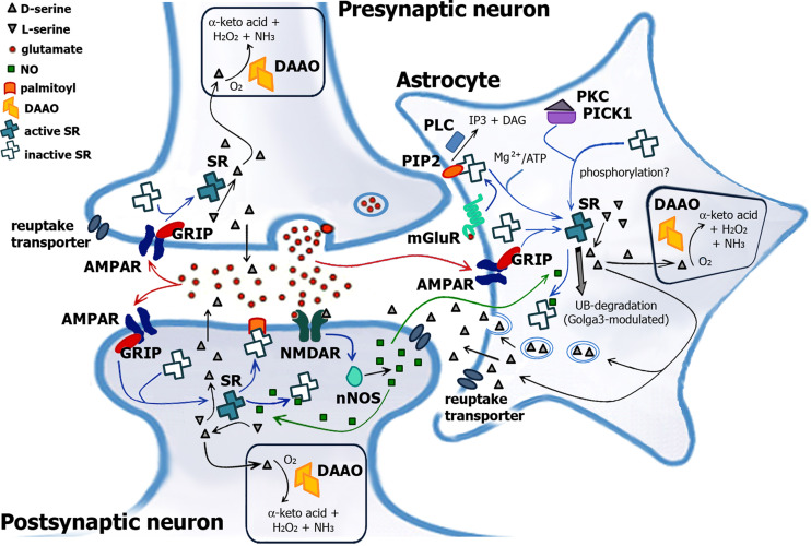
Over the past years, accumulating evidence has indicated that D-serine is the endogenous ligand for the glycine-modulatory binding site on the NR1 subunit of N-methyl-D-aspartate receptors in various brain areas. D-Serine is synthesized in glial cells and neurons by the pyridoxal-5' phosphate-dependent enzyme serine racemase, and it is released upon activation of glutamate receptors. The cellular concentration of this novel messenger is regulated by both serine racemase isomerization and elimination reactions, as well as by its selective degradation catalyzed by the flavin adenine dinucleotide-containing flavoenzyme D-amino acid oxidase. Here, we present an overview of the current knowledge of the metabolism of D-serine in human brain at the molecular and cellular levels, with a specific emphasis on the brain localization and regulatory pathways of D-serine, serine racemase, and D-amino acid oxidase. Furthermore, we discuss how D-serine is involved with specific pathological conditions related to N-methyl-D-aspartate receptors over- or down-regulation.


Figures

Fig. 1
Fig. 1
Model for SR’s and DAAO’s role in d-serine signaling in the brain. Glutamate released from presynaptic neuron acts on the postsynaptic neuron as well as on the surrounding astrocytes (red arrow), activating metabotropic glutamate receptors mGluR5 [and subsequent degradation of PIP2 to inositol triphosphate (IP 3) and diacylglycerol (DAG) by phospholipase C, PLC] and synthesizing d-serine from SR. Activation of glial SR presumably is due to an association with GRIP proteins and/or phosphorylation. Since GRIP-1 is expressed in neurons where the large majority of AMPAR are found, this mechanism might also activate neuronal SR. In the synaptic cleft, d-serine binds to the glycine-binding site on the NMDAR and in conjunction with glutamate results in the opening of the receptor channel. The activity of SR is switched off by different mechanisms: (1) Ca2+ entering the postsynaptic neuron activates neuronal NO synthase, producing NO, which can diffuse into adjacent cells. In astrocytes and neurons, NO gives nitrosylation of SR at Cys113, preventing ATP binding and thus inactivating the enzyme (green arrow). (2) The binding of SR to membrane PIP2 inhibits the enzyme according to a competitive mechanism with ATP. (3) SR is degraded through the ubiquitin-proteasomal system, a process which is modulated by Golga3 (gray arrow). Golga3 interacts with the N-terminal 66 residues of SR, decreasing the ubiquitin/proteasomal degradation of SR, probably interfering with binding of a still unidentified E3-ubiquitin ligase, and increases its steady-state levels (and d-serine synthesis)
Fig. 2
Fig. 2
Reaction mechanism for serine racemization (a) and α,β-elimination (b) by SR [25]. PLP bound to SR through an internal aldimine with Lys56 reacts with l-serine yielding an external aldimine; α-proton abstraction from this intermediate gives a resonance-stabilized carbanion. This intermediate can follow two pathways: for racemization reaction (a), the reprotonation of the intermediate on the opposite face of the planar carbanion generates the external aldimine which releases d-serine via transimination with Lys56; for α,β-elimination reaction (b), the elimination of the β-OH group from the carbanionic intermediate leads to the formation of the aminoacrylate-PLP intermediate; subsequent transimination releases iminopyruvate product, which non-enzymatically hydrolyzes to pyruvate and ammonia
Fig. 3
Fig. 3
a Model of the dimeric structure of hSR as proposed by [37, 38] (reproduced with permission). PLP is shown in blue, Ca2+ in green, and ATP analogue phosphomethylphosphonic acid (ACP) in red. b Dimeric structure of hDAAO in complex with imino-serine (PDB code: 2e49) [52]. FAD cofactor is represented in yellow, ligand (imino serine, SRI) in pink
Fig. 4
Fig. 4
Reaction catalyzed by DAAO [39, 40]. The hydride-transfer of the α-proton of d-serine to the oxidized N(5) FAD position yields imino pyruvic acid and reduced (anionic) flavin. The imino pyruvic acid is then non-enzymatically hydrolyzed to pyruvate and ammonia; the reduced flavin is re-oxidized by dioxygen yielding hydrogen peroxide
Fig. 5
Fig. 5
Effect of pLG72 on hDAAO activity in vitro (a) and in U87 glioblastoma-transfected cells (b) and on d-serine cellular concentration [69]. a Effect of pLG72 binding on hDAAO activity: black bars activity measured without preincubation or gray bars after 30 min of pre-incubation of a fixed amount of hDAAO (0.1 nmol/ml) with increasing amounts of pLG72; (white bars) effect of pLG72 on the reactivity of the hDAAO-bound FAD. In this latter case, the oxidized hDAAO was incubated with a stoichiometric amount of free FAD and increasing amounts of pLG72 and subsequently was anaerobically reduced by adding 1 mM d-serine: the bars report the percentage of reduced flavin. b (Black bars) Summary histogram of the d-/l-serine ratio (percentage) in U87 control cells (CTRL) and in the same cells transfected with hDAAO or pLG72: the change was significant for hDAAO (p = 0.004) and not significant for pLG72. (Gray bars) Summary histogram of the hDAAO activity (arbitrary units) in the same U87 cells: the change in activity with respect to the control was statistically significant for hDAAO (p = 0.012) and not significant for pLG72. The data are reported as mean ± SEM
Fig. 6
Fig. 6
Proposed role of the interaction between hDAAO and pLG72 on the d-serine bioavailability at glutamatergic synapses under normal and pathological (schizophrenia susceptibility) conditions [69]. Up to now, pLG72 was identified only in glial cells, therefore we focused on the modulation of hDAAO activity in astrocytes. According to our hypothesis, pLG72 modulates the amount of active hDAAO acting on the stability of the flavoenzyme (left panel). An abnormal, low expression of pLG72 under pathological conditions could result in hyperactivation of hDAAO and decrease d-serine concentration (right panel). Importantly, the decrease in d-serine concentration results in a lower amount of activated NMDAR and, thus, in a hypofunction of the glutamatergic neurotransmission (see also Fig. 1 for details about the overall mechanisms of d-serine regulation at synapsis)

Similar articles

Cited by

References

    1. Hashimoto A, Oka T, Nishikawa T. Extracellular concentration of endogenous free d-serine in the rat brain as revealed by in vivo microdialysis. Neuroscience. 1995;66:635–643. - PubMed
    1. Wolosker H, Sheth KN, Takahashi M, Mothet JP, Brady RO, Jr, Ferris CD, Snyder SH. Purification of serine racemase: biosynthesis of the neuromodulator d-serine. Proc Natl Acad Sci USA. 1999;96:721–725. - PMC - PubMed
    1. Schell MJ, Molliver ME, Snyder SH. d-serine, an endogenous synaptic modulator: localization to astrocytes and glutamate-stimulated release. Proc Natl Acad Sci USA. 1995;92:3948–3952. - PMC - PubMed
    1. Danysz W, Parsons CG. Glycine and N-methyl-d-aspartate receptors: physiological significance and possible therapeutic applications. Pharmacol Rev. 1998;50:597–664. - PubMed
    1. Hashimoto A, Nishikawa T, Oka T, Takahashi K. Endogenous d-serine in rat brain: N-methyl-d-aspartate receptor-related distribution and aging. J Neurochem. 1993;60:783–786. - PubMed

Publication types

MeSH terms

LinkOut - more resources