. 2009 Sep;13(9B):3668-79.
doi: 10.1111/j.1582-4934.2009.00661.x.

Inhibition of DNA methyltransferase induces G2 cell cycle arrest and apoptosis in human colorectal cancer cells via inhibition of JAK2/STAT3/STAT5 signalling

Affiliations

Inhibition of DNA methyltransferase induces G2 cell cycle arrest and apoptosis in human colorectal cancer cells via inhibition of JAK2/STAT3/STAT5 signalling

Hua Xiong et al. J Cell Mol Med. 2009 Sep.

Abstract

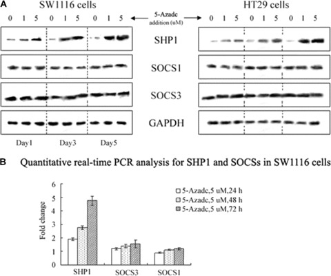
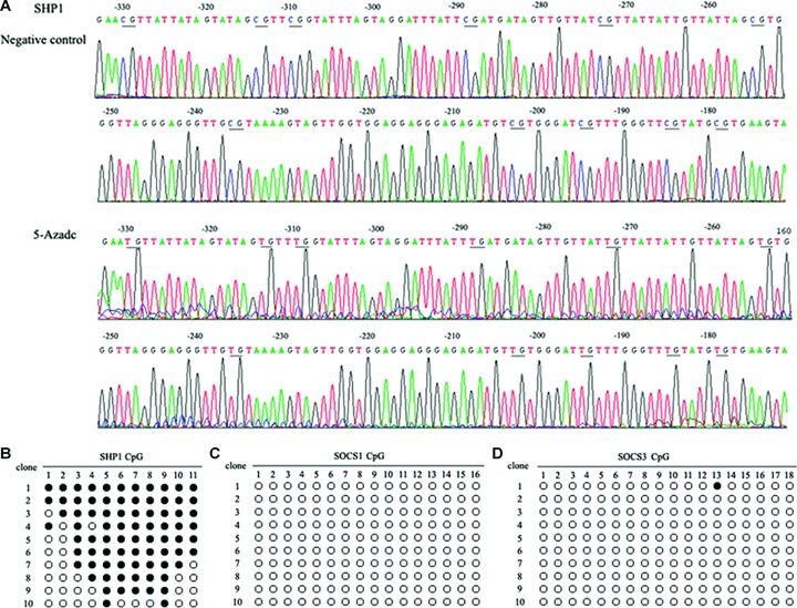
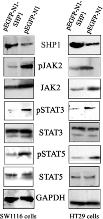
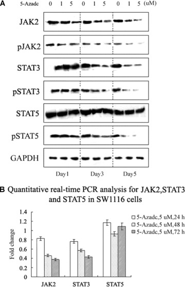
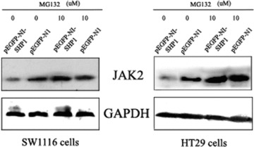
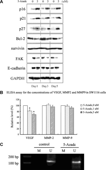
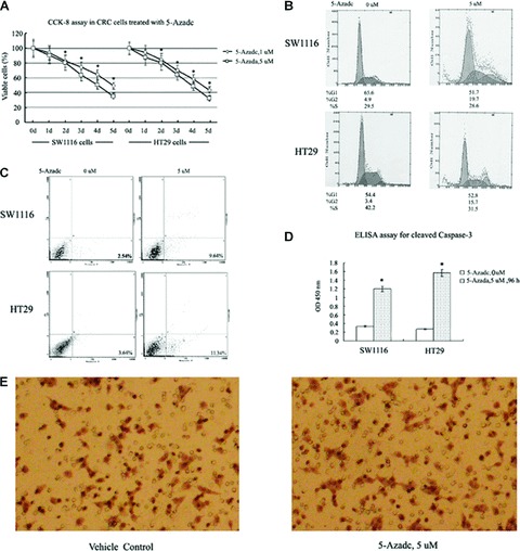
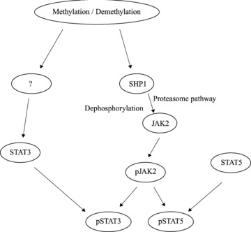
DNA methyltransferase inhibitors (MTIs) have recently emerged as promising chemotherapeutic or preventive agents for cancer, despite their poorly characterized mechanisms of action. The present study shows that DNA methylation is integral to the regulation of SH2-containing protein tyrosine phosphatase 1 (SHP1) expression, but not for regulation of suppressors of cytokine signalling (SOCS)1 or SOCS3 in colorectal cancer (CRC) cells. SHP1 expression correlates with down-regulation of Janus kinase/signal transducers and activators of transcription (JAK2/STAT3/STAT5) signalling, which is mediated in part by tyrosine dephosphorylation events and modulation of the proteasome pathway. Up-regulation of SHP1 expression was achieved using a DNA MTI, 5-aza-2'-deoxycytidine (5-aza-dc), which also generated significant down-regulation of JAK2/STAT3/STAT5 signalling. We demonstrate that 5-aza-dc suppresses growth of CRC cells, and induces G2 cell cycle arrest and apoptosis through regulation of downstream targets of JAK2/STAT3/STAT5 signalling including Bcl-2, p16(ink4a), p21(waf1/cip1) and p27(kip1). Although 5-aza-dc did not significantly inhibit cell invasion, 5-aza-dc did down-regulate expression of focal adhesion kinase and vascular endothelial growth factor in CRC cells. Our results demonstrate that 5-aza-dc can induce SHP1 expression and inhibit JAK2/STAT3/STAT5 signalling. This study represents the first evidence towards establishing a mechanistic link between inhibition of JAK2/STAT3/STAT5 signalling and the anticancer action of 5-aza-dc in CRC cells that may lead to the use of MTIs as a therapeutic intervention for human colorectal cancer.


Figures

Figure 1
Figure 1
5-aza-dc up-regulates SHP1 expression in colorectal cancer (CRC) cells. (A) Western blot analysis of 5-aza-dc-induced time-dependent increases in SHP1 in CRC cells, whereas SOCS1 and SOCS3 showed no detectable changes. The data presented are from a representative experiment and detection of GAPDH was used as a loading control. (B) Quantitative real-time PCR analysis of SHP1 and SOCSs in SW1116 cells. Results are expressed as relative expression compared with untreated cells. Each value is the mean ± S.D. of three experiments.
Figure 2
Figure 2
Bisulphite sequencing of SHP1, SOCS1 and SOCS3 promoters in HT29 cells. (A) Bisulphite sequencing chromatogram of SHP1 in HT29 cells. Bisulphate sequencing studies were performed on DNA extracted from HT29 cells cultured in the absence or presence of 5 μM 5-aza-dc for 96 hrs. Ten individual clones were analysed and the proportion of methylated residues detected ranged from 18.2% (2/11) to 100% (11/11). In this figure, the sequencing results from a clone with 11 demethylated CG pairs identified in the SHP1 promoter at positions −330, −314, −310, −289, −273, −265, −236, −203, −195, −185 and −179. (B) Methylation status of the SHP1, SOCS1 and SOCS3 promoters in HT29 cells (•, methylated cytosine; ○, unmethylated cytosine).
Figure 3
Figure 3
The affects of SHP1 expression on JAK2/STAT3/STAT5 protein levels in CRC cells. CRC cells transfected with pEGFP-N1 or pEGFP-N1-SHP1 were sorted for GFP expression and extracts analysed by Western blot. Substantial decreases in expression of pJAK2, JAK2, pSTAT3 and pSTAT5 were detected. The decrease in pJAK2 levels was greater than that of JAK2. In contrast, no significant changes in STAT3 and STAT5 protein levels were detected. The data shown are from a representative experiment and detection of GAPDH was used as a loading control.
Figure 4
Figure 4
5-aza-dc induces down-regulation of JAK2/STAT3/STAT5 protein levels. (A) Western blot analysis revealed decreases in JAK2 and pJAK2 protein levels for SW1116 cells treated with 5-aza-dc. In the same experiment, decreases in STAT3, pSTAT3 and pSTAT5 were also identified. The data shown are from a representative experiment and detection of GAPDH was used as a loading control. (B) Quantitative real-time PCR analysis of JAK2, STAT3 and STAT5 in SW1116 cells treated with 5-aza-dc. Results are expressed as relative expression compared to untreated cells. Each value is the mean ± S.D. of three experiments.
Figure 5
Figure 5
MG132 prevents SHP1-mediated down-regulation of JAK2. CRC cells were transfected with pEGFP-N1 or pEGFP-N1-SHP1 and sorted for GFP expression. The GFP-positive population was subsequently treated with 10 μM MG132 and extracts were analysed by Western blot. Decreased levels of JAK2 induced by exogenous expression of SHP1 were reversed 10 hrs after the addition of 10 μM MG132 to cell cultures. The data shown are from a representative experiment and detection of GAPDH was used as a loading control.
Figure 6
Figure 6
Disruption of JAK2/STAT3/STAT5 signalling by 5-aza-dc is associated with modulation of downstream STAT targets. (A) Western blot analysis of JAK2/STAT3/STAT5 downstream targets in SW1116 cells following 5-aza-dc treatment. Bcl-2 and FAK were down-regulated, while p16ink4a, p21waf1/cip1 and p27kip1 were up-regulated. Survivin and E-cadherin showed no detectable change. The data shown are from a representative experiment and detection of GAPDH was used as a loading control. (B) Concentrations of VEGF, MMP-2 and MMP-9 in SW1116 cells treated with 5-aza-dc were analysed by ELISA 24 hrs after treatment. A decrease in the secretion of VEGF was detected (*p < 0.05). Results are expressed as relative levels compared with untreated cells. Each value is the mean ± S.D. of three experiments. (C) p27kip1 MSP was performed on DNA from SW1116 cells to specifically detect the methylation status of the promoter (M, methylated-MSP; U, unmethylated-MSP). The data shown are representative of three replicate MSP experiments.
Figure 7
Figure 7
Biological effects of 5-aza-dc in CRC cells. (A) CCK-8 assay of CRC cells treated with 5 μM 5-aza-dc or solvent only as a negative control. The cell numbers of 5-aza-dc-treated cells were normalized to that of the negative control and showed a concentration- and time-dependent decrease in the number of viable CRC cells treated with 5-aza-dc compared to negative control cells (*P < 0.05). (B) Cell cycle analysis was performed 72 hrs after CRC cells were treated with diluent (left) or 5-aza-dc (5 μM, right). Compared to negative control cells, treated cells showed an increased proportion of cells in the G2 phase. Data shown are from a representative experiment. (C) CRC cells were treated with diluent (left) or 5-aza-dc (5 μM, right) for 96 hrs and then analysed for the presence of cell apoptosis by flow cytometric analysis. A 3.80- and 3.12-fold increase in apoptotic cells were detected for SW1116 and HT29 cells treated with 5 μM 5-aza-dc relative to control cells. The data shown are from a representative experiment. (D) Cleavage of caspase-3 in CRC cells treated with 5 μM 5-aza-dc was detected 96 hrs after treatment. Each value is the mean ± S.D. of three experiments (*P < 0.05). (E) An in vitro cell invasion assay was performed using SW1116 cells as described in the ‘Materials and methods’ section. After 24 hrs of treatment with 5 μM 5-aza-dc, no significant difference in cell invasion was detected between treated and vehicle control cells. The data shown are from a representative experiment at 200× magnification.
Figure 8
Figure 8
The possible mechanistic link between JAK2/STAT3/STAT5 signalling and the anticancer action of 5-aza-dc in CRC cells. Using an inhibitor of DNA methytransferase, 5-aza-dc, methylation was shown to be integral to the regulation of SHP1 expression, and SHP1 appears to down-regulate JAK2 by two mechanisms: tyrosine dephosphorylation and the proteasome pathway. The decrease in STAT3 expression observed in CRC cells exposed to 5-aza-dc is unclear at this point and requires additional study, while STAT5 appears to be unaffected by an inhibition of methylation.

Similar articles

Cited by

References

    1. Lubbert M. DNA methylation inhibitors in the treatment of leukemias, myelodysplastic syndromes and hemoglobinopathies: clinical results and possible mechanisms of action. Curr Top Microbiol Immunol. 2000;249:135–64. - PubMed
    1. Momparler RL, Ayoub J. Potential of 5-aza-2’-deoxycytidine (Decitabine) a potent inhibitor of DNA methylation for therapy of advanced non-small cell lung cancer. Lung Cancer. 2001;34:S111–5. - PubMed
    1. Issa JP, Kantarjian H. Azacitidine. Nat Rev Drug Discov. 2005:S6–7. - PubMed
    1. Wijermans P, Lubbert M, Verhoef G, et al. Low-dose 5-aza-2’-deoxycytidine, a DNA hypomethylating agent, for the treatment of high-risk myelodysplastic syndrome: a multicenter phase II study in elderly patients. J Clin Oncol. 2000;18:956–62. - PubMed
    1. Aparicio A, Eads CA, Leong LA, et al. Phase I trial of continuous infusion 5-aza-2′-deoxycytidine. Cancer Chemother Pharmacol. 2003;51:231–9. - PubMed

Publication types

MeSH terms