

Cytopathic bovine viral diarrhea viruses (BVDV): emerging pestiviruses doomed to extinction
- PMID: 20197026
- PMCID: PMC2850149
- DOI: 10.1051/vetres/2010016
Cytopathic bovine viral diarrhea viruses (BVDV): emerging pestiviruses doomed to extinction
Abstract
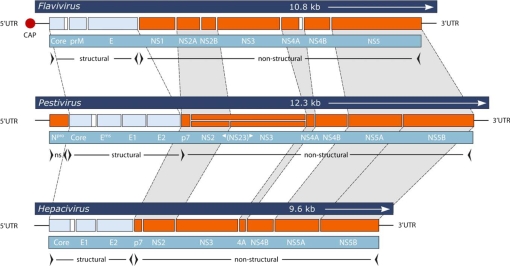
Bovine viral diarrhea virus (BVDV), a Flaviviridae pestivirus, is arguably one of the most widespread cattle pathogens worldwide. Each of its two genotypes has two biotypes, non-cytopathic (ncp) and cytopathic (cp). Only the ncp biotype of BVDV may establish persistent infection in the fetus when infecting a dam early in gestation, a time point which predates maturity of the adaptive immune system. Such fetuses may develop and be born healthy but remain infected for life. Due to this early initiation of fetal infection and to the expression of interferon antagonistic proteins, persistently infected (PI) animals remain immunotolerant to the infecting viral strain. Although only accounting for some 1% of all animals in regions where BVDV is endemic, PI animals ensure the viral persistence in the host population. These animals may, however, develop the fatal mucosal disease, which is characterized by widespread lesions in the gastrointestinal tract. Cp BVD virus, in addition to the persisting ncp biotype, can be isolated from such animals. The cp viruses are characterized by unrestrained genome replication, and their emergence from the persisting ncp ones is due to mutations that are unique in each virus analyzed. They include recombinations with host cell mRNA, gene translocations and duplications, and point mutations. Cytopathic BVD viruses fail to establish chains of infection and are unable to cause persistent infection. Hence, these viruses illustrate a case of "viral emergence to extinction" - irrelevant for BVDV evolution, but fatal for the PI host.
© INRA, EDP Sciences, 2010.
Figures
Similar articles
-
Analysis of Bovine Viral Diarrhea Viruses-infected monocytes: identification of cytopathic and non-cytopathic biotype differences.BMC Bioinformatics. 2010 Oct 7;11 Suppl 6(Suppl 6):S9. doi: 10.1186/1471-2105-11-S6-S9. BMC Bioinformatics. 2010. PMID: 20946620 Free PMC article.
-
[Bovine diarrhea virus: an update].Rev Argent Microbiol. 1997 Jan-Mar;29(1):47-61. Rev Argent Microbiol. 1997. PMID: 9229725 Review. Spanish.
-
Characterization of the cytopathic BVDV strains isolated from 13 mucosal disease cases arising in a cattle herd.Virus Res. 2015 Jan 2;195:141-7. doi: 10.1016/j.virusres.2014.09.015. Epub 2014 Oct 7. Virus Res. 2015. PMID: 25300803
-
Bovine viral diarrhea virus cytopathic and noncytopathic biotypes and type 1 and 2 genotypes in diagnostic laboratory accessions: clinical and necropsy samples from cattle.J Vet Diagn Invest. 2000 Jan;12(1):33-8. doi: 10.1177/104063870001200106. J Vet Diagn Invest. 2000. PMID: 10690773
-
Pathogenesis of mucosal disease, a deadly disease of cattle caused by a pestivirus.Clin Diagn Virol. 1998 Jul 15;10(2-3):121-7. doi: 10.1016/s0928-0197(98)00037-3. Clin Diagn Virol. 1998. PMID: 9741637 Review.
Cited by
-
An Importance of Long-Term Clinical Analysis to Accurately Diagnose Calves Persistently and Acutely Infected by Bovine Viral Diarrhea Virus 2.Viruses. 2021 Dec 3;13(12):2431. doi: 10.3390/v13122431. Viruses. 2021. PMID: 34960700 Free PMC article.
-
Contact-number-driven virus evolution: A multi-level modeling framework for the evolution of acute or persistent RNA virus infection.PLoS Comput Biol. 2023 May 30;19(5):e1011173. doi: 10.1371/journal.pcbi.1011173. eCollection 2023 May. PLoS Comput Biol. 2023. PMID: 37253076 Free PMC article.
-
BVD-2 outbreak leads to high losses in cattle farms in Western Germany.Heliyon. 2015 Sep 21;1(1):e00019. doi: 10.1016/j.heliyon.2015.e00019. eCollection 2015 Sep. Heliyon. 2015. PMID: 27441213 Free PMC article.
-
Transmission of border disease virus from a persistently infected calf to seronegative heifers in early pregnancy.BMC Vet Res. 2015 Feb 22;11:43. doi: 10.1186/s12917-014-0275-7. BMC Vet Res. 2015. PMID: 25889936 Free PMC article.
-
Compartmentalized evolution of Bovine Viral Diarrhoea Virus type 2 in an immunotolerant persistently infected cow.Sci Rep. 2019 Oct 29;9(1):15460. doi: 10.1038/s41598-019-52023-w. Sci Rep. 2019. PMID: 31664116 Free PMC article.
References
-
- Anonymous, Hepatitis C – global prevalence, Wkly Epidemiol. Rec. (1999) 74:421–428 - PubMed
-
- Arnal M.C., Fernández-De-Luco D., Riba L., Maley M., Gilray J., Willoughby K., et al., A novel pestivirus associated with deaths in Pyrenean chamois (Rupicapra pyrenaica pyrenaica), J. Gen. Virol. (2004) 85:3653–3657 - PubMed
-
- Audet S.A., Crim R.L., Beeler J., Evaluation of vaccines, interferons and cell substrates for pestivirus contamination, Biologicals (2000) 28:41–46 - PubMed
-
- Bachofen C., Stalder H., Braun U., Hilbe M., Ehrensperger F., Peterhans E., Co-existence of genetically and antigenically diverse bovine viral diarrhoea viruses in an endemic situation, Vet. Microbiol. (2008) 131:93–102 - PubMed
Publication types
MeSH terms
LinkOut - more resources
Full Text Sources
Other Literature Sources
Research Materials
Miscellaneous