. 2010 Mar 3;98(5):753-61.
doi: 10.1016/j.bpj.2009.10.052.

Two open states with progressive proton selectivities in the branched channelrhodopsin-2 photocycle

Affiliations

Two open states with progressive proton selectivities in the branched channelrhodopsin-2 photocycle

André Berndt et al. Biophys J. .

Abstract

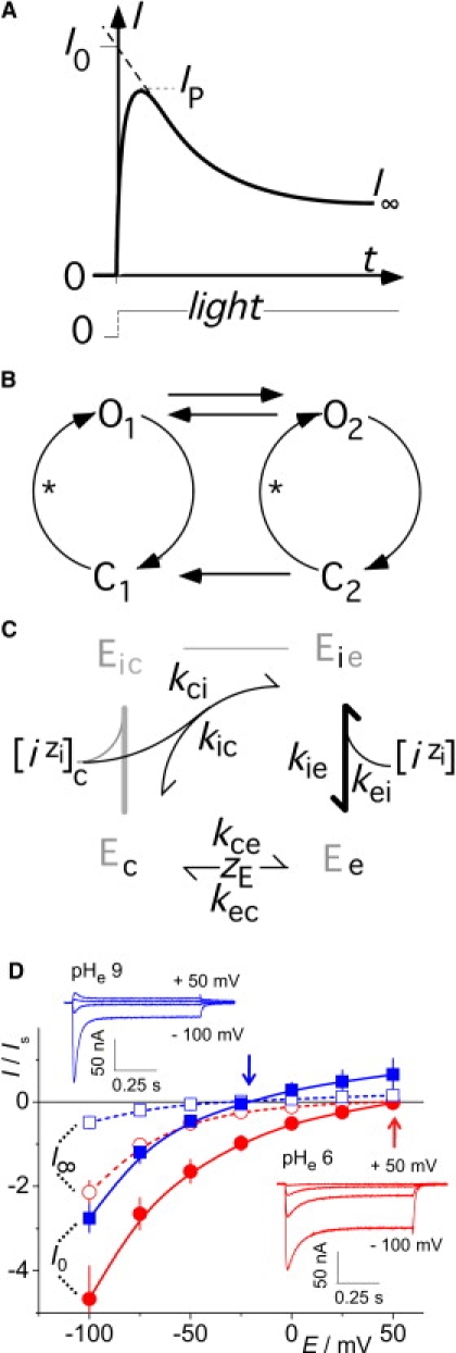
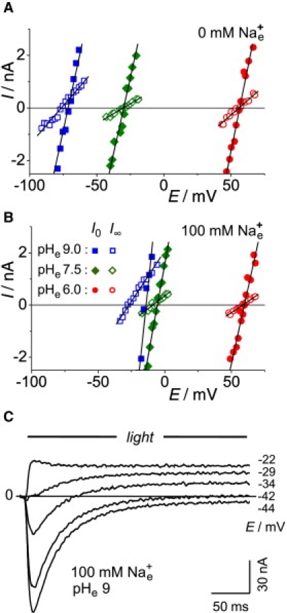
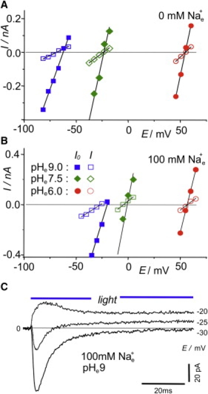
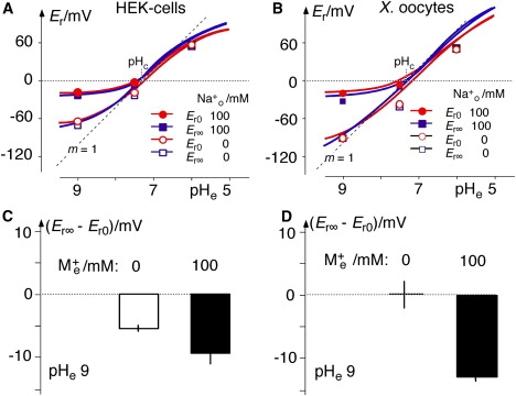
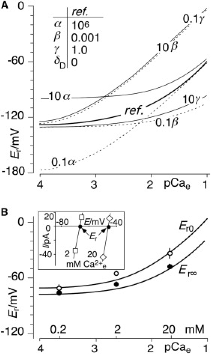
Channelrhodopsins are light-gated ion channels that mediate vision in phototactic green algae like Chlamydomonas. In neurosciences, channelrhodopsins are widely used to light-trigger action potentials in transfected cells. All known channelrhodopsins preferentially conduct H(+). Previous studies have indicated the existence of an early and a late conducting state within the channelrhodopsin photocycle. Here, we show that for channelrhodopsin-2 expressed in Xenopus oocytes and HEK cells, the two open states have different ion selectivities that cause changes in the channelrhodopsin-2 reversal voltage during a light pulse. An enzyme kinetic algorithm was applied to convert the reversal voltages in various ionic conditions to conductance ratios for H(+) and divalent cations (Ca(2+) and/or Mg(2+)), as compared to monovalent cations (Na(+) and/or K(+)). Compared to monovalent cation conductance, the H(+) conductance, alpha, is approximately 3 x 10(6) and the divalent cation conductance, beta, is approximately 0.01 in the early conducting state. In the stationary mixture of the early and late states, alpha is larger and beta smaller, both by a factor of approximately 2. The results suggest that the ionic basis of light perception in Chlamydomonas is relatively nonspecific in the beginning of a light pulse but becomes more selective for protons during longer light exposures.


Figures

Figure 1
Figure 1
Definitions, recapitulation, and reaction schemes of the working hypothesis. (A) Typical time course of ChR2 photocurrent upon a bright, rectangular light pulse: after an early peak (IP), the current relaxes to a stationary level, I; extrapolation of the steepest slope to t = 0 yields the initial current, I0. (B) Simplified photocycle of ChR (6,7) with early and late conducting (open) states O1 and O2, respectively, plus two nonconducting (closed) states, C1 and C2. Asterisks indicate photoisomerization steps. Due to the irreversible step from C2 to C1, the steady-state occupation probabilities, p, of the four states are pC1 = 1, and pO1 = pO2 = pC2 = 0 in the dark; hence, I0 can be assigned to state O1 exclusively. (C) Reaction scheme for experiments with concentration changes of external ions; bold line indicates rapid equilibria. Derivation of three-state model from standard four-state model (gray) for uniport of one substrate i through enzyme E when no internal concentration changes take place. Subscripts c and e represent cytoplasmic and external measures, respectively. In the global model, n ion species, i, with their specific valencies, zi, compete for the empty binding site with charge zE, resulting in n three-state cycles sharing reorientation of the empty binding site between the cytoplasmic and external sides. (D) Current-voltage relationships for current amplitudes of I0 and I versus holding voltage, recorded at pHe 9 and 6. IS is the reference current at pHe 7.5 with 100 mM external Na+, for standardizing results from various preparations. Arrows indicate the estimated reversal voltages, and insets show individual current tracings at the indicated holding voltages.
Figure 2
Figure 2
Typical experiment for determination of reversal voltages Er0 and Er∞ of ChR2 in Xenopus oocytes under various ionic conditions (in mM): internal, ∼120 Na+, ∼40 Cl, pHc 7.3; external, 0.1 CaCl2, 2 MgCl2, and 100 mM NMGCl (A) or 100 mM NaCl (B). Voltage-clamp recordings of initial and stationary currents, I, were carried out at holding voltages, EC, in the vicinity of reversal voltages. Readings of Er are from intersections of regression lines with the zero-current line in the absence of external Na+ (A), and in the presence of 100 mM external Na+ (B) (for statistical evidence, see Fig. 4D; for quantitative analysis, see Table 1). (C) Original photocurrent records, measured at the indicated holding voltages. Note change of current sign in middle tracing (−34 mV), indicating change of reversal voltage and selectivity during a light pulse. Since the experiments in AC were from different oocytes, the current scale in C was adjusted to match A and B.
Figure 3
Figure 3
Typical experiment for determination of reversal voltages Er0 and Er∞ of ChR2 in HEK cells under various ionic conditions (in mM): internal, 120 NaCl, 10 Hepes, 2 MgCl2, 10 EGTA, and 2 CaCl2, pHc 7.3; and external, 2 CaCl2, 2 MgCl2, and 100 mM NMGCl (A) or 100 mM NaCl (B). Voltage-clamp records of initial and stationary currents, I, are shown at holding voltages, EC, in the vicinity of reversal voltages. Readings of Er are from intersections of regression lines with the zero-current line. in the absence of external Na+ (A), and with 100 mM added external Na+ (B). The predominant result was Er∞ < Er0 throughout, indicating increased H+ selectivity in the stationary state compared to the initial state (for statistical evidence, see Fig. 4A, and for quantitative analysis, see Table 1). (C) Original photocurrent records. Note change of current sign in the middle tracing (−25 mV), indicating change of reversal voltage and selectivity during a light pulse. The current scale was adjusted to match A and B.
Figure 4
Figure 4
Initial (red) and stationary (blue) reversal voltages, Er0 and Er∞, of ChR2 as a function of pHe, recorded with and without 100 mM external Na+ in HEK cells and Xenopus oocytes. Values are means of n ≥ 3 independent recordings. (A) Absolute Er values in HEK cells (internal ion concentrations, in mM: 120 NaCl, 10 Hepes, 2 MgCl2, 2 CaCl2, and 10 EGTA, pHc 7.3); small SEs of 0.5–5.7 mV (n = 3) not illustrated. dotted line: theoretical Nernstian EH(pHe) with slope m = 1 (here, rounded 60 mV/pH unit); solid lines: theoretical relationships according to Eq. 10 with mean conductance ratios α and β from Table 1 at pHe 9.0, with and without 100 mM Na+e. (B) Same as A, but for Xenopus oocytes (internal ion concentrations, in mM: 120 Na+, 40 Cl, pHc 7.4; data taken from Zhang and Prigge (4); small SEs of 1.0–5.0 mV (n = 3–7) are not shown. (C and D) Statistically supported differences, Er∞Er0, at pHe 9, for HEK cells and Xenopus oocytes, respectively.
Figure 5
Figure 5
Effect of divalent cations, D2+ (Mg2+ + Ca2+), on Er. (A) Theoretical relations: impact of 10-fold increase and decrease of conductance ratios α (kcH0/kcM0), β (kcD0/kcM0), and γ (kce0/kcM0) on Er([D2+]e) relationships compared to reference configuration (inset). Note that increase of γ and decrease of β are almost equivalent in this configuration ([M+]c = 120 mM (100 mM K+ and 20 mM Na2+), [D2+]c = 2 mM (2 mM Mg2+ and 0.25 μM Ca2+), pHc 7.3, pHe [M+]e = 0, [D2+]c as marked by pCae on abscissa). To circumvent corresponding fluctuations in fits, γ was arbitrarily fixed at γ = 1 for numerical analysis. (B) Experimental results (symbols, means ± SE) and fits (solid lines) of Er at various external [Ca2+]; cytoplasmic ions, γ = 1, [M+]e = 0, pHe 9.0, as in A. To obtain fair fits, an ad hoc correction, δD, was introduced to account for the impact of external Mg2+: [D2+]e = δD + [Ca2+]e (see numerical results in Table 2). (Inset) Example of original current-voltage data for ChR2 in HEK cells (pHe 9, [M+]e = 0, internal solution, as in main figure) showing [Ca2+]e dependency of Er.

References

    1. Hegemann P. Algal sensory photoreceptors. Annu. Rev. Plant Biol. 2008;59:167–189. - PubMed
    1. Zhang F., Aravanis A.M., Deisseroth K. Circuit-breakers: optical technologies for probing neural signals and systems. Nat. Rev. Neurosci. 2007;8:577–581. - PubMed
    1. Ernst O.P., Sánchez Murcia P.A., Hegemann P. Photoactivation of channelrhodopsin. J. Biol. Chem. 2008;283:1637–1643. - PubMed
    1. Zhang F., Prigge M., Deisseroth K. Red-shifted optogenetic excitation: a tool for fast neural control derived from Volvox carteri. Nat. Neurosci. 2008;11:631–633. - PMC - PubMed
    1. Tsunoda S.P., Hegemann P. Glu 87 of channelrhodopsin-1 causes pH-dependent color tuning and fast photocurrent inactivation. Photochem. Photobiol. 2009;85:564–569. - PubMed

Publication types