

Effect of membrane thickness on conformational sampling of phospholamban from computer simulations
- PMID: 20197034
- PMCID: PMC2830431
- DOI: 10.1016/j.bpj.2009.11.015
Effect of membrane thickness on conformational sampling of phospholamban from computer simulations
Abstract
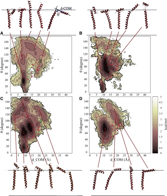
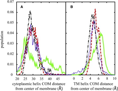
The conformational sampling of monomeric, membrane-bound phospholamban is described from computer simulations. Phospholamban (PLB) plays a key role as a regulator of sarcoplasmic reticulum calcium ATPase. An implicit membrane model is used in conjunction with replica exchange molecular dynamics simulations to reach mus-ms timescales. The implicit membrane model was also used to study the effect of different membrane thicknesses by scaling the low-dielectric region. The conformational sampling with the membrane model mimicking dipalmitoylphosphatidylcholine bilayers is in good agreement overall with experimental measurements, but consists of a wide variety of different conformations including structures not described previously. The conformational ensemble shifts significantly in the presence of thinner or thicker membranes. This has implications for the structure and dynamics of PLB in physiological membranes and offers what we believe to be a new interpretation of previous experimental measurements of PLB in detergents and microsomal membrane.
2010 Biophysical Society. Published by Elsevier Inc. All rights reserved.
Figures
Similar articles
-
Molecular dynamics simulations of biological membranes and membrane proteins using enhanced conformational sampling algorithms.Biochim Biophys Acta. 2016 Jul;1858(7 Pt B):1635-51. doi: 10.1016/j.bbamem.2015.12.032. Epub 2016 Jan 5. Biochim Biophys Acta. 2016. PMID: 26766517 Free PMC article. Review.
-
Structure and dynamics of phospholamban in solution and in membrane bilayer: computer simulations.Biochemistry. 2005 Feb 15;44(6):1780-92. doi: 10.1021/bi0488404. Biochemistry. 2005. PMID: 15697203
-
Role of conformational sampling of Ser16 and Thr17-phosphorylated phospholamban in interactions with SERCA.Biochim Biophys Acta. 2013 Feb;1828(2):577-85. doi: 10.1016/j.bbamem.2012.08.017. Epub 2012 Aug 29. Biochim Biophys Acta. 2013. PMID: 22959711
-
Effects of CMAP and electrostatic cutoffs on the dynamics of an integral membrane protein: the phospholamban study.J Biomol Struct Dyn. 2008 Aug;26(1):17-34. doi: 10.1080/07391102.2008.10507220. J Biomol Struct Dyn. 2008. PMID: 18533723
-
Direct spectroscopic detection of molecular dynamics and interactions of the calcium pump and phospholamban.Ann N Y Acad Sci. 1998 Sep 16;853:186-94. doi: 10.1111/j.1749-6632.1998.tb08266.x. Ann N Y Acad Sci. 1998. PMID: 10603946 Review.
Cited by
-
Molecular dynamics simulations of biological membranes and membrane proteins using enhanced conformational sampling algorithms.Biochim Biophys Acta. 2016 Jul;1858(7 Pt B):1635-51. doi: 10.1016/j.bbamem.2015.12.032. Epub 2016 Jan 5. Biochim Biophys Acta. 2016. PMID: 26766517 Free PMC article. Review.
-
Effect of flanking residues on the conformational sampling of the internal fusion peptide from Ebola virus.Proteins. 2011 Apr;79(4):1109-17. doi: 10.1002/prot.22947. Epub 2011 Jan 18. Proteins. 2011. PMID: 21246633 Free PMC article.
-
Discrimination of Native-like States of Membrane Proteins with Implicit Membrane-based Scoring Functions.J Chem Theory Comput. 2017 Jun 13;13(6):3049-3059. doi: 10.1021/acs.jctc.7b00254. Epub 2017 May 11. J Chem Theory Comput. 2017. PMID: 28475346 Free PMC article.
-
Dynamic Heterogeneous Dielectric Generalized Born (DHDGB): An implicit membrane model with a dynamically varying bilayer thickness.J Chem Theory Comput. 2013 Mar 12;9(3):1709-1719. doi: 10.1021/ct300975k. J Chem Theory Comput. 2013. PMID: 23585740 Free PMC article.
-
CHARMM at 45: Enhancements in Accessibility, Functionality, and Speed.J Phys Chem B. 2024 Oct 17;128(41):9976-10042. doi: 10.1021/acs.jpcb.4c04100. Epub 2024 Sep 20. J Phys Chem B. 2024. PMID: 39303207 Free PMC article. Review.
References
-
- Post R.L., Hegyvary C., Kume S. Activation by adenosine triphosphate in the phosphorylation kinetics of sodium and potassium ion transport adenosine triphosphatase. J. Biol. Chem. 1972;247:6530–6540. - PubMed
-
- Simmerman H.K.B., Jones L.R. Phospholamban: protein structure, mechanism of action, and role in cardiac function. Physiol. Rev. 1998;78:921–947. - PubMed
-
- Autry J.M., Jones L.R. Functional Co-expression of the canine cardiac Ca2+ pump and phospholamban in Spodoptera frugiperda (Sf21) cells reveals new insights on ATPase regulation. J. Biol. Chem. 1997;272:15872–15880. - PubMed
-
- Bilezikjian L.M., Kranias E.G., Schwartz A. Studies on phosphorylation of canine cardiac sarcoplasmic reticulum by calmodulin-dependent protein kinase. Circ. Res. 1981;49:1356–1362. - PubMed
-
- Li J.H., Bigelow D.J., Squier T.C. Phosphorylation by cAMP-dependent protein kinase modulates the structural coupling between the transmembrane and cytosolic domains of phospholamban. Biochemistry. 2003;42:10674–10682. - PubMed
Publication types
MeSH terms
Substances
Grants and funding
LinkOut - more resources
Full Text Sources
