. 2010 Jul;7(7):942-50.
doi: 10.1016/j.hrthm.2010.02.036. Epub 2010 Mar 1.

Epicardial neural ganglionated plexus of ovine heart: anatomic basis for experimental cardiac electrophysiology and nerve protective cardiac surgery

Affiliations

Epicardial neural ganglionated plexus of ovine heart: anatomic basis for experimental cardiac electrophysiology and nerve protective cardiac surgery

Inga Saburkina et al. Heart Rhythm. 2010 Jul.

Abstract

Background: Sheep are routinely used in experimental cardiac electrophysiology and surgery.

Objective: The purpose of this study was to (1) ascertain the topography and architecture of the ovine epicardial neural plexus (ENP), (2) determine the relationships of ENP with vagal and sympathetic cardiac nerves and ganglia, and (3) evaluate gross anatomic differences and similarities of ENP in humans, sheep, and other species.

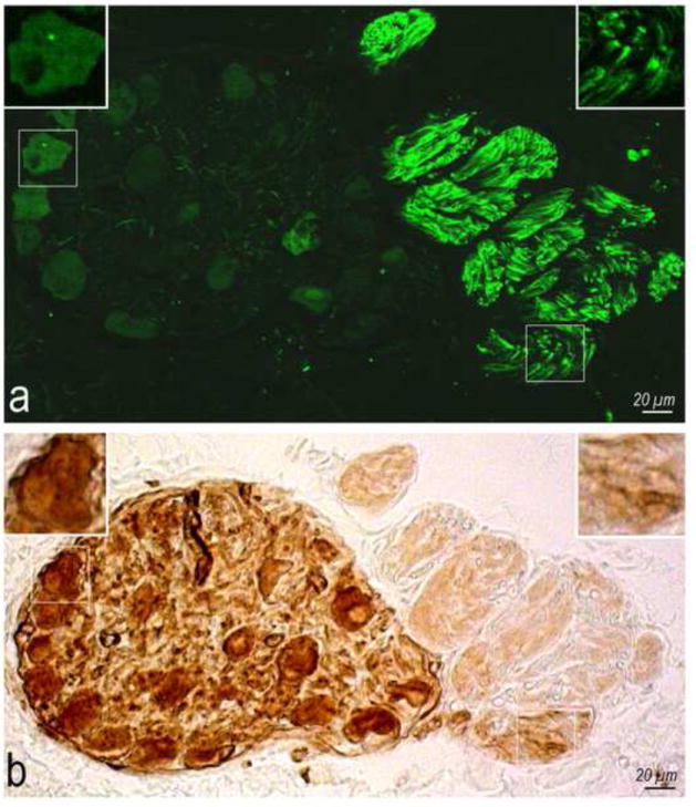
Methods: Ovine ENP and extrinsic sympathetic and vagal nerves were stained histochemically for acetylcholinesterase in whole heart and/or thorax-dissected preparations from 23 newborn lambs, with subsequent examination by stereomicroscope.

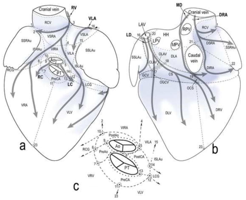
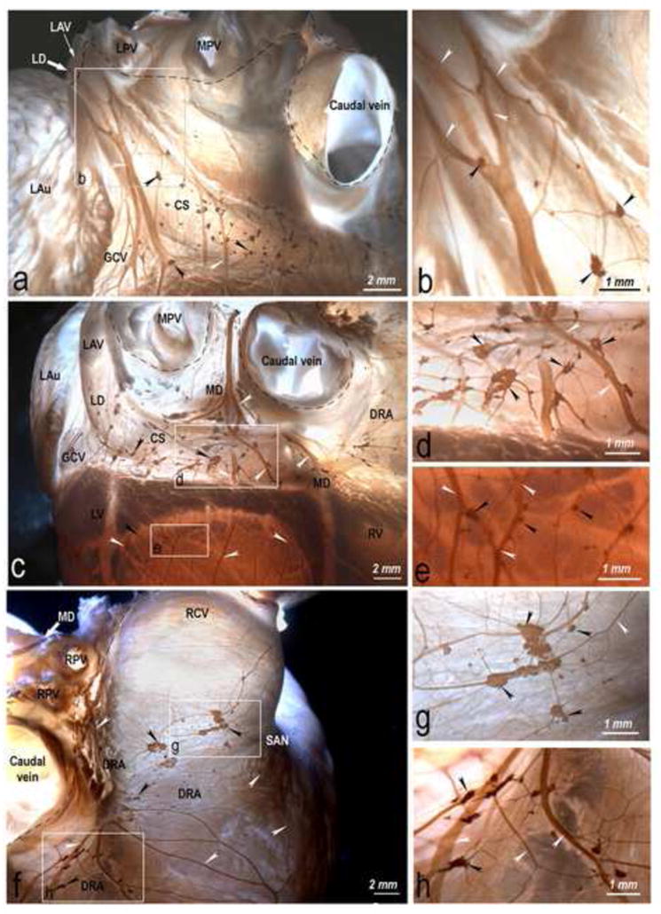
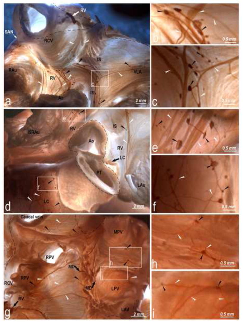
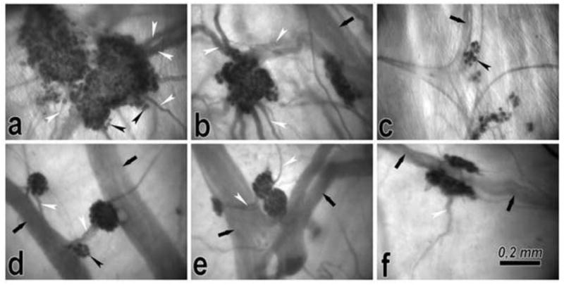
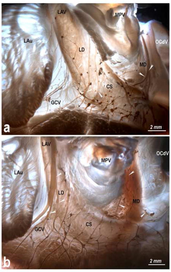
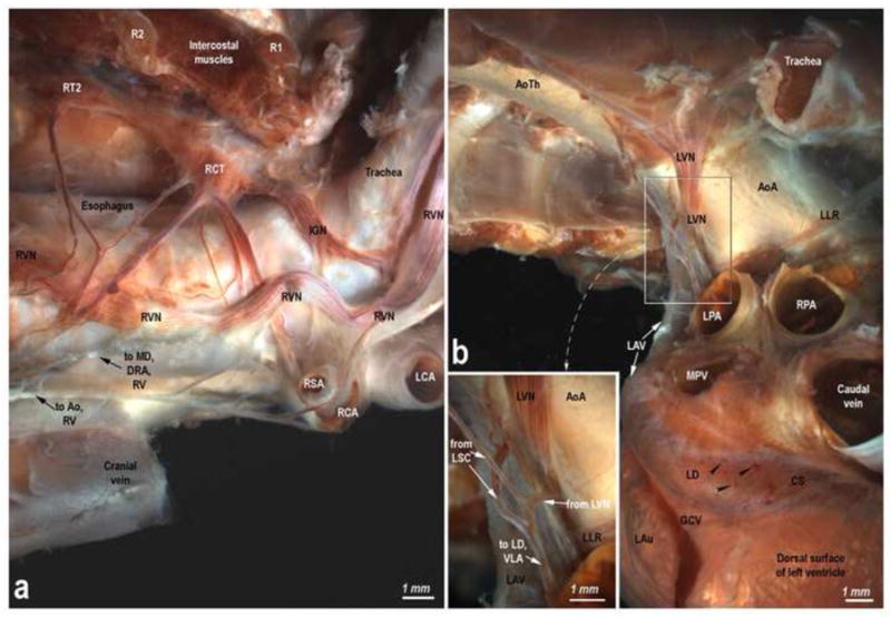
Results: Intrinsic cardiac nerves extend from the venous part of the ovine heart hilum along the roots of the cranial (superior) caval and left azygos veins to both atria and ventricles via five epicardial routes: dorsal right atrial, middle dorsal, left dorsal, right ventral, and ventral left atrial nerve subplexuses. Intrinsic nerves proceeding from the arterial part of the heart hilum along the roots of the aorta and pulmonary trunk extend exclusively into the ventricles as the right and left coronary subplexuses. The dorsal right atrial, right ventral, and middle dorsal subplexuses receive the main extrinsic neural input from the right cervicothoracic and right thoracic sympathetic T(2) and T(3) ganglia as well as from the right vagal nerve. The left dorsal is supplied by sizeable extrinsic nerves from the left thoracic T(4)-T(6) sympathetic ganglia and the left vagal nerve. Sheep hearts contained an average of 769 +/- 52 epicardial ganglia. Cumulative areas of epicardial ganglia on the root of the cranial vena cava and on the wall of the coronary sinus were the largest of all regions (P <.05).

Conclusion: Despite substantial interindividual variability in the morphology of ovine ENP, right-sided epicardial neural subplexuses supplying the sinoatrial and atrioventricular nodes are mostly concentrated at a fat pad between the right pulmonary veins and the cranial vena cava. This finding is in sharp contrast with a solely left lateral neural input to the human atrioventricular node, which extends mainly from the left dorsal and middle dorsal subplexuses. The abundance of epicardial ganglia distributed widely along the ovine ventricular nerves over respectable distances below the coronary groove implies a distinctive neural control of the ventricles in human and sheep hearts.


Conflict of interest statement

No Conflicts

Figures

Fig 1
Fig 1
Coexistence of TH positive nerve fibers (a) with AChE (b). Boxed areas on both images are enlarged as top insets to show the adrenergic nerve fibers (a) surrounded by brown diffuse precipitates ensued by AChE histochemical reaction (b).
Fig 2
Fig 2
Schematic ventral (a) and dorsal (b) views of the pressure-inflated lamb heart. The heart surface is subdivided into regions for quantitative analysis of ganglia and representation of the course and innervation regions of the seven epicardial subplexuses. Inset (c), cardiac zone around the roots of the aorta and the pulmonary trunk. Dotted lines, limits of the HH; thick grey semitransparent arrows, course of neural subplexuses; blue shadowed areas, main areas with the highest density of subplexal ganglia. Ao – aorta; PT – pulmonary trunk; OCS – opening of coronary sinus; OGCV – opening of great cardiac vein; OLAV – opening of left azygos vein; veins: GCV – great cardiac; LPV – left pulmonary; MPV – middle pulmonary; RPV – right pulmonary; LAV – left azygos; subplexuses: DRA – dorsal right atrial; LC – left coronary; LD – left dorsal; MD – middle dorsal; RC – right coronary; VLA – ventral LA; RV – right ventral; regions: CLAV – cardiac portion of the LAV; CS – coronary sinus; DIRA – dorsal inferior right atrial; DLA – dorsal LA; DLV – dorsal left ventricular; DRV – dorsal right ventricular; DSRA – dorsal superior right atrial; HH – heart hilum; ISLAu – inferior surface of left auricle; ISRAu - inferior surface of right auricle; LCG – left side of coronary groove; PostAo – post-aortic; PreAo – pre-aortic; PostCA – region laying behind the conus arteriosus; PreCA – region laying in front of the conus arteriosus; RCG – right side of coronary groove; RCV – root of the cranial caval vein; SSLAu – superior surface of left auricle; SSRAu - superior surface of right auricle; VILA – ventral inferior LA; VIRA – ventral inferior right atrial; VLV – ventral left ventricular; VRV – ventral right ventricular; VSLA – ventral superior LA; VSRA – ventral superior right atrial; points (1-23): (1) approximate mid-point of terminal groove; (2) cranial end of free margin of right auricle; (3) cranial dorsal bend of ventral interatrial groove; (4) middle of ventral interatrial groove; (5) base of ventral interatrial groove; (6) mid-point of the aorta and pulmonary trunk on the level of HH; (7) ventral edge of the bed of the root of the right coronary artery; (8) branching site of the first branches from the right coronary artery to the conus arteriosus; (9) right lateral edge of aorta; (10) dorsal edge of the bed right coronary artery root; (11) basal midst of conus arteriosus; (12) cranial end of ventral interventricular groove or ventral edge of the left coronary artery root; (13) left lateral edge of pulmonary trunk; (14) dorsal edge of the bed of the left coronary artery root; (15) ventral end of the free margin of left auricle; (16) lateral edge of left azygos vein; (17) point of juncture of left azygos vein and great cardiac vein; (18) point of juncture of left azygos vein and coronary sinus; (19) lower edge of caudal caval vein; (20) medial edge of left azygos vein; (21) upper edge of caudal caval vein; (22) caudal end of free margin of right auricle; (23) notch of heart apex.
Fig 3
Fig 3
Topography and structure of the dorsal subplexuses of the lamb heart, i.e. left (a, b) and middle (c, d, e) dorsal subplexuses and dorsal right atrial (f, g, h) subplexus, stained for acetylcholinesterase. Boxed areas in a, c and f are enlarged on the left as b, d, e, g and h to illustrate details of plexus. Black arrowheads, epicardial ganglia: white arrowheads, subplexal nerves: white solid arrows, preganglionated nerves entering epicardium through the HH, and the dashed line shows the limits of the HH (broken lines). Note in panel c numerous ventricular ganglia scattered on the dorsal side of the left ventricle. Abbreviations: LAu – left auricle; LV – left ventricle; RV – right ventricle; SAN – sinuatrial nodal zone; other abbreviations as in Fig 2.
Fig 4
Fig 4
Right ventral, ventral LA (a, b, c, d, e), and left coronary (d, f) epicardial subplexuses and intrinsic ganglionated nerves in the ovine HH (g, h, i). Boxed areas in a, d and g are enlarged as b, c, e, f, h and i panels to show detail. Black arrowheads, epicardial ganglia; white arrowheads, subplexal nerves; white and black solid arrows, preganglionated nerves entering epicardium through the HH; dashed line, limits of the HH. Abbreviations: IS – ventral groove of the interatrial septum; RAu – right auricle; other abbreviations as in Figs 2 and 3.
Fig 5
Fig 5
Morphologic pattern of atrial (a-c) and ventricular (d-f) epicardial ganglia. Some regions of ganglia are out of sharp focus because of their deep location in the epicardium. Black arrows, longitudinally oriented epicardial nerves; black arrowheads, ganglionic cells; white arrowheads, interganglionic nerves. Atrial ganglia are irregular in shape due to their extensions at the sites where nerves connect to a ganglion; ventricular ganglia are oval or spindle-shaped and situated along the epicardial nerves.
Fig 6
Fig 6
Variability in morphology of the left and the middle dorsal subplexuses in hearts from newborn lambs. Black arrowheads, left dorsal subplexal nerves; white arrowheads, middle dorsal subplexal nerves. Note the distinct branching patterns of epicardial nerves coursing along the left azygos vein. Abbreviations: OCdV – orifice of the caudal vein; others – as in Figs 2-4.
Fig 7
Fig 7
Lamb thorax-dissected preparations showing the origin and course of the right (a) and left (b) extrinsic cardiac nerves. Abbreviations: Ao – aorta; AoA – aortic arch; AoTh – thoracic aorta; IGN – interganglionated nerve; LCA – left common carotid artery; LLR – left laryngeal recurrent nerve; LPA – left pulmonary artery; LSC – left sympathetic chain; LV – left vagus; R – rib; RCA – right common carotid artery; RCT – right cervicothoracic ganglion; RPA – right pulmonary artery; RSA – right subclavian artery; RT – right thoracic ganglion; RV – right vagus; other abbreviations as for Figs 2-6.

Comment in

References

    1. Gorman MW, Tune JD, Richmond KN, Feigl EO. Quantitative analysis of feedforward sympathetic coronary vasodilation in exercising dogs. J Appl Physiol. 2000;89:1903–1911. - PubMed
    1. Tsuboi M, Furukawa Y, Nakajima K, Kurogouchi F, Chiba S. Inotropic, chronotropic, and dromotropic effects mediated via parasympathetic ganglia in the dog heart. Am J Physiol Heart Circ Physiol. 2000;279:H1201–1207. - PubMed
    1. Cifelli C, Rose RA, Zhang H, et al. RGS4 regulates parasympathetic signaling and heart rate control in the sinoatrial node. Circ Res. 2008;103:527–535. - PubMed
    1. Levy MN, Warner MR. Parasympathetic effects on cardiac function. In: Armour JA, Ardell JL, editors. Neurocardiology. New York: Oxford University Press; 1994. pp. 53–76.
    1. Armour JA, Kember GC. Cardiac sensory neurons. In: Armour JA, Ardell JL, editors. Basic and clinical neurocardiology. New York: Oxford University Press; 2004. pp. 79–117.

Publication types

MeSH terms

Substances