. 2010 Apr 19;28(18):3106-11.
doi: 10.1016/j.vaccine.2010.02.063. Epub 2010 Mar 1.

Rotavirus enterotoxin NSP4 has mucosal adjuvant properties

Affiliations

Rotavirus enterotoxin NSP4 has mucosal adjuvant properties

Owen V Kavanagh et al. Vaccine. .

Abstract

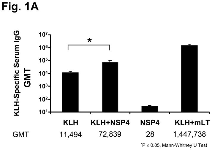
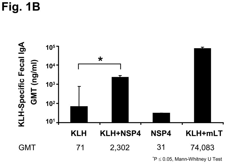
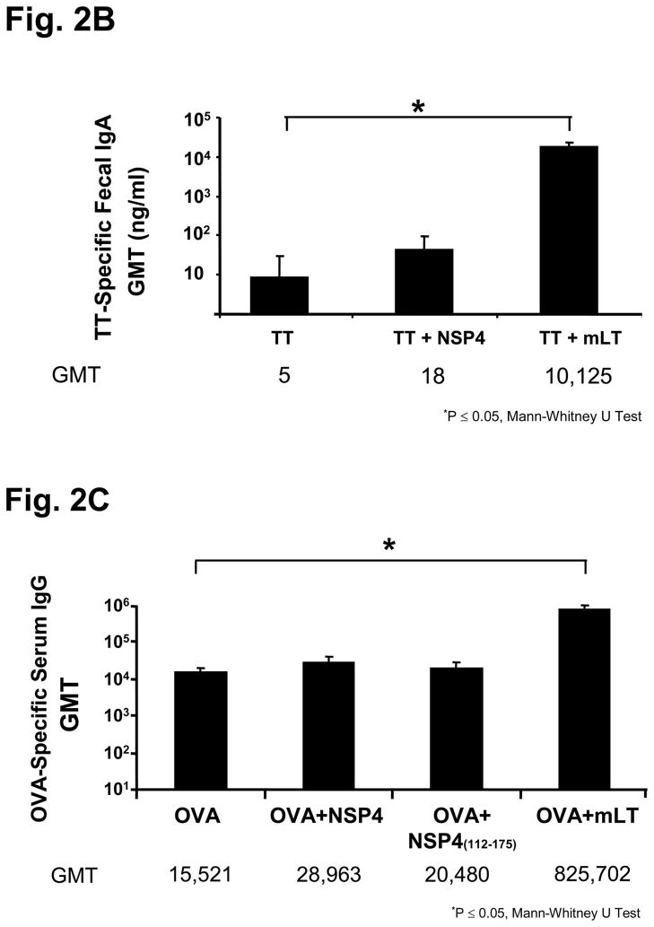
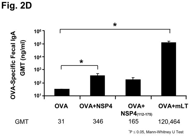
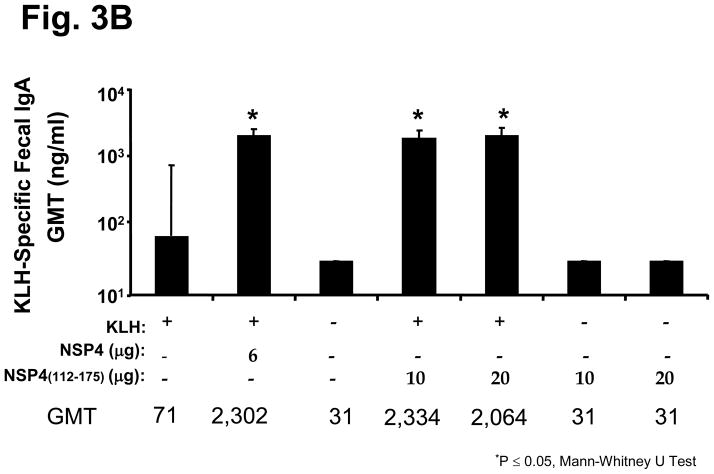
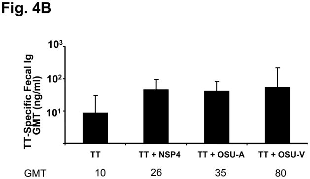
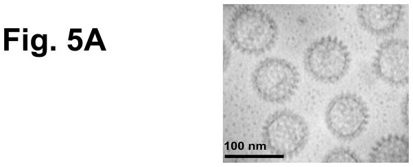
Rotavirus nonstructural protein 4 (NSP4) is a protein with pleiotropic properties. It functions in rotavirus morphogenesis, pathogenesis, and is the first described viral enterotoxin. Since many bacterial toxins function as potent mucosal adjuvants, we evaluated whether baculovirus-expressed recombinant simian rotavirus SA11 NSP4 possesses adjuvant activity by co-administering NSP4 with keyhole limpet hemocyanin (KLH), tetanus toxoid (TT) or ovalbumin (OVA) as model antigens in mice. Following intranasal immunization, NSP4 significantly enhanced both systemic and mucosal immune responses to model immunogens, as compared to the control group, in an antigen-specific manner. Both full-length and a cleavage product of SA11 NSP4 had adjuvant activity, localizing this activity to the C-terminus of the protein. NSP4 forms from virulent and avirulent porcine rotavirus OSU strain, and SA11 NSP4 localized within a 2/6-virus-like particle (VLP) also exhibited adjuvant effects. These studies suggest that the rotavirus enterotoxin NSP4 can function as an adjuvant to enhance immune responses for a co-administered antigen.


Figures

Figure 1
Figure 1
KLH-specific serum and fecal antibody response profiles of mice intranasally inoculated with KLH and SA11 NSP4. Animals (n=5) were administered two times, three weeks apart, with KLH alone, KLH + NSP4, NSP4 alone, or KLH + mLT as the immunostimulatory control. Samples were collected 14 days post second inoculation and assayed by ELISA. Panel (a) shows the KLH-specific serum IgG results in Geometric Mean Titers (GMT) and panel (b) shows the level of KLH-specific fecal IgA responses expressed as the ratio of nanograms of KLH-specific IgA per microgram of total IgA among various immunized groups of mice.
Figure 1
Figure 1
KLH-specific serum and fecal antibody response profiles of mice intranasally inoculated with KLH and SA11 NSP4. Animals (n=5) were administered two times, three weeks apart, with KLH alone, KLH + NSP4, NSP4 alone, or KLH + mLT as the immunostimulatory control. Samples were collected 14 days post second inoculation and assayed by ELISA. Panel (a) shows the KLH-specific serum IgG results in Geometric Mean Titers (GMT) and panel (b) shows the level of KLH-specific fecal IgA responses expressed as the ratio of nanograms of KLH-specific IgA per microgram of total IgA among various immunized groups of mice.
Figure 2
Figure 2
Serum and fecal antibody profiles of mice intranasally inoculated with SA11 NSP4 and either tetanus toxoid (TT) or ovalbumin (OVA). Animals (n=5) were administered with model antigen alone, co-administered with full-length NSP4 or with mLT as an immunostimulatory control. Samples were collected 14 days post final inoculation and assayed by ELISA. Panel (a) shows the KLH-specific serum IgG and total immunoglobulin results in Geometric Mean Titers (GMT), panel (b) shows the level of KLH-specific fecal IgA responses, panel (c) shows OVA-specific serum IgG levels and panel (d) shows OVA-specific IgA fecal levels among the various immunized groups of mice.
Figure 2
Figure 2
Serum and fecal antibody profiles of mice intranasally inoculated with SA11 NSP4 and either tetanus toxoid (TT) or ovalbumin (OVA). Animals (n=5) were administered with model antigen alone, co-administered with full-length NSP4 or with mLT as an immunostimulatory control. Samples were collected 14 days post final inoculation and assayed by ELISA. Panel (a) shows the KLH-specific serum IgG and total immunoglobulin results in Geometric Mean Titers (GMT), panel (b) shows the level of KLH-specific fecal IgA responses, panel (c) shows OVA-specific serum IgG levels and panel (d) shows OVA-specific IgA fecal levels among the various immunized groups of mice.
Figure 3
Figure 3
KLH-specific serum and fecal antibody response profiles of mice intranasally inoculated with KLH and SA11 NSP4 cleavage product. Full-length NSP4 as well as the cleavage product NSP4(112-175) were given intranasally two times three weeks apart and KLH-specific serum IgG expressed as GMT (a) and fecal IgA expressed as ratio of ng of KLH-specific IgA per μg of total IgA (b) were evaluated in all immunized groups of animals 14 days after the second inoculation.
Figure 3
Figure 3
KLH-specific serum and fecal antibody response profiles of mice intranasally inoculated with KLH and SA11 NSP4 cleavage product. Full-length NSP4 as well as the cleavage product NSP4(112-175) were given intranasally two times three weeks apart and KLH-specific serum IgG expressed as GMT (a) and fecal IgA expressed as ratio of ng of KLH-specific IgA per μg of total IgA (b) were evaluated in all immunized groups of animals 14 days after the second inoculation.
Figure 4
Figure 4
TT-specific serum and fecal antibody response profiles of mice intranasally co-administered TT with full length NSP4 from the rotavirus strains SA11, OSU-v and OSU-a. Animals (n=5) were given TT alone or co-administered with either of the full-length NSP4 forms three times two weeks apart. Samples were collected 14 days post final inoculation and assayed by ELISA. TT-specific serum total immunoglobulins expressed as GMT (a) and fecal IgA in ng specific IgA per μg total IgA (b).
Figure 4
Figure 4
TT-specific serum and fecal antibody response profiles of mice intranasally co-administered TT with full length NSP4 from the rotavirus strains SA11, OSU-v and OSU-a. Animals (n=5) were given TT alone or co-administered with either of the full-length NSP4 forms three times two weeks apart. Samples were collected 14 days post final inoculation and assayed by ELISA. TT-specific serum total immunoglobulins expressed as GMT (a) and fecal IgA in ng specific IgA per μg total IgA (b).
Figure 5
Figure 5
Characterization of 2/6-VLP containing NSP4 and TT-specific serum and fecal antibody response profiles of mice intranasally co-administered TT with rotavirus 2/6 VLP containing NSP4. To test another form of NSP4 obtained from a system other than the baculovirus-expressed and affinity-purified SA11 NSP4-containing 2/6 VLPs were used as immunogens. (a) Negative stain (1% uranyl acetate, pH4.5) electron micrographs of NSP4-2/6 VLP produced in insect Sf9 cells. Magnification bar equals 100nm. Animals (n=5) were given TT alone or co-administered with either 2/6 VLP alone or 2/6 VLP containing NSP4 three times two weeks apart. Samples were collected 14 days post final inoculation and assayed by ELISA TT-specific serum IgG (b) and fecal IgA (c) were evaluated for enhanced responses compared to the antigen alone group.
Figure 5
Figure 5
Characterization of 2/6-VLP containing NSP4 and TT-specific serum and fecal antibody response profiles of mice intranasally co-administered TT with rotavirus 2/6 VLP containing NSP4. To test another form of NSP4 obtained from a system other than the baculovirus-expressed and affinity-purified SA11 NSP4-containing 2/6 VLPs were used as immunogens. (a) Negative stain (1% uranyl acetate, pH4.5) electron micrographs of NSP4-2/6 VLP produced in insect Sf9 cells. Magnification bar equals 100nm. Animals (n=5) were given TT alone or co-administered with either 2/6 VLP alone or 2/6 VLP containing NSP4 three times two weeks apart. Samples were collected 14 days post final inoculation and assayed by ELISA TT-specific serum IgG (b) and fecal IgA (c) were evaluated for enhanced responses compared to the antigen alone group.

Similar articles

Cited by

References

    1. Hasegawa H, et al. Development of mucosal adjuvants for intranasal vaccine for H5N1 influenza viruses. Ther Clin Risk Manag. 2009;5(1):125–32. - PMC - PubMed
    1. Stanley M, Gissmann L, Nardelli-Haefliger D. Immunobiology of human papillomavirus infection and vaccination - implications for second generation vaccines. Vaccine. 2008;26(Suppl 10):K62–7. - PubMed
    1. Lehner T, et al. A rational basis for mucosal vaccination against HIV infection. Immunol Rev. 1999;170:183–96. - PubMed
    1. Schnitzler AC, Burke JM, Wetzler LM. Induction of cell signaling events by the cholera toxin B subunit in antigen-presenting cells. Infect Immun. 2007;75(6):3150–9. - PMC - PubMed
    1. van Ginkel FW, et al. Cutting edge: the mucosal adjuvant cholera toxin redirects vaccine proteins into olfactory tissues. J Immunol. 2000;165(9):4778–82. - PubMed

Publication types

MeSH terms