. 2010 May 14;285(20):15178-15186.
doi: 10.1074/jbc.M110.103150. Epub 2010 Mar 2.

Engineered interfaces of an AAA+ ATPase reveal a new nucleotide-dependent coordination mechanism

Affiliations

Engineered interfaces of an AAA+ ATPase reveal a new nucleotide-dependent coordination mechanism

Nicolas Joly et al. J Biol Chem. .

Abstract

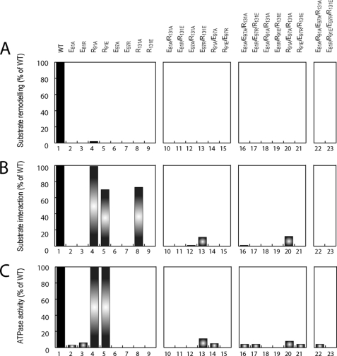
Homohexameric ring AAA(+) ATPases are found in all kingdoms of life and are involved in all cellular processes. To accommodate the large spectrum of substrates, the conserved AAA(+) core has become specialized through the insertion of specific substrate-binding motifs. Given their critical roles in cellular function, understanding the nucleotide-driven mechanisms of action is of wide importance. For one type of member AAA(+) protein (phage shock protein F, PspF), we identified and established the functional significance of strategically placed arginine and glutamate residues that form interacting pairs in response to nucleotide binding. We show that these interactions are critical for "cis" and "trans" subunit communication, which support coordination between subunits for nucleotide-dependent substrate remodeling. Using an allele-specific suppression approach for ATPase and substrate remodeling, we demonstrate that the targeted residues directly interact and are unlikely to make any other pairwise critical interactions. We then propose a mechanistic rationale by which the nucleotide-bound state of adjacent subunits can be sensed without direct involvement of R-finger residues. As the structural AAA(+) core is conserved, we propose that the functional networks established here could serve as a template to identify similar residue pairs in other AAA(+) proteins.


Figures

FIGURE 1.
FIGURE 1.
Structural basis for putative PspF dimer cis and trans communication. Center panel shows the overlay of a putative PspF1–275 dimer (extracted from the hexamer model (Rappas et al. (25)) bound with MgATP (in light red, Protein Data Bank 2C9C) or with ADP (light blue, Protein Data Bank 2C98). Residues Glu81, Arg95, Glu97, Arg98, Glu130, and Arg131 are shown as sticks. The six subunits of the hexamer are numbered from ”n“ to ”n + 5.“ L1n represents loop 1 in subunit n; L1n+1 loop 1 in subunit ”n + 1.“ L2n represents loop 2 in subunit n; L2n+1 loop 2 in subunit n + 1. Zoom in subunit n shows the side chain orientations of residues Glu81, Arg91, Glu97, and Arg131 in the presence of MgATP (A) or ADP (B). Zoom in at the level of the putative interface shows the side chains orientation of the residues Arg95, Arg98, and Glu130 in the presence of MgATP (C) or ADP (D). Clear side chain orientation differences dependent on the nucleotide present are observed. Figure was generated using PyMOL.
FIGURE 2.
FIGURE 2.
cis variant functional activities. For each variant potentially involved in cis subunit communication, we tested the following: A, remodeling activity (transcription activation, as % of WT; maximal error is 14%); B, substrate interaction activity (binding to σ54 using ADP-AlFx, as % of WT; maximal error is 15%); and C, ATPase activity (as % of WT; maximal error is 10%).
FIGURE 3.
FIGURE 3.
trans variant functional activities. For each variant potentially involved in trans subunit communication, we tested the following: A, remodeling activity (transcription activation, as % of WT; maximal error is 14%); B, substrate interaction activity (binding to σ54 using ADP-AlFx, as % of WT; maximal error is 15%); and C, ATPase activity (as % of WT; maximal error is 10%).
FIGURE 4.
FIGURE 4.
E97R complements R131E activity in cis. Doping experiments using E97R and R131E are shown. A, native gel autoradiograph shows in lane 1 ADP-AlFx-dependent formation of a stable complex between PspF and 32P-σ54. Lanes 2–8 show the absence of this complex with σ54 using different E97R:R131E ratios as indicated. Lane 9 shows the signal obtained with the double variant E97R/R131E. Radioactivity was quantified, and the quantity of complex formed for each mixture is expressed as percent of complex formed with WT. B, same protein mixtures were used in substrate remodeling activities (black bars, transcription activation, as % of WT; maximal error is 14%) and ATPase activity (gray bars, as % of WT; maximal error is 10%).
FIGURE 5.
FIGURE 5.
R95E or R98E complement E130R activity in trans. Doping experiments using R95E and E130R or R98E and E130R are shown. The native gel autoradiograph shows the formation of a stable complex between PspF and 32P-σ54 in the presence of ADP-AlFx. Lane 1 is the positive control showing the complex formed with WT. Lanes 2–8 show the complex formation using different R95E:E130R (A) or R98E:E130R (C) ratios (as indicated). Lane 9 shows the signal obtained with the double variant corresponding to R95E/E130R or R98E/E130R. Radioactivity was quantified, and the quantity of complex formed for each mixture is expressed as percentage of complex formed with WT. The same protein mixtures for R95E/E130R (B) or R98E/E130R (D) were used in substrate remodeling activities (black bars, transcription activation, as % of WT; maximal error is 14%) and ATPase activity (gray bars, as % of WT; maximal error is 10%).
FIGURE 6.
FIGURE 6.
Model of cis and trans communication pathways for coordinated activity. From our experimental data and the previous structural model of the interface between PspF subunits, we propose a mechanism where cis and trans communication pathways are linked together. When ATP is bound in subunit n, Glu97 interacts with Arg131 (in L2n) and Glu130 (just adjacent to Arg131 in L2n) with transArg98 (adjacent to Glu97n+1) allowing the exposure of L1n. These interactions will fully change in the presence of bound ADP. At this point, Glu97 interacts with Arg91 (L1n), whereas Glu81 interacts with Arg131 (L2n), which is expected to change the presentation of L1n from an exposed to a more buried conformation. At the same time the bound nucleotide signal will be propagated to the adjacent subunit by the new interaction made between Glu130 and transArg95.

Similar articles

Cited by

References

    1. Konakova M., Pulst S. M. (2005) J. Mol. Neurosci. 25, 105–117 - PubMed
    1. Guinto J. B., Ritson G. P., Taylor J. P., Forman M. S. (2007) Acta Neuropathol. 114, 55–61 - PubMed
    1. Kimonis V. E., Watts G. D. (2005) Alzheimer Dis. Assoc. Disord. 19, S44–S47 - PubMed
    1. Watts G. D., Mehta S. G., Zhao C., Ramdeen S., Hamilton S. J., Novack D. V., Mumm S., Whyte M. P., Mc Gillivray B., Kimonis V. E. (2005) Hum. Genet. 118, 508–514 - PubMed
    1. Watts G. D., Thomasova D., Ramdeen S. K., Fulchiero E. C., Mehta S. G., Drachman D. A., Weihl C. C., Jamrozik Z., Kwiecinski H., Kaminska A., Kimonis V. E. (2007) Clin. Genet. 72, 420–426 - PubMed

Publication types

Substances