. 2010 Apr;24(4):790-9.
doi: 10.1210/me.2009-0345. Epub 2010 Mar 2.

The transcription factor ATF4 promotes skeletal myofiber atrophy during fasting

Affiliations

The transcription factor ATF4 promotes skeletal myofiber atrophy during fasting

Scott M Ebert et al. Mol Endocrinol. 2010 Apr.

Abstract

Prolonged fasting alters skeletal muscle gene expression in a manner that promotes myofiber atrophy, but the underlying mechanisms are not fully understood. Here, we examined the potential role of activating transcription factor 4 (ATF4), a transcription factor with an evolutionarily ancient role in the cellular response to starvation. In mouse skeletal muscle, fasting increases the level of ATF4 mRNA. To determine whether increased ATF4 expression was required for myofiber atrophy, we reduced ATF4 expression with an inhibitory RNA targeting ATF4 and found that it reduced myofiber atrophy during fasting. Likewise, reducing the fasting level of ATF4 mRNA with a phosphorylation-resistant form of eukaryotic initiation factor 2alpha decreased myofiber atrophy. To determine whether ATF4 was sufficient to reduce myofiber size, we overexpressed ATF4 and found that it reduced myofiber size in the absence of fasting. In contrast, a transcriptionally inactive ATF4 construct did not reduce myofiber size, suggesting a requirement for ATF4-mediated transcriptional regulation. To begin to determine the mechanism of ATF4-mediated myofiber atrophy, we compared the effects of fasting and ATF4 overexpression on global skeletal muscle mRNA expression. Interestingly, expression of ATF4 increased a small subset of five fasting-responsive mRNAs, including four of the 15 mRNAs most highly induced by fasting. These five mRNAs encode proteins previously implicated in growth suppression (p21(Cip1/Waf1), GADD45alpha, and PW1/Peg3) or titin-based stress signaling [muscle LIM protein (MLP) and cardiac ankyrin repeat protein (CARP)]. Taken together, these data identify ATF4 as a novel mediator of skeletal myofiber atrophy during starvation.


Figures
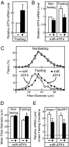
Figure 1
Figure 1
An artificial micro-RNA targeting ATF4 (miR-ATF4) reduces skeletal muscle ATF4 mRNA and myofiber atrophy during fasting. A, Mice were allowed ad libitum access to food or fasted for 24 h, and then ATF4 mRNA levels in the TA muscles were determined by qPCR. Data are means ± sem from four experiments, each using pooled RNA from three mice. *, P < 0.01. B–E, Mouse TA muscles were transfected on d 0. On d 14, mice were subjected to nonfasting or fasting conditions for 24 h, as indicated, before the TA muscles were harvested. B, The left TA was transfected with 20 μg empty pU6 vector, and the right TA was transfected with 20 μg pU6-miR-ATF4. TA muscle ATF4 mRNA levels were determined by qPCR and normalized to levels in the left TA of nonfasted mice. Data are means ± sem from four experiments and a total of seven transfected muscles per condition. *, P < 0.02. C and D, TA muscles were transfected with 5 μg pCMV-GFP plus either 20 μg empty pU6 vector (left TA) or 20 μg pU6-miR-ATF4 (right TA). C, Fiber size distribution curves from a representative experiment. D, Mean diameters of transfected myofibers ± sem from five experiments each using one mouse. *, P < 0.01. E, TA muscles were transfected as described in B. TA muscle mRNA levels were determined by qPCR, and levels under fasting conditions were normalized to levels in TA muscles transfected with empty pU6 vector and harvested under nonfasting conditions, which are indicated by the dashed line. Data are means ± sem from four experiments and a total of seven transfected muscles per condition. *, P < 0.02. Additional micro-RNA controls may be found in Supplemental Fig. 1 (published on The Endocrine Society’s Journals Online web site at http://mend.endojournals.org).
Figure 2
Figure 2
A phosphorylation-resistant form of eIF2α (eIF2α-S51A) reduces skeletal muscle ATF4 mRNA and myofiber atrophy during fasting. A–E, Mouse TA muscles were transfected on d 0. On d 10, mice were subjected to nonfasting or fasting conditions for 24 h, as indicated, before the TA muscles were harvested. A and B, The left TA was transfected with 25 μg pcDNA3, and the right TA was transfected with 25 μg pCMV-eIF2α-S51A-FLAG; A, TA muscle protein extracts were subjected to immunoprecipitation with anti-FLAG monoclonal IgG, followed by SDS-PAGE and immunoblot analysis with anti-eIF2α polyclonal antiserum; B, TA muscle ATF4 mRNA levels were determined by qPCR and normalized to levels in the left TA of nonfasted mice, which were set at 1. Data are means ± sem from five experiments each using pooled RNA from three mice. *, P < 0.01. C and D, Mouse TA muscles were transfected with 2 μg pCMV-eGFP plus either 25 μg pcDNA3 (left TA) or 25 μg pCMV-eIF2α-S51A-FLAG (right TA); C, fiber size distribution curves from a representative experiment; D, mean diameters of transfected myofibers ± sem from three experiments using a total of seven mice. *, P < 0.01. E, TA muscles were transfected as described in A and B. TA muscle mRNA levels were determined by qPCR, and levels under fasting conditions were normalized to levels in TA muscles transfected with pcDNA3 and harvested under nonfasting conditions, which are indicated by the dashed line. Data are means ± sem from five experiments, each using pooled RNA from three mice. *, P < 0.01.
Figure 3
Figure 3
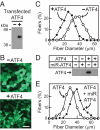
Overexpression of ATF4 reduces myofiber size in the absence of fasting. A, Mouse TA muscles were transfected with 20 μg empty pcDNA3 vector (left TA) or 20 μg pCMV-ATF4-FLAG (right TA) and then harvested under nonfasting conditions 11 d later. Skeletal muscle protein extracts were subjected to immunoprecipitation with anti-ATF4 polyclonal antiserum, followed by SDS-PAGE and immunoblot analysis with anti-FLAG monoclonal IgG. B and C, Mouse TA muscles were transfected with 5 μg pCMV-GFP plus either 20 μg empty pcDNA3 vector (left TA) or 20 μg pCMV-ATF4-FLAG (right TA) and then harvested under nonfasting conditions 11 d later; B, representative images in the absence or presence of pCMV-ATF4-FLAG (bar, 100 μm); C, representative fiber size distribution curves from one of eight experiments. D, Mouse TA muscles were transfected with 15 μg empty pcDNA3 vector or pCMV-ATF4-FLAG with or without 20 μg pU6 vector or pU6-miR-ATF4, as indicated. Muscles were harvested under nonfasting conditions 9 d after transfection, and skeletal muscle protein extracts were analyzed as in A. E, Mouse TA muscles were transfected with 5 μg pCMV-GFP plus 15 μg pCMV-ATF4-FLAG plus either 20 μg pU6 vector (left TA) or 20 μg pU6-miR-ATF4 (right TA) and then harvested under nonfasting conditions 12 d after transfection. Data are representative fiber size distribution curves from one of three experiments.
Figure 4
Figure 4
ATF4-mediated reduction in myofiber size requires an intact bZIP domain. A, Mouse TA muscles were transfected with 20 μg empty pcDNA3 vector (left TA) or 20 μg pCMV-ATF4ΔbZIP-FLAG (right TA) and then harvested under nonfasting conditions 11 d later. Skeletal muscle protein extracts were subjected to immunoprecipitation with anti-ATF4 polyclonal antiserum, followed by SDS-PAGE and immunoblot analysis with anti-FLAG monoclonal IgG. B and C, Left-sided mouse TA muscles were transfected with 5 μg pCMV-GFP plus 20 μg empty pcDNA3 vector. Right-sided TA muscles were transfected with 5 μg pCMV-GFP plus either 20 μg pCMV-ATF4-FLAG or 20 μg pCMV-ATF4ΔbZIP-FLAG, as indicated. Muscles were harvested under nonfasting conditions 11 d later. B, Representative fiber size distribution curves in the presence of pCMV-ATF4-FLAG or pCMV-ATF4ΔbZIP-FLAG from one of six experiments. C, Mean fiber diameters ± sem from at least six experiments per ATF4 construct. *, P < 0.01 using paired t tests.
Figure 5
Figure 5
ATF4 increases a subset of five fasting-responsive skeletal muscle mRNAs. A, To determine the effect of fasting on skeletal muscle mRNA expression, mice were allowed ad libitum access to food or fasted for 24 h before TA muscle RNA was harvested. RNA from three mice per group was pooled and then subjected to exon expression array analysis. mRNA levels under fasting conditions were normalized to levels under nonfasting conditions, which were set at 1. To determine the effect of ATF4 overexpression on skeletal muscle mRNA expression, mouse TA muscles were transfected with 25 μg empty pcDNA3 vector (left TA) or 25 μg pCMV-ATF4-FLAG (right TA). TA muscle RNA was harvested under nonfasting conditions 11 d later and then subjected to exon expression array analysis. mRNA levels in the presence of ATF4 (right TA) were normalized to levels in the absence of ATF4 (left TA), which were set at 1. Both experiments were performed four times to generate the final data. The figure shows the 15 mRNAs most increased by fasting as well as the effect of ATF4 on these mRNAs. Data are the mean fold changes induced by fasting or ATF4. For all mRNAs shown, the effect of fasting was statistically significant (P ≤ 0.01). The red boldface denotes transcripts that were also increased by ATF4 (P ≤ 0.01). B, Mice were subjected to nonfasting or fasting conditions for 24 h, and then TA muscle mRNA was harvested and analyzed by qPCR. mRNA levels under fasting conditions were normalized to mRNA levels under nonfasting conditions, which were set at 1 and are indicated by the dashed line. Data are the means ± sem from four experiments. P ≤ 0.03 for all mRNAs shown. C and D, Mouse TA muscles were transfected with 25 μg empty pcDNA3 vector (left TA) or 25 μg pCMV-ATF4-FLAG (right TA). TA muscles were harvested under nonfasting conditions 11 d later. C, TA muscle mRNA was analyzed by qPCR. mRNA levels in the presence of ATF4 (right TA) were normalized to mRNA levels in the absence of ATF4 (left TA), which were set at 1 and are indicated by the dashed line. Data are the means ± sem from seven experiments. P ≤ 0.01 for all mRNAs shown. D, An equal amount (100 μg) of total muscle protein extract from each condition was subjected to SDS-PAGE, followed by immunoblot analysis with anti-p21 monoclonal IgG or anti-MLP polyclonal antiserum. The membranes were stained with Ponceau S to confirm equal sample loading. The graph shows average densitometry signals ± sem from three experiments. Levels in the presence of ATF4 (right TA) were normalized to levels in the absence of ATF4 (left TA), which were set at 1 and are indicated by the dashed line.

Similar articles

Cited by

References

    1. Harding HP, Zhang Y, Zeng H, Novoa I, Lu PD, Calfon M, Sadri N, Yun C, Popko B, Paules R, Stojdl DF, Bell JC, Hettmann T, Leiden JM, Ron D 2003 An integrated stress response regulates amino acid metabolism and resistance to oxidative stress. Mol Cell 11:619–633 - PubMed
    1. Yamaguchi S, Ishihara H, Yamada T, Tamura A, Usui M, Tominaga R, Munakata Y, Satake C, Katagiri H, Tashiro F, Aburatani H, Tsukiyama-Kohara K, Miyazaki J, Sonenberg N, Oka Y 2008 ATF4-mediated induction of 4E-BP1 contributes to pancreatic β-cell survival under endoplasmic reticulum stress. Cell Metab 7:269–276 - PubMed
    1. Wek RC, Cavener DR 2007 Translational control and the unfolded protein response. Antioxid Redox Signal 9:2357–2371 - PubMed
    1. Proud CG 2005 eIF2 and the control of cell physiology. Semin Cell Dev Biol 16:3–12 - PubMed
    1. Kaufman RJ, Davies MV, Pathak VK, Hershey JW 1989 The phosphorylation state of eucaryotic initiation factor 2 alters translational efficiency of specific mRNAs. Mol Cell Biol 9:946–958 - PMC - PubMed

Publication types

Substances