. 2010 Apr;24(4):846-58.
doi: 10.1210/me.2009-0392. Epub 2010 Mar 2.

RUNX2 transcription factor regulates gene expression in luteinizing granulosa cells of rat ovaries

Affiliations

RUNX2 transcription factor regulates gene expression in luteinizing granulosa cells of rat ovaries

Eun-Sil Park et al. Mol Endocrinol. 2010 Apr.

Abstract

The LH surge promotes terminal differentiation of follicular cells to become luteal cells. RUNX2 has been shown to play an important role in cell differentiation, but the regulation of Runx2 expression and its function in the ovary remain to be determined. The present study examined 1) the expression profile of Runx2 and its partner CBFbeta during the periovulatory period, 2) regulatory mechanisms of Runx2 expression, and 3) its potential function in the ovary. Runx2 expression was induced in periovulatory granulosa cells of human and rodent ovaries. RUNX2 and core binding factor-beta (CBFbeta) proteins in nuclear extracts and RUNX2 binding to a consensus binding sequence increased after human chorionic gonadotropin (hCG) administration. This in vivo up-regulation of Runx2 expression was recapitulated in vitro in preovulatory granulosa cells by stimulation with hCG. The hCG-induced Runx2 expression was reduced by antiprogestin (RU486) and EGF-receptor tyrosine kinase inhibitor (AG1478), indicating the involvement of EGF-signaling and progesterone-mediated pathways. We also found that in the C/EBPbeta knockout mouse ovary, Runx2 expression was reduced, indicating C/EBPbeta-mediated expression. Next, the function of RUNX2 was investigated by suppressing Runx2 expression by small interfering RNA in vitro. Runx2 knockdown resulted in reduced levels of mRNA for Rgc32, Ptgds, Fabp6, Mmp13, and Abcb1a genes. Chromatin immunoprecipitation analysis demonstrated the binding of RUNX2 in the promoter region of these genes, suggesting that these genes are direct downstream targets of RUNX2. Collectively, the present data indicate that the LH surge-induced RUNX2 is involved in various aspects of luteal function by directly regulating the expression of diverse luteal genes.


Figures
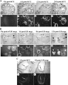
Figure 1
Figure 1
In situ localization of Runx2 mRNA in rat ovaries obtained from gonadotropin-primed immature rats (A) and naturally cycling rats (B). Representative bright-field (A, a–d; B, a–d; and C, a and b) and corresponding dark-field (A, e–h; B, e–h; and C, c and d) photomicrographs are depicted. Ovaries were collected at the indicated time points before or after hCG injection (A or C) or the LH surge (B or C). Arrows indicate Runx2 mRNA expression in periovulatory follicles (PF). Arrowheads indicate nCL expressing Runx2 mRNA. Asterisks indicate CL generated during previous estrous cycles (pCL). Wavy arrows indicate Runx2 expression in cumulus cells in C. F, Follicle. Original magnification of all slides in A and B is ×40. Magnification of all slides in C is ×100.
Figure 2
Figure 2
Ovarian expression of Runx2 and Cbfβ during the periovulatory period. Ovaries were collected before or at indicated hours (h) after hCG injection from PMSG-primed immature rats (n = 4 animals per time point). A and B, Whole ovarian levels of Runx2 (A) and Cbfβ (B) mRNA in rats. C, RUNX2 and CBFβ proteins in whole-cell extracts or nuclear extracts of rat ovaries obtained at indicated time points after hCG injection. D, The DNA binding activity of RUNX2 in nuclear extracts collected at indicated time points in rats using a TransAM kit. The levels of Runx2 mRNA and Cbfβ mRNA were measured by real-time PCR and Northern blot analysis, respectively. The levels of target genes were normalized to the L32 value in each sample. For the Western blot analysis, each lane was loaded with 50 μg protein extracts from ovaries of each animal. The membrane was reprobed with a monoclonal antibody against TATA binding protein (TBP) as a nuclear loading control and β-actin as a whole cell loading control. Bars with no common superscripts are significantly different (P < 0.05).
Figure 3
Figure 3
Granulosa cells (GC) or COC expression of Runx1 or Runx2 in rats or humans during the periovulatory period. A and B, Granulosa cells or COCs were collected from periovulatory ovaries obtained before or at indicated hours (h) after hCG injection from PMSG-primed immature rats (n = 4 animals per time point). COCs were also collected from the oviduct at 24 h after hCG. C and D, Granulosa cells were isolated from periovulatory follicles collected before (PEO, preovulatory phase) or indicated times (EO, early ovulatory phase; LO, late ovulatory phase) after rhCG injection from women (n = 5 patients per time point). The levels of Runx1 and Runx2 mRNA were measured by real-time PCR and normalized to the L32 (rat) and GAPDH (human) value in each sample. Bars with no common superscripts are significantly different (P < 0.05).
Figure 4
Figure 4
Regulation of Runx2 and Cbfβ expression in periovulatory granulosa cells. A and B, Granulosa cells obtained from rat preovulatory ovaries (48 h after PMSG) were cultured for 0, 6, 12, 24, or 48 h in medium alone (control) or with hCG (1 IU/ml). The levels of Runx2 mRNA (A) and Runx1 mRNA (B) were measured using real-time PCR (mean ± sem; n = 3 independent experiments). C, Granulosa cells isolated from rat ovaries were cultured in medium alone (C) or with hCG (1 IU/ml) for 0, 8, 24, or 48 h. RUNX1, RUNX2, and CBFβ proteins in nuclear extracts and CBFβ protein in whole-cell extracts were detected by Western blot analyses. Each lane was loaded with 40 μg protein extracts. The membranes were reprobed with a monoclonal antibody against TATA binding protein (TBP) for nuclear loading control or β-actin for whole cell loading control (n = 4 independent experiments). D, Granulosa cells were cultured with hCG or without (Cont) for 48 h. The direct interaction of RUNX2 and CBFβ was determined by immunoprecipitation (IP) assays. Cytoplasmic and nuclear fractions (100 μg) were immunoprecipitated with RUNX2 antibody. The resulting precipitates were analyzed for CBFβ protein by Western blot (WB) analysis. E, Preovulatory granulosa cells were cultured for 48 h in medium alone (Cont) or with hCG (1 IU/ml), FSK (10 μm), or PMA (20 nm). The levels of Runx2 mRNA were measured using real-time PCR (mean ± sem; n = 4 independent culture experiments). F, Preovulatory granulosa cells were cultured for 24 h in medium alone (Cont) or with AG1479 (AG, 1 μm), NS-398 (NS, 1 μm), RU486 (RU, 10 μm), hCG (1 IU/ml), or hCG plus inhibitor. The levels of Runx2 mRNA were measured using real-time PCR (mean ± sem; n = 3 independent culture experiments). In A, B, E, and F, bars with no common superscripts are significantly different (P < 0.05). G and H, Granulosa cells were isolated from ovaries collected before or at indicated hours after hCG injection from PMSG-primed immature wild-type (WT) and C/EBPβ knockout (KO) mice. Levels of mRNA for Runx2 (G) and Runx1 mRNA (H) were measured by real-time PCR. For mean comparison between KO vs. WT: *, P < 0.05.
Figure 5
Figure 5
Reduction in Runx2 expression by Runx2 siRNA in cultured granulosa cells. Granulosa cells obtained from rat preovulatory ovaries (48 h after PMSG) were transfected without (vehicle) or with negative control scrambled siRNA (NC siRNA) and Runx2 siRNA, and then treated with FSK for 48 h. The levels of mRNA for Runx2 (A) and Cyp11a1 (C) were measured by real-time PCR and normalized to the L32 in each sample (mean ± sem; n = 4 independent culture experiments). Bars with no common superscripts are significantly different (P < 0.05). Western blot (B) shows RUNX2 and CBFβ proteins in nuclear and cytoplasmic fractions isolated from siRNA-transfected cells. Each lane was loaded with 50 μg protein extracts. The membrane was reprobed with a monoclonal antibody against TATA binding protein (TBP) and β-actin for the nuclear and cytoplasmic loading control, respectively. The blots are representatives of three separate experiments. Concentrations of progesterone (D) were measured in granulosa cell culture media collected at 48 h after FSK treatment (mean ± sem; n = 6 independent culture experiments). Bars with no common superscripts are significantly different (P < 0.05).
Figure 6
Figure 6
RUNX2 regulation of Rgc32, Spp1, Mmp13, Ptgds, Fabp6, and Abcb1a expression in luteinizing granulosa cells in vitro. Granulosa cells obtained from rat preovulatory ovaries (48 h after PMSG) were transfected without (vehicle) or with FSK plus negative control scrambled siRNA (NC siRNA) and FSK plus Runx2 siRNA for 48 h. The levels of mRNA for Rgc32 (A), Spp1 (B), Mmp13 (C), Ptgds (D), Fabp6 (E), and Abcb1a (F) were measured by real-time PCR and normalized to the L32 value in each sample (mean ± sem; n = 4 independent culture experiments). Bars with no common superscripts are significantly different (P < 0.05).
Figure 7
Figure 7
hCG stimulation of Rgc32, Mmp13, Ptgds, Fabp6, and Abcb1a expression in luteinizing granulosa cells in vitro. Granulosa cells obtained from rat preovulatory ovaries (48 h after PMSG) were cultured for 0, 6, 24, or 48 h in medium alone (control) or with hCG (1 IU/ml). Levels of Rgc32 (A), Mmp13 (B), Ptgds (C), Fabp6 (D), Abcb1a (E), and Spp1 (F) mRNA were measured using real-time PCR and normalized to the L32 value in each sample (mean ± sem; n = 3 independent culture experiments). Bars with no common superscripts are significantly different (P < 0.05).
Figure 8
Figure 8
ChIP analysis for RUNX2 transcription factor binding to the Rgc32, Mmp13, Ptgds, Fabp6, and Abcb1a promoter regions in luteinizing granulosa cells. RUNX binding sites were predicted by a TFSEARCH program and numbered from transcription start site at +1 in A–E. ChIP assays were performed using granulosa cells cultured with hCG (1 IU/ml) for 48 h. DNAs were analyzed by PCR using primers listed in Supplemental Table 2 and represented as arrows in A–E. Amplified DNA fragments containing RUNX transcription factor binding sites are represented as black boxes with the indicated PCR product size. Experiments were repeated at least three times, each with different cultured luteinizing granulosa cell samples.
Figure 9
Figure 9
Effects of PGD2 on granulosa cell viability. Granulosa cells obtained from rat preovulatory ovaries (48 h after PMSG) were cultured without (control) or with DMSO (0.025%), hCG (1 IU/ml), or synthetic PGD2 (0.1, 0.5, 1, 2.5, or 5 μm) for 24 h. Granulosa cell viability was measured using a MTS kit (mean ± sem; n = 3 independent culture experiments). Bars with no common superscripts are significantly different (P < 0.05).

Similar articles

Cited by

References

    1. Murphy BD 2000 Models of luteinization. Biol Reprod 63:2–11 - PubMed
    1. Richards JS, Russell DL, Ochsner S, Espey LL 2002 Ovulation: new dimensions and new regulators of the inflammatory-like response. Annu Rev Physiol 64:69–92 - PubMed
    1. Richards JS 2005 Ovulation: new factors that prepare the oocyte for fertilization. Mol Cell Endocrinol 234:75–79 - PubMed
    1. Richards JS 2007 Genetics of ovulation. Semin Reprod Med 25:235–242 - PubMed
    1. Hernandez-Gonzalez I, Gonzalez-Robayna I, Shimada M, Wayne CM, Ochsner SA, White L, Richards JS 2006 Gene expression profiles of cumulus cell oocyte complexes during ovulation reveal cumulus cells express neuronal and immune-related genes: does this expand their role in the ovulation process? Mol Endocrinol 20:1300–1321 - PubMed

Publication types

MeSH terms