. 2010 Mar;9(3):653-60.
doi: 10.1158/1535-7163.MCT-09-0812. Epub 2010 Mar 2.

ABT-869 inhibits the proliferation of Ewing Sarcoma cells and suppresses platelet-derived growth factor receptor beta and c-KIT signaling pathways

Affiliations

ABT-869 inhibits the proliferation of Ewing Sarcoma cells and suppresses platelet-derived growth factor receptor beta and c-KIT signaling pathways

Alan K Ikeda et al. Mol Cancer Ther. 2010 Mar.

Abstract

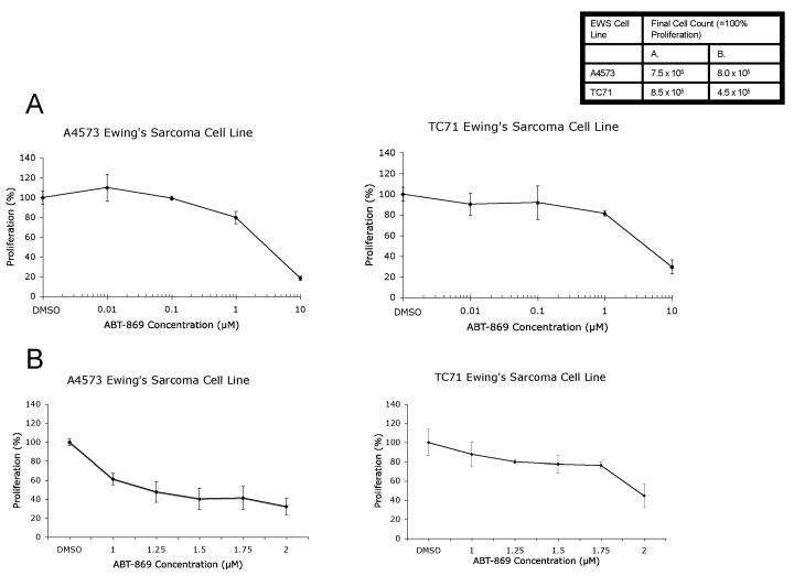
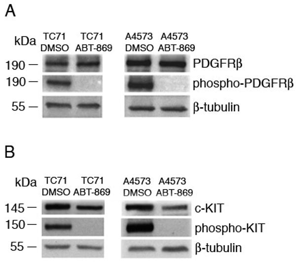
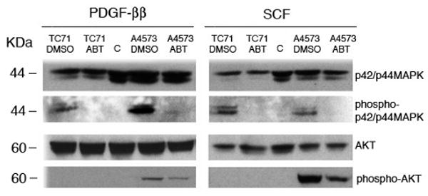
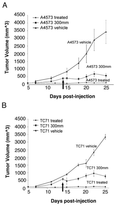
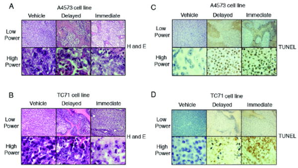
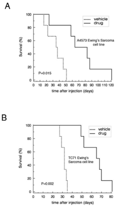
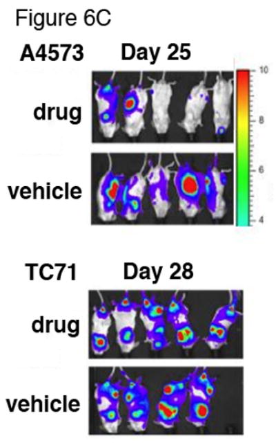
The Ewing Sarcoma (EWS) family of tumors is one of the most common tumors diagnosed in children and adolescents and is characterized by a translocation involving the EWS gene. Despite advances in chemotherapy, the prognosis of metastatic EWS is poor with an overall survival of <30% after 5 years. EWS tumor cells express the receptor tyrosine kinases, platelet-derived growth factor receptor (PDGFR) and c-KIT. ABT-869 is a multitargeted small-molecule inhibitor that targets Fms-like tyrosine kinase-3, c-KIT, vascular endothelial growth receptors, and PDGFRs. To determine the potential therapeutic benefit of ABT-869 in EWS cells, we examined the effects of ABT-869 on EWS cell lines and xenograft mouse models. ABT-869 inhibited the proliferation of two EWS cell lines, A4573 and TC71, at an IC(50) of 1.25 and 2 mumol/L after 72 h of treatment, respectively. The phosphorylation of PDGFRbeta, c-KIT, and extracellular signal-regulated kinases was also inhibited. To examine the effects of ABT-869 in vivo, the drug was given to mice injected with EWS cells. We observed inhibition of growth of EWS tumor cells in a xenograft mouse model and prolonged survival in a metastatic mouse model of EWS. Therefore, our in vitro and in vivo studies show that ABT-869 inhibits proliferation of EWS cells through inhibition of PDGFRbeta and c-KIT pathways.


Figures

Figure 1
Figure 1. ABT-869 inhibits growth of EWS cells
A4573 or TC71 cells were seeded at a density of 1 × 10⁵ cells/ml. Next day, the media was replaced and drug was added at concentrations ranging from (A) 0.01 to 10 μM or (B) 1 to 2 μM. After 72 hours, cells were counted using trypan blue exclusion method and proliferation was calculated relative to the vehicle control (shown as mean ± SD). The IC50 ± asymptotic SE of ABT-869 was estimated by inverse regression to be 1.25 ± 0.10 μM and 1.96± 0.04 μM for the A4573 and TC71 cell lines, respectively.
Figure 2
Figure 2. ABT-869 treatment of TC71 and A4573 EWS cells inhibits the phosphorylation of PDGFRβ and c-KIT receptor tyrosine kinases
(A) TC71 cells were seeded at a density of 1 × 10⁵ cells/ml and grown for three days in the presence or absence of ABT-869. Prior to lysis, cells were stimulated with PDGF-BB (100 μM) for 10 minutes. Immunoprecipitations were perform with anti-PDGFβ antibody. Pretreatment with ABT-869 completely inhibits the phosphorylation of PDGFRB, indicating that the drug inhibits activation of the receptor. (B) A4573 cells were seeded at a density of 1 × 10⁵ cells/ml. Prior to lysis, cells were stimulated with SCF (100 μM) for 10 minutes. Immunoprecipitations were performed with anti-c-KIT antibody. Pretreatment with ABT-869 completely inhibits the phosphorylation of c-KIT. β-tubulin antisera were used as the internal loading control.
Figure 3
Figure 3. ABT-869 treatment of TC71 and A4573 EWS cells inhibits the activation of p42/p44 MAPK/ERKs and AKT
Cells were seeded at a density of 1 × 10⁵ cells/ml and media was replaced the following day. Cells were grown for three days in ABT-869. Prior to lysis, cells were stimulated with 100 μM of PDGF-BB (left) or SCF (right) for 10 minutes. Western blot analysis was performed with anti-phosphorylated p42/p44 MAPK/ERK or AKT antibodies. The same blots were reprobed with unphosphorylated p42/p44MAPK/ERK or AKT antisera. Pretreatment with ABT-869 inhibited the activation of (A) p42/44 MAPK/ERK in both cells and (B) AKT in A4573 cells.
Figure 4
Figure 4. ABT-869 treatment in a xenograft model of EWS suppresses tumor growth in vivo
Mean ± SE tumor volumes in SCID mice (3-5 per group) injected with 5 × 10⁶ A4573 cells (A) or TC71 (B) in the flank with matrigel. Mice were given ABT-869 40 mg/kg/day orally every day. Mice gavaged with corn oil vehicle alone had a 28 to 50-fold greater tumor volume at 25 days compared to mice treated at the time of injection. In mice treated when tumor volume was 300 mm3 (see arrow), log linear tumor growth rates tended to be slower than control after the start of this delayed treatment.
Figure 5
Figure 5. A4573 and TC71 EWS tumors stained for with H&E and TUNEL after ABT-869 treatment show increased necrosis
Tumors from NOD/SCID mice were removed at the time that tumor volume of the vehicle controls reached the maximum allowed volume of 2.5 cm3. A4573 (A) and TC71 (B) tumors were smaller and the slides stained with H&E showed prominent areas of necrosis in the immediate (right) and delayed treatment groups (middle). TUNEL staining of A4573 (C) and TC71 (D) tumors showed increased apoptosis in the immediate (right) and delayed (middle) treated groups compared to the vehicle control groups (left). Upper rows are visualized at 10× magnification and the lower rows are at 100×. Experiments were repeated twice.
Figure 6
Figure 6. Treatment of ABT-869 in vivo inhibits the metastatic progression of EWS cells in NOD/SCID mice and prolongs survival
EWS cell lines A4573 and TC71 transduced with GFP and luciferase (5×10⁶) were injected through the tail vein. Six mice per group were either treated with ABT-869 40 mg/kg/day or corn oil vehicle by daily gavage feeds. Metastatic disease was confirmed with bioluminescent imaging. Kaplan-Meier analysis was performed to analyze survival of mice injected with either A4573 (A) or TC71 (B) cells (5×10⁶) treated with ABT-869 vs. control. Median survival for treated mice was 29 days and 33 days longer than control for A4573 (p=0.015 by logrank test) and TC71 (p=0.002), respectively. (C) EWS cell lines A4573 and TC71 transduced with GFP and luciferase (5×10⁶) were injected through the tail vein and the mice were monitored with bioluminescent imaging at least once per week. The mice were either treated with ABT-869 40 mg/kg/day or corn oil vehicle by daily gavage feeds. The treated mice grossly showed slower progression of signal compared to vehicle control treated mice. Metastatic disease was confirmed with bioluminescent imaging.

References

    1. Arvand A, Denny CT. Biology of EWS/ETS fusions in Ewing's family tumors. Oncogene. 2001;20(40):5747–54. - PubMed
    1. Denny C. Defining small round cell tumors of childhood: when is a rose really a rose. J Pediatr Hematol Oncol. 2001;23(6):338–9. - PubMed
    1. Potikyan G, France KA, Carlson MR, Dong J, Nelson SF, Denny CT. Genetically defined EWS/FLI1 model system suggests mesenchymal origin of Ewing's family tumors. Lab Invest. 2008 - PubMed
    1. Potikyan G, Savene RO, Gaulden JM, et al. EWS/FLI1 regulates tumor angiogenesis in Ewing's sarcoma via suppression of thrombospondins. Cancer Res. 2007;67(14):6675–84. - PubMed
    1. Rodriguez-Galindo C, Liu T, Krasin MJ, et al. Analysis of prognostic factors in ewing sarcoma family of tumors: review of St. Jude Children's Research Hospital studies. Cancer. 2007;110(2):375–84. - PubMed

Publication types

MeSH terms