. 2010 Apr 1;123(Pt 7):1050-9.
doi: 10.1242/jcs.066258. Epub 2010 Mar 2.

Changes in Bni4 localization induced by cell stress in Saccharomyces cerevisiae

Affiliations

Changes in Bni4 localization induced by cell stress in Saccharomyces cerevisiae

Jennifer R Larson et al. J Cell Sci. .

Abstract

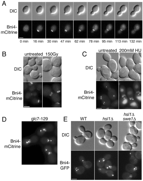
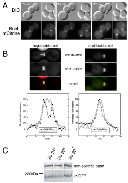
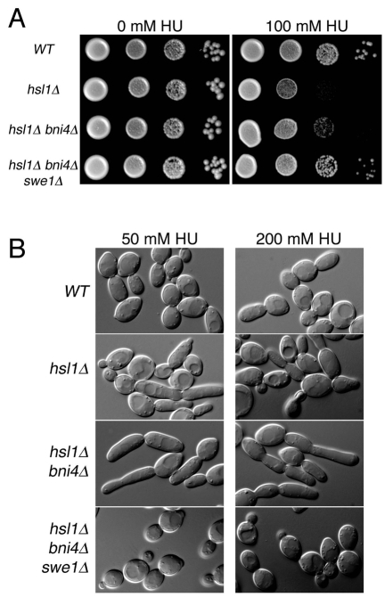
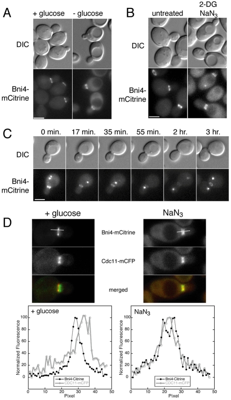
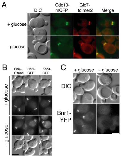
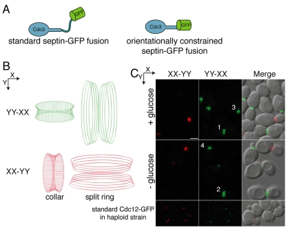
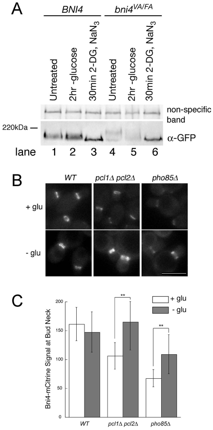
Septin complexes at the bud neck in Saccharomyces cerevisiae serve as a scaffold for proteins involved in signaling, cell cycle control, and cell wall synthesis. Many of these bind asymmetrically, associating with either the mother- or daughter-side of the neck. Septin structures are inherently apolar so the basis for the asymmetric binding remains unknown. Bni4, a regulatory subunit of yeast protein phosphatase type 1, Glc7, binds to the outside of the septin ring prior to bud formation and remains restricted to the mother-side of the bud neck after bud emergence. Bni4 is responsible for targeting Glc7 to the mother-side of the bud neck for proper deposition of the chitin ring. We show here that Bni4 localizes symmetrically, as two distinct rings on both sides of the bud neck following energy depletion or activation of cell cycle checkpoints. Our data indicate that loss of Bni4 asymmetry can occur via at least two different mechanisms. Furthermore, we show that Bni4 has a Swe1-dependent role in regulating the cell morphogenesis checkpoint in response to hydroxyurea, which suggests that the change in localization of Bni4 following checkpoint activation may help stabilize the cell cycle regulator Swe1 during cell cycle arrest.


Figures

Fig. 1.
Fig. 1.
Bni4 is targeted to mother- and daughter-sides of the bud neck following cell cycle arrest. (A) DIC and fluorescence microscopy showing Bni4-mCitrine localization throughout the cell cycle. Strain JRL265 (BNI4-mCitrine) was grown to mid-log phase in synthetic complete (SC) medium then examined at the indicated times. (B) DIC and fluorescence images of Bni4-mCitrine in strain JRL264. Cells were grown to mid-log phase and either left untreated or exposed to 150 Gy γ-radiation. Cells were examined after a 3 hour incubation at 30°C following irradiation. (C) DIC and fluorescence images of cells from diploid strain JRL1000 (BNI4-mCitrine/BNI4-mCitrine SPC42-GFP(3X)/SPC42). Cells were grown to mid-log phase in SC medium and incubated for 2 hours in the presence of 200 mM hydroxyurea (HU). (D) DIC and fluorescence images of cells from strain KT2592 (BNI4-mCitrine SPC42-GFP(3X) glc7-129) grown to log phase at 24°C in synthetic medium. (E) DIC and fluorescence images of cells from strains JRL240 (BNI4-GFP SPC42-GFP(3X)), JRL236 (hsl1Δ BNI4-GFP SPC42-GFP(3X)), and JRL238 (hsl1Δ swe1Δ BNI4-GFP SPC42-GFP(3X)). Cells were grown to mid-log phase in SC medium. Scale bars: 3 μm.
Fig. 2.
Fig. 2.
Bni4-mCitrine localization in large budded cells. (A) Time-lapse imaging of DIC and fluorescence images of cells from strain KT2575 (BNI4-mCitrine cdc13-1) after a shift from 24°C to 32°C. The time indicated in each panel is the time after the temperature shift. Note the small budded cells at the 90 minute and 120 minute time points still retain Bni4-mCitrine on the daughter-side of the bud neck. (B) Upper panels: fluorescence images of septin (Cdc11-mCFP) and Bni4-mCitrine in diploid strain JRL265/JRL574 (BNI4-mCitrine homozygous CDC11-mCFP heterozygous). Cells were grown to mid-log phase in synthetic medium and then incubated for 1.5 hours in 150 mM hydroxyurea. Lower panels: the relative fluorescence of the CFP and mCitrine was measured through the middle of the bud neck along the mother-bud axis for 3 μm as illustrated by the bars in the upper panels. (C) Immunoblot analysis of Bni4-mCitrine using α-GFP. Strain KT2575 (BNI4-mCitrine cdc13-1) was grown to mid-log phase in YPD at 24°C and then incubated for the indicated times at either 24°C or 30°C. A non-specific band was used as a load control.
Fig. 3.
Fig. 3.
Genetic interactions between BNI4 and HSL1. (A) Cultures of diploid strains KT1112xKT1113 (WT), JRL1002xJRL1005 (hsl1Δ), JRL1003xJRL1004 (hsl1Δ bni4Δ), KT2801xKT2803 (hsl1Δ bni4Δ swe1Δ) were serially diluted onto SC medium or SC medium containing 100 mM hydroxyurea (HU) and incubated at 33°C for 72 hours before imaging. (B) DIC images of cells from strains listed in A. Cells were grown to mid-log phase in SC medium and then incubated for 5 hours in SC medium containing the designated concentrations of HU.
Fig. 4.
Fig. 4.
Localization of Bni4 following energy depletion. (A) DIC and fluorescence microscopy of cells from strains JRL264 (BNI4-mCitrine). Cells were grown to mid-log phase in synthetic medium then placed in synthetic medium lacking glucose for 1.5 hours. Scale bar: 3 μm. (B) DIC and fluorescence microscopy analysis of homozygous diploid strain JRL876 (BNI4-mCitrine). Cells were grown to mid-log phase in synthetic medium then incubated in synthetic medium lacking glucose and containing 100 mM 2-deoxyglucose/10 mM NaN3 for 30 minutes. Scale bar: 3 μm. (C) Time-lapse microscopy of strain JRL326 [BNI4-mCitrine SPC42-GFP(3X)]. Cells were grown to mid-log phase in synthetic medium then placed in synthetic medium lacking glucose for 1.5 hours. Cells were then followed by time-lapse on synthetic medium containing 2% glucose. The Spc42-GFP protein marks the spindle pole body, which duplicates and separates during S phase. In many images only one spindle pole body is visible in the focal plane. Scale bar: 3 μm. (D) Upper panels: fluorescence images of Cdc11-mCFP and Bni4-mCitrine in diploid strain JRL265/JRL574 growing in synthetic medium (left panels) or incubated for 30 minutes in 10 mM sodium azide (right panels). Lower panels: the relative fluorescence of the mCFP and mCitrine for 3 μm through the middle of the bud neck along the mother-bud axis as illustrated by the bars in the upper panels.
Fig. 5.
Fig. 5.
Energy deprivation changes the location of some, but not all, bud neck-associated proteins. (A) DIC and fluorescence images of septin (Cdc10-mCFP) and Glc7-tdimer2 in diploid strain JRL884/KT2450. Cells were grown to mid-log phase in synthetic medium then incubated for 1.5 hours in synthetic medium with or without 2% glucose. (B) DIC and fluorescence images of cells from strains JRL264 (BNI4-mCitrine), YLK173 (HSL1-GFP) and YLK179 (KCC4-GFP). Cells were treated as in A. (C) DIC and fluorescence images of cells from strain JRL443 (BNR1-YFP). Cells were treated as in A. Scale bars: 3 μm.
Fig. 6.
Fig. 6.
Loss of Bni4 asymmetry is not due to changes in septin architecture. (A) The orientationally constrained septin-GFP fusion protein. The standard septin-GFP fusions contain a flexible linker connecting the septin and GFP portions of the fusion protein (left). In the orientationally constrained septin-GFP fusion, the last α-helix of the coiled-coil domain of Cdc3 was fused to the first α-helix of GFP, creating a single α-helix. Consequently, the orientation of Cdc3 dictates the orientation of GFP (right). (B) Diagram of septin filaments and their orientation as visualized by polarized fluorescence microscopy. Filaments oriented in the direction of the Y-axis are colored green and filaments oriented in the X-axis are colored red. (C) Polarized fluorescence images of strain JRL794 (CDC3-GFP heterozygous diploid). Strain YLK68 (CDC12-GFP), which does not contain an orientationally constrained GFP-tag, was used as a negative control. Cells were treated as in Fig. 4A and then imaged by polarized fluorescence microscopy. The X-axis signal subtracted from the Y-axis signal (YY-XX) and the Y-axis signal subtracted from the X-axis signal (XX-YY) are pseudocolored green and red, respectively, and correspond to the orientations shown in B. Scale bar, 3 μm.
Fig. 7.
Fig. 7.
Bni4 and Bni4V831A/F833A become hypophosphorylated following energy depletion. (A) Immunoblot of Bni4-GFP and Bni4V831A/F833A-GFP using α-GFP. Strains JRL369 (BNI4-GFP) (lanes 1-3) and YLK85 (bni4V831A/F833A-GFP) (lanes 4-6) were grown to mid-log phase in synthetic medium containing glucose. Cells were then left untreated (lanes 1 and 4) or incubated in synthetic media lacking glucose (lanes 2 and 5) with the addition of 100 mM 2-deoxyglucose and 10 mM NaN3 (lanes 3 and 6) for the indicated times. A non-specific band was used as a load control. (B) Fluorescence microscopy of Bni4-mCitrine at the neck in wild-type (JRL265), pcl1Δ pcl2Δ (JRL685) and pho85Δ (JRL653) cells. Cells were imaged at mid-log phase in synthetic medium (+ glu) or after incubation for 1.5 hours in synthetic medium without glucose (− glu). Scale bar: 5 μm. (C) Quantitative analysis of relative fluorescent signal at the bud neck of the cells in B (n>50). Double asterisks denote significantly different levels. **P<0.0001, according to the unpaired Student's t-test.

Similar articles

Cited by

References

    1. Barral Y., Parra M., Bidlingmaier S., Snyder M. (1999). Nim1-related kinases coordinate cell cycle progression with the organization of the peripheral cytoskeleton in yeast. Gene. Dev. 13, 176-187 - PMC - PubMed
    1. Bertin A., McMurray M. A., Grob P., Park S. S., Garcia G., 3rd, Patanwala I., Ng H. L., Alber T., Thorner J., Nogales E. (2008). Saccharomyces cerevisiae septins: supramolecular organization of heterooligomers and the mechanism of filament assembly. Proc. Natl. Acad. Sci. USA 105, 8274-8279 - PMC - PubMed
    1. Bharucha J. P., Larson J. R., Gao L., Daves L. K., Tatchell K. (2008). Ypi1, a Positive Regulator of Nuclear Protein Phosphatase Type 1 Activity in Saccharomyces cerevisiae. Mol. Biol. Cell 19, 1032-1045 - PMC - PubMed
    1. Bloecher A., Tatchell K. (1999). Defects in Saccharomyces cerevisiae protein phosphatase type I activate the spindle/kinetochore checkpoint. Gene. Dev. 13, 517-522 - PMC - PubMed
    1. Bloecher A., Tatchell K. (2000). Dynamic localization of protein phosphatase type 1 in the mitotic cell cycle of Saccharomyces cerevisiae. J. Cell Biol. 149, 125-140 - PMC - PubMed

Publication types

MeSH terms