

A novel clinically relevant animal model for studying galectin-3 and its ligands during colon carcinogenesis
- PMID: 20197492
- PMCID: PMC2874187
- DOI: 10.1369/jhc.2010.955237
A novel clinically relevant animal model for studying galectin-3 and its ligands during colon carcinogenesis
Abstract
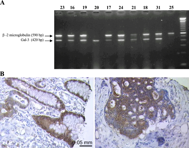
Galectin-3 (Gal-3) is a multifunctional protein that plays different roles in cancer biology. To better understand the role of Gal-3 and its ligands during colon carcinogenesis, we studied its expression in tumors induced in rats treated with 1,2-dimethylhydrazine (DMH) and in human tissues. Normal colon from untreated rats showed no staining using two specific monoclonal antibodies. In contrast, morphologically normal colon from DMH-treated rats and dysplastic aberrant crypt foci were strongly stained, indicating that increased Gal-3 expression is an early event during the neoplastic transformation in colon cells. Gal-3 was weakly expressed in adenocarcinomas. Overall, the Gal-3 expression pattern observed in the DMH rat model closely resembles that displayed by human colon stained with the same antibodies. We also found that Gal-3 phosphorylation diminishes in serines while increasing in tyrosines during rat colon carcinogenesis. Finally, we showed that Gal-3-ligands expression is strikingly similar in rat and human malignant colon and in non-malignant tissues. In conclusion, the DMH-induced rat colon cancer model displays expression patterns of Gal-3 and its ligands very similar to those observed in human samples. This animal model should contribute to clarifying the role of Gal-3 in colon carcinogenesis and also to finding effective preventive cancer agents based on Gal-3 targeting.
Figures


Similar articles
-
Phosphotyrosine, p62 c-myc and p21 c-Ha-ras proteins in colonic epithelium of normal and dimethylhydrazine-treated rats: an immunohistochemical analysis.Anticancer Res. 1995 Jan-Feb;15(1):211-8. Anticancer Res. 1995. PMID: 7537485
-
Cell surface overexpression of galectin-3 and the presence of its ligand 90k in the blood plasma as determinants in colon neoplastic lesions.Glycobiology. 2004 Sep;14(9):783-92. doi: 10.1093/glycob/cwh092. Epub 2004 May 12. Glycobiology. 2004. PMID: 15140826
-
Overexpression of cyclin D1 and cyclin E in 1,2-dimethylhydrazine dihydrochloride-induced rat colon carcinogenesis.J Vet Sci. 2000 Dec;1(2):121-6. J Vet Sci. 2000. PMID: 14614307
-
Aberrant crypt foci and colon cancer: comparison between a short- and medium-term bioassay for colon carcinogenesis using dimethylhydrazine in Wistar rats.Braz J Med Biol Res. 2002 Mar;35(3):351-5. doi: 10.1590/s0100-879x2002000300010. Braz J Med Biol Res. 2002. PMID: 11887213
-
[Effects of combined use of curcumin and catechin on cyclooxygenase-2 mRNA expression in dimethylhydrazine-induced rat colon carcinogenesis].Di Yi Jun Yi Da Xue Xue Bao. 2005 Jan;25(1):48-52. Di Yi Jun Yi Da Xue Xue Bao. 2005. PMID: 15683997 Chinese.
Cited by
-
Could Galectin 3 Be a Good Prognostic Factor in Endometrial Cancer?Diagnostics (Basel). 2020 Aug 26;10(9):635. doi: 10.3390/diagnostics10090635. Diagnostics (Basel). 2020. PMID: 32859099 Free PMC article.
-
Although with intact mucosa at colonoscopy, chagasic megacolons have an overexpression of Gal-3.Einstein (Sao Paulo). 2020 Mar 6;18:eAO5105. doi: 10.31744/einstein_journal/2020AO5105. eCollection 2020. Einstein (Sao Paulo). 2020. PMID: 32159607 Free PMC article.
-
Galectin-3 up-regulation in hypoxic and nutrient deprived microenvironments promotes cell survival.PLoS One. 2014 Nov 4;9(11):e111592. doi: 10.1371/journal.pone.0111592. eCollection 2014. PLoS One. 2014. PMID: 25369297 Free PMC article.
-
Functions of galectins as 'self/non-self'-recognition and effector factors.Pathog Dis. 2017 Jul 31;75(5):ftx046. doi: 10.1093/femspd/ftx046. Pathog Dis. 2017. PMID: 28449072 Free PMC article. Review.
-
Galectin-3 accelerates the progression of oral tongue squamous cell carcinoma via a Wnt/β-catenin-dependent pathway.Pathol Oncol Res. 2013 Jul;19(3):461-74. doi: 10.1007/s12253-013-9603-7. Epub 2013 Mar 22. Pathol Oncol Res. 2013. PMID: 23519607
References
-
- Abdel-Aziz HO, Murai Y, Takasaki I, Tabuchi Y, Zheng HC, Nomoto K, Takahashi H, et al. (2008) Targeted disruption of the galectin-3 gene results in decreased susceptibility to NNK-induced lung tumorigenesis: an oligonucleotide microarray study. J Cancer Res Clin Oncol 134:777–788 - PubMed
-
- Ahmed H, Banerjee PP, Vasta GR (2007) Differential expression of galectins in normal, benign and malignant prostate epithelial cells: silencing of galectin-3 expression in prostate cancer by its promoter methylation. Biochem Biophys Res Commun 358:241–246 - PubMed
-
- Ahmed H, Bianchet MA, Amzel LM, Hirabayashi J, Kasai K, Giga-Hama Y, Tohda H, et al. (2002) Novel carbohydrate specificity of the 16-kDa galectin from Caenorhabditis elegans: binding to blood group precursor oligosaccharides (type 1, type 2, Talpha, and Tbeta) and gangliosides. Glycobiology 12:451–461 - PubMed
-
- Ahmed H, Pohl J, Fink NE, Strobel F, Vasta GR (1996) The primary structure and carbohydrate specificity of a beta-galactosyl-binding lectin from toad (Bufo arenarum Hensel) ovary reveal closer similarities to the mammalian galectin-1 than to the galectin from the clawed frog Xenopus laevis. J Biol Chem 271:33083–33094 - PubMed
-
- Allegrucci C, Liguori L, Minelli A (2001) Stimulation by N6-cyclopentyladenosine of A1 adenosine receptors, coupled to galphai2 protein subunit, has a capacitative effect on human spermatozoa. Biol Reprod 64:1653–1659 - PubMed
Publication types
MeSH terms
Substances
LinkOut - more resources
Full Text Sources
