. 2010 Apr 29;115(17):3589-97.
doi: 10.1182/blood-2009-04-215848. Epub 2010 Mar 2.

Conditional expression of heterozygous or homozygous Jak2V617F from its endogenous promoter induces a polycythemia vera-like disease

Affiliations

Conditional expression of heterozygous or homozygous Jak2V617F from its endogenous promoter induces a polycythemia vera-like disease

Hajime Akada et al. Blood. .

Abstract

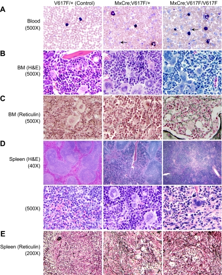
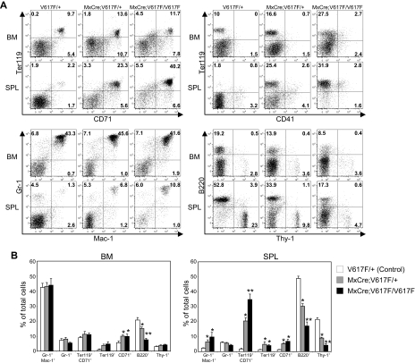
A somatic point mutation (V617F) in the JAK2 tyrosine kinase was found in a majority of patients with polycythemia vera (PV), essential thrombocythemia, and primary myelofibrosis. However, contribution of the JAK2V617F mutation in these 3 clinically distinct myeloproliferative neoplasms (MPNs) remained unclear. To investigate the role of JAK2V617F in the pathogenesis of these MPNs, we generated an inducible Jak2V617F knock-in mouse, in which the expression of Jak2V617F is under control of the endogenous Jak2 promoter. Expression of heterozygous mouse Jak2V617F evoked all major features of human polycythemia vera (PV), which included marked increase in hemoglobin and hematocrit, increased red blood cells, leukocytosis, thrombocytosis, splenomegaly, reduced serum erythropoietin (Epo) levels and Epo-independent erythroid colonies. Homozygous Jak2V617F expression also resulted in a PV-like disease associated with significantly greater reticulocytosis, leukocytosis, neutrophilia and thrombocytosis, marked expansion of erythroid progenitors and Epo-independent erythroid colonies, larger spleen size, and accelerated bone marrow fibrosis compared with heterozygous Jak2V617F expression. Biochemical analyses revealed Jak2V617F gene dosage-dependent activation of Stat5, Akt, and Erk signaling pathways. Our conditional Jak2V617F knock-in mice provide an excellent model that can be used to further understand the molecular pathogenesis of MPNs and to identify additional genetic events that cooperate with Jak2V617F in different MPNs.


Figures

Figure 1
Figure 1
Generation of inducible Jak2V617F knock-in mice. (A) The targeted allele contains the floxed PGK-Neo-Stop cassette and the V617F mutation. This allele is transcriptionally silent, but can be induced in the presence of Cre to generate the activated Jak2V617F allele. (B) Constitutive phosphorylation of Stat5 in the BM of induced MxCre;V617F/+ and MxCre;V617F/V617F mice confirm expression of the mutant Jak2V617F protein. (C) Expression of total Jak2 mRNA was measured in the BM of WT, V617F/+, MxCre;V617F/+ (heterozygous), and MxCre;V617F/+V617F (homozygous) mice (n = 4) by real-time PCR. Total Jak2 mRNA expression was significantly reduced in the BM of both heterozygous and homozygous Jak2V617F mice compared with WT mice (P < .05 between WT and heterozygous Jak2V617F, P < .05 between WT and homozygous Jak2V617F; unpaired t test), whereas there were no significant differences between V617F/+ and heterozygous or V617F/+ and homozygous Jak2V617F mice. (D) A standard curve made from known ratios of accurately measured pMSCV-IRES-GFP plasmids containing mouse Jak2WT and mouse Jak2V617F showing the linearity and accuracy of the measurement of T/G fluorescence ratio (T-peak identifies the mutant, G-peak identifies the WT allele) for determination of allelic ratio. (E) Allelic ratio of the mutant Jak2V617F to WT Jak2 mRNA was determined by the T/G ratio after direct sequencing of the real-time PCR products from the BM of heterozygous Jak2V617F mice (n = 8). (F) The chromatograms of 3 sequenced real-time PCR products from the BM of heterozygous Jak2V617F mice are shown.
Figure 2
Figure 2
Mice expressing Jak2V617F develop MPN. Peripheral blood hematocrit (A), hemoglobin (B), and RBC (C) counts were significantly increased in heterozygous and homozygous Jak2V617F mice compared with controls (V617F/+). Mean corpuscular volume (MCV; D) was significantly reduced in both heterozygous and homozygous Jak2V617F mice compared with controls (V617F/+). WBC (E), neutrophil (F), and platelet (G) counts were also significantly increased in both heterozygous and homozygous Jak2V617F mice compared with controls. However, the WBC, neutrophil, and platelet counts were much greater in peripheral blood of Jak2V617F homozygous mice compared with heterozygous Jak2V617F mice. Blood counts at 4, 8, 12, 16, and 20 weeks after induction with pI:pC are shown. (n = 30 at all time points for V617F/+ control; n = 30 at all time points for heterozygous Jak2V617F; n = 10 at 4, 8, 12 weeks, n = 6 at 16 and 20 weeks for homozygous Jak2V617F mice). (H) Reticulocyte counts were markedly increased in the peripheral blood of homozygous Jak2V617F mice. (I) Serum Epo level was significantly reduced in both heterozygous (n = 9) and homozygous Jak2V617F (n = 6) mice compared with controls (n = 9). (J) BM cellularity (total BM cell count; was significantly reduced (12 to 16 weeks after induction) in mice expressing homozygous Jak2V617F. (K) Spleen weight/size was significantly increased in Jak2V617F heterozygous (n = 20) and homozygous (n = 9) mice compared with controls (n = 20; 12 to 16 weeks after induction). *Significance between control and heterozygous or between control and homozygous; **significance between control and homozygous as well as between heterozygous and homozygous Jak2V617F mice; P < .05 determined by unpaired, 2-tailed Student t test.
Figure 3
Figure 3
Histopathologic characterization of the MPN induced by Jak2V617F. (A) Peripheral blood smears show increased RBCs, leukocytes, and platelets in induced heterozygous and homozygous Jak2V617F mice (12 weeks after induction). Leukocytosis and thrombocytosis were more pronounced in homozygous Jak2V617F mice compared with heterozygous Jak2V617F animals. Arrows point to reticulocytes. (B) BM sections from induced Jak2V617F heterozygous and homozygous mice show trilineage hyperplasia (hematoxylin and eosin staining, ×500). (C) Reticulin staining on the BM sections (×500) shows mild fibrosis (grade 1) in older Jak2V617F heterozygous mice (24 weeks after induction), whereas homozygous mice show more reticulin fibrosis (grade 2) than heterozygous mice. (D) Spleens from heterozygous and homozygous Jak2V617F mice display extensive destruction of normal splenic architecture (×40 and ×500) with attenuated white pulp and markedly expanded red pulp, increased numbers of megakaryocytes, and clusters of immature erythroid precursors. (E) Reticulin staining of the heterozygous spleen shows increased fibrosis of the white pulp and slight reticulin fibrosis of the red pulp. Spleens from homozygous mice show pronounced reticulin fibrosis in the white pulp and also increased fibrosis in the red pulp compared with heterozygous mice.
Figure 4
Figure 4
Flow cytometric analysis of BM and spleen from mice expressing Jak2V617F. (A) Dot plots demonstrate a marked increase in the Ter-119/CD71–postive populations in the spleens of heterozygous and homozygous mice compared with control (V617F/+) mice. Modest increases in Gr-1/Mac-1–positive cells in the spleen of heterozygous and homozygous Jak2V617F mice were observed. However, B-cell populations (B220-positive) were proportionately decreased in the BM and spleens of heterozygous and homozygous Jak2V617F mice compared with control animals. (B) Percentages of myeloid, erythroid, B- and T-lymphoid populations are shown in histograms as mean ± SEM. Data are presented as percentage of total cells (control, n = 11; heterozygous, n = 11; homozygous, n = 5). *Significance between control and heterozygous or between control and homozygous; **significance between control and homozygous as well as between heterozygous and homozygous; significant difference at P < .05.
Figure 5
Figure 5
Effects of Jak2V617F on hematopoietic progenitors. (A) Flow cytometric analysis of the LSK compartment (LinSca1+c-kit+) and subsets of myeloid progenitors including CMP (LinSca1c-kit+CD34+FcγRII/IIIlo), GMP (LinSca1c-kit+CD34+FcγRII/IIIhigh), and MEP (LinSca1c-kit+CD34FcγRII/III) in the BM and spleen from control (n = 8), heterozygous (n = 8), and homozygous Jak2V617F (n = 5) mice. (B) Representative contour plots are shown. The percentage of LSK, CMP, GMP, and MEP is shown in histograms as mean ± SEM. Data are presented as percentage of total cells. *Significance between control and heterozygous or between control and homozygous; **significance between control and homozygous as well as between heterozygous and homozygous Jak2V617F mice; significant difference of P < .05. (C) Hematopoietic progenitor colonies. BM (2 × 10⁴) and spleen (1 × 10⁵) cells from control (n = 6), heterozygous (n = 6), and homozygous Jak2V617F (n = 5) mice were plated in complete methylcellulose (Methocult M3434) medium. BFU-E, CFU-GM, and CFU-GEMM colonies were counted on day 7. (D) Epo-independent CFU-E colonies. Spleen cells (1 × 10⁵) from control, heterozygous, and homozygous Jak2V617F mice were plated in methylcellulose medium without any cytokine. CFU-E colonies were counted after 2 days.
Figure 6
Figure 6
Signaling effects of heterozygous and homozygous Jak2V617F in erythroid progenitors. Primary erythroblasts were derived from the BM and spleen of control (V617F/+), heterozygous, and homozygous Jak2V617F mice. For signaling studies, erythroblasts were starved in Iscove modified Dulbecco medium plus 0.5% BSA for 4 hours. Cell lysates were prepared in radioimmunoprecipitation assay (RIPA) buffer and subjected to immunoblotting with anti-phosphotyrosine (4G10) antibody (A) or phospho-specific antibodies against Stat5, Akt, and Erk1/2 (B).

Similar articles

Cited by

References

    1. James C, Ugo V, Le Couedic JP, et al. A unique clonal JAK2 mutation leading to constitutive signaling causes polycythemia vera. Nature. 2005;434(7037):1144–1148. - PubMed
    1. Levine RL, Wadleigh M, Cools J, et al. Activating mutation in the tyrosine kinase JAK2 in polycythemia vera, essential thrombocythemia, and myeloid metaplasia with myelofibrosis. Cancer Cell. 2005;7(4):387–397. - PubMed
    1. Baxter EJ, Scott LM, Campbell PJ, et al. Acquired mutation of the tyrosine kinase JAK2 in human myeloproliferative disorders. Lancet. 2005;365(9464):1054–1061. - PubMed
    1. Kralovics R, Passamonti F, Buser AS, et al. A gain-of-function mutation of JAK2 in myeloproliferative disorders. N Engl J Med. 2005;352(17):1779–1790. - PubMed
    1. Zhao R, Xing S, Li Z, et al. Identification of an acquired JAK2 mutation in polycythemia vera. J Biol Chem. 2005;280(24):22788–22792. - PMC - PubMed

Publication types

MeSH terms