. 2010 Apr 22;115(16):3304-13.
doi: 10.1182/blood-2009-07-233304. Epub 2010 Mar 2.

Acquired resistance to ABT-737 in lymphoma cells that up-regulate MCL-1 and BFL-1

Affiliations

Acquired resistance to ABT-737 in lymphoma cells that up-regulate MCL-1 and BFL-1

Derek Yecies et al. Blood. .

Abstract

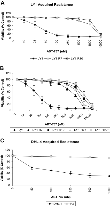
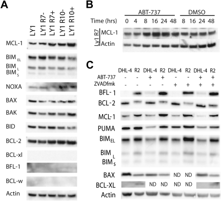
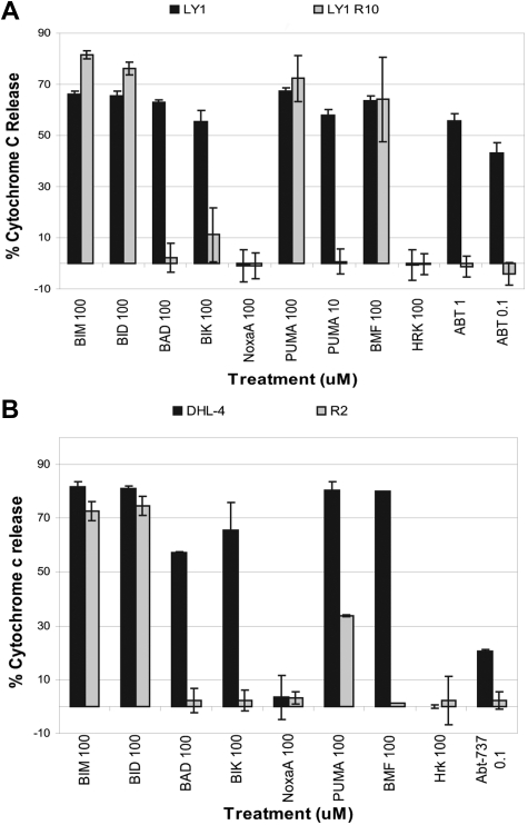
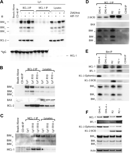
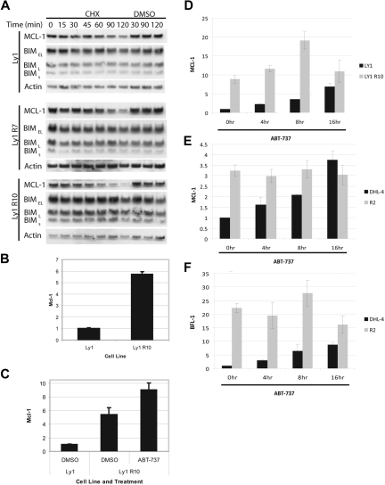
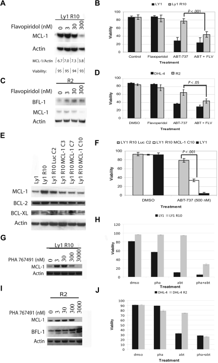
ABT-737 is a small-molecule antagonist of BCL-2 currently under evaluation in clinical trials in the oral form of ABT-263. We anticipate that acquired resistance to this promising drug will inevitably arise. To study potential mechanisms of resistance to ABT-737, we derived resistant lines from initially sensitive OCI-Ly1 and SU-DHL-4 lymphoma cell lines via long-term exposure. Resistance was based in the mitochondria and not due to an inability of the drug to bind BCL-2. Resistant cells had increased levels of BFL-1 and/or MCL-1 proteins, which are not targeted by ABT-737. Proapoptotic BIM was displaced from BCL-2 by ABT-737 in both parental and resistant cells, but in resistant cells, BIM was sequestered by the additional BFL-1 and/or MCL-1. Decreasing MCL-1 levels with flavopiridol, PHA 767491, or shRNA restored sensitivity to ABT-737 resistant cells. MCL-1 was up-regulated not by protein stabilization but rather by increased transcript levels. Surprisingly, in addition to stable increases in MCL-1 transcript and protein in resistant cells, there was a dynamic increase within hours after ABT-737 treatment. BFL-1 protein and transcript levels in resistant cells were similarly dynamically up-regulated. This dynamic increase suggests a novel mechanism whereby modulation of antiapoptotic protein function communicates with nuclear transcriptional machinery.


Figures

Figure 1
Figure 1
B-cell lymphoma cells acquire resistance to ABT-737 after long-term exposure. (A) OCI-Ly1 cells were treated with increasing doses of ABT-737 for 48 hours and stained with annexin-V–FITC for flow cytometry analysis. Before treatments, OCI-Ly1 R7 cells were cultured in media containing 320nM ABT-737 and OCI-Ly1 R10 cells in media with 1μM ABT-737. ABT-737 was withdrawn 24 hours before dose response treatment. Viability is shown as a percentage of DMSO-treated control cells. SD is of quadruplicates and is indicated by error bars. (B) OCI-Ly1 R7 and OCI-Ly1 R10 cells were withdrawn (−) from or continued treatment (+) with ABT-737 for 3 weeks. Cells were then washed and treated with increasing doses of ABT-737 for 48 hours, stained with annexin-V–FITC, and assayed by flow cytometry analysis. Viability is shown as a percentage of DMSO-treated control cells. SD is of quadruplicates and is indicated by error bars. (C) SU-DHL-4 and SU-DHL-4 R2 cells were treated with increasing doses of ABT-737 for 12 hours. After treatment, cells were stained with annexin-V–FITC and PI for flow cytometry analysis. Before treatment, SU-DHL-4 R2 cells were cultured in media containing 1μM ABT-737. ABT-737 was withdrawn 3 weeks before dose response treatment. Viability is shown as a percentage of DMSO-treated control cells. SE is of triplicates and is indicated by error bars.
Figure 2
Figure 2
BFL-1 and/or MCL-1 proteins are up-regulated in resistant cell lines and are inducible upon treatment with ABT-737. (A) Immunoblot analysis of OCI-LY1, OCI-LY1 R7, and OCI-LY1 R10 cell line lysates with the indicated antibodies. − indicates that ABT-737 was withdrawn for 3 weeks; +, continuous culture with ABT-737. Unpictured antibody controls confirmed the efficacy of the BCL-XL and BFL-1 antibodies used in this assay. These results are representative of 3 independent experiments. (B) OCI-Ly1 R7 cells were removed from ABT-737–containing media for 3 weeks. ABT-737 was added back to culture for the indicated time frame at 320nM. Whole-cell lysates were made after treatment and analyzed by immunoblot. DMSO was used as a solvent-only negative control. These results are representative of 3 independent experiments. (C) SU-DHL-4 R2 cells were removed from ABT-737–containing media for 3 weeks. SU-DHL-4 and SU-DHL-4 R2 cells were treated with 1μM ABT-737 or DMSO for 12 hours after pretreatment with 10μM ZVAD.fmk or DMSO for 1 hour as indicated. Whole-cell lysates were made after treatment and analyzed by immunoblot. These results are representative of 3 independent experiments. ND indicates that these samples were not analyzed.
Figure 3
Figure 3
BH3 profiling reveals a mitochondrial basis for acquired resistance to ABT-737. (A) Mitochondria were isolated from OCI-Ly1 and OCI-Ly1 R10 cells and treated with different concentrations of peptides. Cytochrome c release was measured by ELISA. Shown is the average and SE of 3 independent experiments. (B) Mitochondria were isolated from SU-DHL-4 and SU-DHL-4 R2 cells and treated with different peptides. Cytochrome c release was measured by ELISA. Shown is the average and SE of 3 independent experiments. In both cases, ABT-737 was removed from culture medium for at least 1 week.
Figure 4
Figure 4
ABT-737 reaches its target, BCL-2, in sensitive and resistant cells, and increased MCL-1 and BFL-1 sequester BIM displaced from BCL-2 by ABT-737. (A) OCI-LY1 cells were treated with 1μM ABT-737 for 4 hours after pretreatment with 10μM ZVAD.fmk for 30 minutes as indicated. Cells were lysed with 1% CHAPS lysis buffer. Lysates were immunoprecipitated using BCL-2– and MCL-1–specific antibodies. The resulting immunoprecipitated and coimmunoprecipitated proteins were analyzed by immunoblot alongside whole-cell lysates. *IgG heavy chain. (B) OCI-Ly1 R10 cells were treated with DMSO or 1μM ABT-737 (+) for 4 hours. Cells were lysed with 1% CHAPS lysis buffer. A total of 100 μg of lysate were immunoprecipitated with the BCL-2 (6C8) antibody. The resulting immunoprecipitated and coimmunoprecipitated proteins were analyzed by immunoblot alongside whole-cell lysates. These results are representative of 3 independent experiments. (C) OCI-Ly1 R10 cells were treated and lysed as in panel B. A total of 100 μg of lysate were immunoprecipitated with an MCL-1–specific antibody. The resulting immunoprecipitated and coimmunoprecipitated proteins were analyzed by immunoblot alongside whole-cell lysates. These results are representative of 3 independent experiments. (D) SU-DHL-4 cells were treated with DMSO or 1μM ABT-737 (+) for 12 hours after pretreatment with 10μM ZVAD.fmk for 1 hour. SU-DHL-4 R2 cells were treated with DMSO or 1μM ABT-737 (+) for 12 hours. Cells were lysed with 1% CHAPS lysis buffer. Lysates were immunoprecipitated using a BCL-2–specific antibody (6C8). The resulting immunoprecipitated and coimmunoprecipitated proteins were analyzed by immunoblot. (E) SU-DHL-4 and SU-DHL-4 R2 cells were treated and lysed as in panel D. Lysates were immunoprecipitated using a BIM-specific antibody. The resulting immunoprecipitated and coimmunoprecipitated proteins were analyzed by immunoblot. (F) Immunoblot analysis of the whole-cell lysates used for immunoprecipitation in panels D and E.
Figure 5
Figure 5
MCL-1 and BFL-1 are transcriptionally up-regulated in resistant cells. (A) OCI-Ly1, OCI-Ly1 R7, and OCI-LY1 R10 cells were treated with 20 μg/mL CHX for the time indicated. Cells were lysed immediately after treatment and subject to immunoblot analysis. These results are representative of 3 independent experiments. (B) Cells were cultured in the absence of ABT-737 for 2 weeks. RNA was isolated and MCL-1 fold change was analyzed by quantitative PCR. (C) Cells were cultured in the absence of ABT-737 for 2 weeks and then treated with DMSO or 250nM ABT-737 for 16 hours. RNA was isolated and MCL-1 fold change was analyzed by quantitative PCR. (D) OCI-Ly1 and OCI-Ly1 R10 cells were cultured in the absence of ABT-737 for 3 weeks. Cells were then treated with 20μM ZVAD.fmk for 45 minutes before treatment with 1μM ABT-737 for the time indicated. RNA was isolated and MCL-1 fold change was analyzed by quantitative PCR. (E) SU-DHL-4 and SU-DHL-4 R2 cells were cultured in the absence of ABT-737 for 3 weeks and treated and analyzed as in panel D. (F) RNA from panel E was used to assay BFL-1 fold change by quantitative PCR. Error bars in panels B through F indicate the SEM. RNA levels from the untreated parental cell lines were set to 1.
Figure 6
Figure 6
CDK9 inhibition or shRNA decrease MCL-1 levels and restore sensitivity to ABT-737. (A) Immunoblot analysis of OCI-LY1 R10 whole-cell lysates after 4 hours of flavopiridol treatment with different doses. Viability was assayed by annexin-V–FITC staining and flow cytometry. (B) OCI-LY1 and OCI-LY1 R10 cells were treated for 4 hours with DMSO or 300nM flavopiridol in combination with 0 or 250nM ABT-737, stained with annexin-V–FITC, and analyzed by flow cytometry. This experiment was performed in quadruplicate. (C) Immunoblot analysis of SU-DHL-4 R2 whole-cell lysates after 4 hours of flavopiridol treatment with different doses. (D) SU-DHL-4 and SU-DHL-4 R2 cells were treated for 12 hours with DMSO or 500nM ABT-737. During the last 4 hours of ABT-737 treatment, cells were treated with DMSO or 300nM flavopiridol. Cells were then stained with annexin-V–FITC and PI and analyzed by flow cytometry. The experiment was performed in triplicate. (E) Immunoblot analysis of MCL-1 knockdown by shRNA in single-cell clones. (F) Clones were chosen based on knockdown in panel E and tested for sensitivity by treatment with 500nM ABT-737 or DMSO. This experiment was performed in quadruplicate. All P values were determined by 2-tailed t tests. (G) Immunoblot analysis of OCI-LY1 R10 whole-cell lysates after 4 hours of PHA 767491 treatment with different doses. (H) OCI-LY1 and OCI-LY1 R10 cells were treated for 4 hours with DMSO or 3000nM PHA 767491 in combination with 0 or 1μM ABT-737, stained with annexin-V–FITC, and analyzed by flow cytometry. (I) Immunoblot analysis of SU-DHL-4 R2 whole-cell lysates after 4 hours of PHA 767491 treatment with different doses. (J) SU-DHL-4 and SU-DHL-4 R2 cells were treated for 12 hours with DMSO or 1μM ABT-737. During the last 4 hours of ABT-737 treatment, cells were treated with DMSO or 3000nM PHA 76741. Cells were then stained with annexin-V–FITC and PI and analyzed by flow cytometry. Error bars for panels B, D, and F represent SEM of independent quadruplicate, triplicate, and quadruplicate experiments, respectively. Error bars in panels H and J are representative of technical replicates, and the graphs presented are representative of 2 independent experiments.
Figure 7
Figure 7
Proposed mechanism of acquired resistance. In sensitive cells, BIM is displaced from BCL-2, inducing BAX activation and apoptosis. In resistant cells, BIM is still displaced, but is captured by BFL-1 and/or MCL-1, preventing BAX activation and maintaining survival.

References

    1. Bakhshi A, Jensen JP, Goldman P, et al. Cloning the chromosomal breakpoint of t(14;18) human lymphomas: clustering around JH on chromosome 14 and near a transcriptional unit on 18. Cell. 1985;41(3):899–906. - PubMed
    1. Cleary ML, Sklar J. Nucleotide sequence of a t(14;18) chromosomal breakpoint in follicular lymphoma and demonstration of a breakpoint-cluster region near a transcriptionally active locus on chromosome 18. Proc Natl Acad Sci U S A. 1985;82(21):7439–7443. - PMC - PubMed
    1. Tsujimoto Y, Cossman J, Jaffe E, Croce CM. Involvement of the bcl-2 gene in human follicular lymphoma. Science. 1985;228(4706):1440–1443. - PubMed
    1. Strasser A, Harris AW, Bath ML, Cory S. Novel primitive lymphoid tumours induced in transgenic mice by cooperation between myc and bcl-2. Nature. 1990;348(6299):331–333. - PubMed
    1. McDonnell TJ, Korsmeyer SJ. Progression from lymphoid hyperplasia to high-grade malignant lymphoma in mice transgenic for the t(14; 18). Nature. 1991;349(6306):254–256. - PubMed

Publication types

MeSH terms