. 2010 Mar;120(3):827-39.
doi: 10.1172/JCI36362. Epub 2010 Feb 8.

The von Hippel-Lindau Chuvash mutation promotes pulmonary hypertension and fibrosis in mice

Affiliations

The von Hippel-Lindau Chuvash mutation promotes pulmonary hypertension and fibrosis in mice

Michele M Hickey et al. J Clin Invest. 2010 Mar.

Abstract

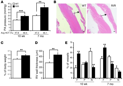
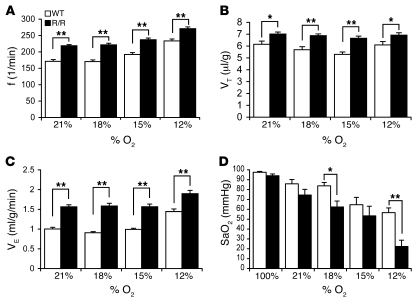
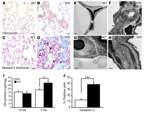
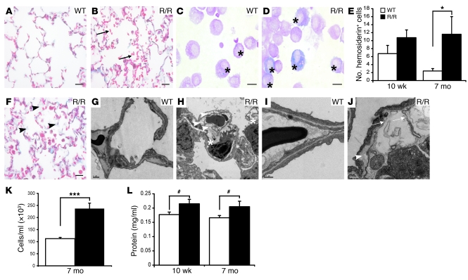
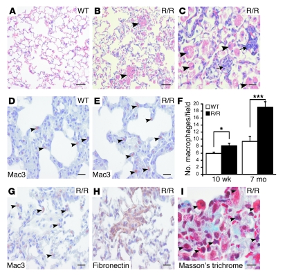
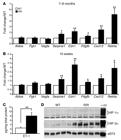
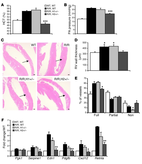
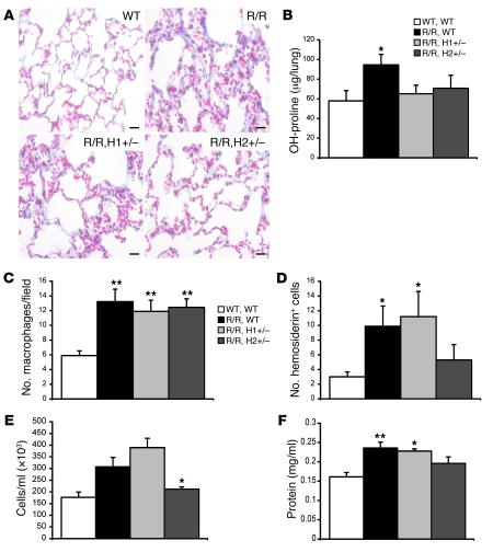
Mutation of the von Hippel-Lindau (VHL) tumor suppressor protein at codon 200 (R200W) is associated with a disease known as Chuvash polycythemia. In addition to polycythemia, Chuvash patients have pulmonary hypertension and increased respiratory rates, although the pathophysiological basis of these symptoms is unclear. Here we sought to address this issue by studying mice homozygous for the R200W Vhl mutation (VhlR/R mice) as a model for Chuvash disease. These mice developed pulmonary hypertension independently of polycythemia and enhanced normoxic respiration similar to Chuvash patients, further validating VhlR/R mice as a model for Chuvash disease. Lungs from VhlR/R mice exhibited pulmonary vascular remodeling, hemorrhage, edema, and macrophage infiltration, and lungs from older mice also exhibited fibrosis. HIF-2alpha activity was increased in lungs from VhlR/R mice, and heterozygosity for Hif2a, but not Hif1a, genetically suppressed both the polycythemia and pulmonary hypertension in the VhlR/R mice. Furthermore, Hif2a heterozygosity resulted in partial protection against vascular remodeling, hemorrhage, and edema, but not inflammation, in VhlR/R lungs, suggesting a selective role for HIF-2alpha in the pulmonary pathology and thereby providing insight into the mechanisms underlying pulmonary hypertension. These findings strongly support a dependency of the Chuvash phenotype on HIF-2alpha and suggest potential treatments for Chuvash patients.


Figures

Figure 1
Figure 1. VhlR/R (R/R) mice develop pulmonary hypertension.
(A) Systolic PA pressure was increased 1.5-fold in older, polycythemic VhlR/R mice (7 months of age) compared with WT controls (n = 12–13 per genotype). Elevated PA pressure was already present in 10-week-old VhlR/R mice (1.4-fold greater, n = 20–25), prior to any significant increase in hematocrit (HCT) levels and the onset of polycythemia (**P < 0.001, ***P < 0.0004). (B) VhlR/R hearts displayed RV hypertrophy (arrows) by 7 months of age. (C and D) Heart/body weight ratios (C, n = 18–21) and RV wall thickness (D, n = 11–17) were both significantly increased in older VhlR/R mice (1.2-fold and 1.5-fold greater, respectively, **P < 0.009). (E) The distribution of fully muscularized (F), partially muscularized (P), and nonmuscularized (N) vessels was shifted in VhlR/R lungs at both ages, with an increased proportion of fully muscularized vessels and a decrease in nonmuscularized vessels compared with WT animals (n = 4–5 per genotype at each age, **P < 0.006). Scale bars: 300 μm.
Figure 2
Figure 2. Baseline respiration is elevated in VhlR/R mice.
(AC) Respiration was assessed in 7- to 8-month-old VhlR/R mice and WT controls via whole body plethysmography under normoxia (21% O2) and various levels of hypoxia (18%, 15%, or 12% O2). The ventilatory rate (f, A) and tidal volume (VT, B) increased approximately 1.2-fold in VhlR/R mice under both normoxic and hypoxic conditions, corresponding to a 1.5-fold elevation in minute ventilation (VE, C) (n = 42–48, *P < 0.05, **P < 0.001). In both WT and VhlR/R mice, enhanced respiration was only observed under more severe hypoxia (12% O2). (D) SaO2 was decreased in VhlR/R mice under hypoxic conditions, significantly at 18% (1.3-fold) and 12% O2 (2.5-fold), indicative of impaired oxygen uptake (n = 4 mice per genotype with 3 measurements per O2 level; *P < 0.02, **P < 0.005).
Figure 3
Figure 3. Fibrosis develops in older VhlR/R lungs.
(A and B) Fibronectin deposition (brown) was dysregulated and enhanced in VhlR/R lungs at 7 months of age (B). (C and D) These VhlR/R lungs also displayed accumulation of collagen fibers (shown in blue), as visualized with Masson’s trichrome. Abundant collagen staining was often localized adjacent to areas of hemorrhage (D, arrows) and mononuclear infiltration (D, arrowheads). (EH) EM analysis also revealed an increase in collagen in older VhlR/R lungs (F and H, asterisks), compared with WT controls (E and G). (I) Although there was no change in hydroxyproline levels in younger VhlR/R lungs (10 weeks, n = 4), collagen deposition was increased (1.4-fold) in older mutant lungs (7 months, n = 5, *P < 0.02), indicating the presence of fibrosis. (J) The number of tenascin C–positive cells was significantly greater (2.6-fold) in VhlR/R lungs, suggesting an increase in fibroblasts compared with WT mice (n = 5, ***P < 0.0008). Scale bars: 20 μm (AD), 2 μm (E and F), 0.5 μm (G and H).
Figure 4
Figure 4. The R200W mutation causes pulmonary hemorrhage and edema.
(A and B) Areas of mild to moderate hemorrhage were detected in VhlR/R (B, arrows) compared with WT lungs (A) lungs at 7 months of age. (CE) Hemorrhage was quantitated by scoring hemosiderin-positive cells in cytospins of BAL fluid (C and D, asterisks). The number of positive cells in VhlR/R lungs was slightly increased (1.6-fold) at 10 weeks and significantly greater (4.8-fold) at 7 months of age (E, n = 10 per genotype, *P < 0.05). (F) Mutant lungs also displayed edema, evidenced by thickening of alveolar walls (arrowheads). (GJ) EM revealed marked thickening of alveolar walls and expansion of the interstitial space, confirming the presence of edema in VhlR/R lungs (H and J, arrowheads) compared with WT controls (G and I). Furthermore, VhlR/R endothelium was sometimes irregular and discontinuous (J, arrows). Asterisks in H indicate collagen deposition. (K and L) The number of cells (K, 2.1-fold) was increased in VhlR/R lungs, as was the total BAL protein concentration at both ages (L, 1.2-fold) (n = 5–10; ***P < 0.0001; #P < 0.08, trending toward significance). Sections in A, B, and F were stained with Masson’s trichrome. Scale bars: 20 μm (A, B, and F), 10 μm (C and D), 2 μm (G and H), 0.5 μm (I and J).
Figure 5
Figure 5. Macrophage infiltration is increased in VhlR/R lungs.
(AC) Compared with WT lungs (A), VhlR/R lungs displayed patches of infiltrating macrophages at 7 months of age (B and C, arrowheads). These cells were sometimes associated with lymphocytes (C, arrows) and contained vacuoles suggesting active phagocytosis (C). (DF) Mac-3 staining (D and E, brown, arrowheads) demonstrated increased macrophages in VhlR/R lungs at both 10 weeks and 7 months (E), as confirmed by quantitation (F, 1.4- and 2-fold increase, respectively, n = 5 per genotype, *P < 0.02, ***P < 0.001). (GI) Macrophage infiltration in VhlR/R lungs (G and I, arrowheads) often colocalized with areas of enhanced fibronectin deposition (H, brown) and collagen accumulation (I, blue staining with Masson’s trichrome). Scale bars: 50 μm (A and B), 20 μm (CE, G, and H), 10 μm (I).
Figure 6
Figure 6. HIF target gene expression is increased in VhlR/R lungs.
(A and B) Pulmonary expression of HIF targets was determined by TaqMan real-time PCR. Many HIF target genes likely to contribute to the VhlR/R pulmonary phenotype were upregulated, including Serpine1, Edn1, Pdgfb, and Cxcl12, in both older (7–8 months, A) and younger (10 weeks, B) mice. The expression of HIF-1α–specific targets Aldoa and Pgk1 was unchanged in older VhlR/R compared with WT lungs, whereas the mRNA levels of HIF-2α–preferential genes such as Serpine1 were significantly elevated in VhlR/R lungs (A and B) (*P < 0.03, **P < 0.007, #P < 0.06). (C) ET-1 protein expression was also increased in older VhlR/R lungs, as determined by ELISA (3.2-fold, **P < 0.004). (D) HIF-2α protein expression was increased in VhlR/R lungs compared with WT controls; in contrast, HIF-1α was undetectable in both WT and VhlR/R lungs.
Figure 7
Figure 7. The polycythemic and pulmonary hypertension phenotypes are dependent on increased HIF-2α activity.
(A) Loss of 1 allele of Hif2a, but not Hif1a, restored hematocrit levels in VhlR/R mice to WT levels (n = 5–8, ***P < 0.0001 compared with VhlR/R mice). H1+/–, Hif1a+/–; H2+/–, Hif2a+/–. (B) Systolic PA pressure was partially but significantly decreased in VhlR/RHif2a+/– mice (n = 20–23, ***P < 0.0001 compared with VhlR/R mice). (C and D) RV hypertrophy was also decreased in VhlR/RHif2a+/– mice (C, arrows, and D, n = 4–6, *P < 0.05 compared with WT hearts). (E) Only heterozygosity for Hif2a resulted in a significant decrease in fully muscularized pulmonary vessels (1.2-fold) and a concomitant increase in nonmuscularized vessels to WT levels (1.6-fold) (n = 4–5 per genotype, *P < 0.03 compared with VhlR/R lungs). (F) The expression of HIF target genes in VhlR/R mice was reduced to nearly WT levels by heterozygosity for Hif2a, but not Hif1a (n = 5, *P < 0.03, **P < 0.008 compared with VhlR/R lungs). Scale bars: 300 μm.
Figure 8
Figure 8. Some, but not all, VhlR/R pulmonary phenotypes, are dependent on HIF-2α activity.
(A) Representative lung images from each genotype suggest that neither Hif1a nor Hif2a heterozygosity completely restored WT pulmonary architecture (Masson’s trichrome staining). (B) Hydroxyproline content was decreased in both VhlR/R Hif1a+/– and VhlR/RHif2a+/– lungs compared with VhlR/R (n = 4–6 per genotype, *P < 0.02 compared with WT controls). (C) Macrophage infiltration was not significantly affected in either VhlR/R Hif1a+/– or VhlR/RHif2a+/– lungs (n = 5, **P < 0.007 compared with WT). (D) In contrast, only Hif2a heterozygosity resulted in a partial rescue of the hemorrhage phenotype, as quantitated by hemosiderin staining (n = 5–7, *P < 0.01 compared with WT controls). (E and F) Similarly, the number of cells in BAL from VhlR/RHif2a+/– lungs was restored to near WT levels, suggesting a rescue of edema (E, n = 5, *P < 0.05 compared with VhlR/R lungs). There was also a trend toward decreased BAL protein concentration in VhlR/RHif2a+/– lungs (F, n = 5, *P < 0.01, **P < 0.003 as compared with WT lungs). Scale bars: 20 μm.

Similar articles

Cited by

References

    1. Semenza GL. Hypoxia-inducible factor 1: oxygen homeostasis and disease pathophysiology. Trends Mol Med. 2001;7(8):345–350. doi: 10.1016/S1471-4914(01)02090-1. - DOI - PubMed
    1. Hu CJ, Wang LY, Chodosh LA, Keith B, Simon MC. Differential roles of hypoxia-inducible factor 1alpha (HIF-1alpha) and HIF-2alpha in hypoxic gene regulation. Mol Cell Biol. 2003;23(24):9361–9374. doi: 10.1128/MCB.23.24.9361-9374.2003. - DOI - PMC - PubMed
    1. Rankin EB, et al. Hypoxia-inducible factor-2 (HIF-2) regulates hepatic erythropoietin in vivo. J Clin Invest. 2007;117(4):1068–1077. doi: 10.1172/JCI30117. - DOI - PMC - PubMed
    1. Gruber M, Hu CJ, Johnson RS, Brown EJ, Keith B, Simon MC. Acute postnatal ablation of Hif-2alpha results in anemia. Proc Natl Acad Sci U S A. 2007;104(7):2301–2306. - PMC - PubMed
    1. Kim WY, et al. Failure to prolyl hydroxylate hypoxia-inducible factor alpha phenocopies VHL inactivation in vivo. EMBO J. 2006;25(19):4650–4662. doi: 10.1038/sj.emboj.7601300. - DOI - PMC - PubMed

Publication types

MeSH terms

Substances