. 2010 Mar;120(3):768-77.
doi: 10.1172/JCI39569. Epub 2010 Feb 8.

Loss of the BMP antagonist USAG-1 ameliorates disease in a mouse model of the progressive hereditary kidney disease Alport syndrome

Affiliations

Loss of the BMP antagonist USAG-1 ameliorates disease in a mouse model of the progressive hereditary kidney disease Alport syndrome

Mari Tanaka et al. J Clin Invest. 2010 Mar.

Abstract

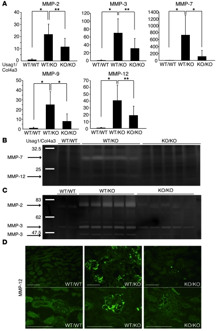
The glomerular basement membrane (GBM) is a key component of the filtering unit in the kidney. Mutations involving any of the collagen IV genes (COL4A3, COL4A4, and COL4A5) affect GBM assembly and cause Alport syndrome, a progressive hereditary kidney disease with no definitive therapy. Previously, we have demonstrated that the bone morphogenetic protein (BMP) antagonist uterine sensitization-associated gene-1 (USAG-1) negatively regulates the renoprotective action of BMP-7 in a mouse model of tubular injury during acute renal failure. Here, we investigated the role of USAG-1 in renal function in Col4a3-/- mice, which model Alport syndrome. Ablation of Usag1 in Col4a3-/- mice led to substantial attenuation of disease progression, normalization of GBM ultrastructure, preservation of renal function, and extension of life span. Immunohistochemical analysis revealed that USAG-1 and BMP-7 colocalized in the macula densa in the distal tubules, lying in direct contact with glomerular mesangial cells. Furthermore, in cultured mesangial cells, BMP-7 attenuated and USAG-1 enhanced the expression of MMP-12, a protease that may contribute to GBM degradation. These data suggest that the pathogenetic role of USAG-1 in Col4a3-/- mice might involve crosstalk between kidney tubules and the glomerulus and that inhibition of USAG-1 may be a promising therapeutic approach for the treatment of Alport syndrome.


Figures

Figure 1
Figure 1. Usag1–/–Col4a3–/– mice showed less glomerular and tubular injury.
(A) Representative histological findings in Usag1+/+Col4a3–/– mice (WT/KO) and Usag1–/–Col4a3–/– mice (KO/KO) at 6 weeks and 10 weeks of age. Scale bars: 100 μm. (B and C) Quantitative assessment of the number of glomeruli, percentages of sclerotic and hemorrhagic glomeruli, and tubulointerstitial fibrosis score in Usag1+/+Col4a3–/– mice (WT/KO) and Usag1–/–Col4a3–/– mice (KO/KO) at 6 weeks (B, n = 5) and 10 weeks of age (C, n = 10). Bars indicate the mean ± SD. **P < 0.01; ***P < 0.05. (D) Electron microphotographs in Usag1+/+Col4a3–/– mice (WT/KO) and Usag1–/–Col4a3–/– mice (KO/KO) at 4 weeks and 10 weeks of age. Arrows indicate the splitting of GBM. Scale bars: 5 μm. (E) Immunostaining for α1(IV) and α3(IV) collagen in the glomeruli of WT littermates (WT/WT), Usag1+/+Col4a3–/– mice (WT/KO), and Usag1–/–Col4a3–/– mice (KO/KO) at 6 weeks of age. Podocin was used as a podocyte marker. Note the positive staining for α1(IV) collagen along with the GBM of Usag1+/+Col4a3–/– mice (WT/KO) and Usag1–/–Col4a3–/– mice (KO/KO), while the staining is restricted to mesangial areas in the glomeruli of WT littermates.
Figure 2
Figure 2. Usag1–/–Col4a3–/– mice showed less albuminuria and preserved renal function.
(A) Urinary albumin excretion normalized by urinary creatinine in WT littermates (WT/WT, n = 4), Usag1+/+Col4a3–/– mice (WT/KO, n = 8), Usag1+/–Col4a3–/– mice (HE/KO, n = 5), and Usag1–/–Col4a3–/– mice (KO/KO, n = 7) at 6 weeks of age. Open circles represent mean value of each column, while closed circles represent individual mice. (B) Plasma creatinine and blood urea nitrogen (BUN) levels in WT littermates (WT/WT, n = 20), Usag1+/+Col4a3–/– mice (WT/KO, n = 18), Usag1+/–Col4a3–/– mice (HE/KO, n = 34), and Usag1–/–Col4a3–/– mice (KO/KO, n = 17) at 10 weeks of age. Bars indicate mean ± SD. Open circles represent mean value of each column, while closed circles represent individual mice.
Figure 3
Figure 3. The expression of inflammatory cytokines significantly decreased in the kidneys of Usag1–/–Col4a3–/– mice.
(A) Real-time RT-PCR analysis of inflammatory cytokine mRNA in the kidneys of WT littermates (WT/WT), Usag1+/+Col4a3–/– mice (WT/KO), and Usag1–/–Col4a3–/– mice (KO/KO) at 10 weeks of age. The expression levels were normalized to those of GAPDH and expressed relative to those of WT littermates (n = 10). Bars indicate the mean ± SD. *P < 0.001; **P < 0.01; ***P < 0.05. (B) Representative immunostaining for MCP-1 in the kidneys of WT littermates (WT/WT), Usag1+/+Col4a3–/– mice (WT/KO), and Usag1–/–Col4a3–/– mice (KO/KO) at 10 weeks of age. Scale bars: 100 μm.
Figure 4
Figure 4. Smad1/5/8 phosphorylation was enhanced in Usag1+/+Col4a3–/– mice, but not in Usag1–/–Col4a3–/– mice.
(A) Representative immunoblotting for Smad phosphorylation in the kidneys of WT littermates (WT/WT), Usag1+/+Col4a3–/– mice (WT/KO), and Usag1–/–Col4a3–/– mice (KO/KO) at 4 and 10 weeks of ages. (B) A panel of cell lines were tested for the ability of TGF-β to induce phosphorylation of Smad1/5/8 in addition to Smad2. Cells were either left untreated or stimulated with 4 ng/ml of TGF-β for 1 hour. Whole-cell extracts were analyzed by immunoblotting using antibodies against phosphorylated Smad1/5/8 (pSmad1/5/8), pSmad2, and GAPDH as a loading control. (C) Representative immunoblotting showing the effect of various concentrations of TGF-β on the phosphorylation of Smad1/5/8 in MDCK cells. (D) Correlation between the phosphorylation levels of Smad1/5/8 in the kidneys of Usag1+/+Col4a3–/– mice and mRNA expression of TGF-β and BMP-7 and serum creatinine (n = 14). The levels of phosphorylation of Smad1/5/8 were determined using the LAS image analysis system.
Figure 5
Figure 5. Usag1–/–Col4a3–/– mice showed less expression and activity of MMPs in the kidneys.
(A) Real-time RT-PCR analysis of MMP mRNA in the kidneys of WT littermates (WT/WT), Usag1+/+Col4a3–/– mice, (WT/KO) and Usag1–/–Col4a3–/– mice (KO/KO) at 10 weeks of age. The expression levels were normalized to that of GAPDH and expressed relative to that in WT littermates (n = 10). Bars indicate the mean ± SD. *P < 0.001; **P < 0.01. (B) Casein zymography analyzing the kidneys of WT littermates (WT/WT), Usag1+/+Col4a3–/– mice (WT/KO), and Usag1–/–Col4a3–/– mice (KO/KO) at 10 weeks of age. (C) Gelatin zymography analyzing the kidneys of WT littermates (WT/WT), Usag1+/+Col4a3–/– mice (WT/KO), and Usag1–/–Col4a3–/– mice (KO/KO) at 10 weeks of age. (D) Representative immunostaining for MMP-12 in the kidneys of WT littermates (WT/WT), Usag1+/+Col4a3–/– mice (WT/KO), and Usag1–/–Col4a3–/– mice (KO/KO) at 10 weeks of age. Scale bars: 100 μm.
Figure 6
Figure 6. USAG-1 colocalizes with BMP-7 in the macula densa and inhibits the action of BMP-7 in mesangial cells.
(A) In situ hybridization for USAG-1 mRNA in the kidneys of 10-week-old Col4a3–/– mice. Scale bars: 100 μm. G, glomerulus. (B) A schematic illustration of the juxtaglomerular apparatus. The macula densa is a part of distal tubules contacting with its glomerulus of origin and adjacent to mesangial cells. (C) β-Gal staining as well as immunostaining with anti-LacZ antibody colocalized with immunostaining with anti-nNOS (a marker for macula densa) antibody in the kidneys of Usag1+/LacZ mice and Bmp7+/LacZ mice. Kidney section from WT mice was also treated in the same way with the sections from Usag1+/LacZ mice and Bmp7+/LacZ mice and demonstrated no β-gal staining. Scale bars: 100 μm. (D) Real-time RT-PCR analysis of MMP-12 mRNA in cultured mesangial cells treated with inflammatory cytokines. The expression levels were normalized to those of GAPDH and expressed relative to those in controls. TGF-β markedly increased MMP-12 mRNA expression in mesangial cells. The graph reflects data that are representative for results of 4 independent experiments. (E) Real-time RT-PCR analysis of MMP-12 mRNA in cultured mesangial cells that were incubated with TGF-β, BMP-7, and USAG-1. BMP-7 suppressed TGF-β–induced MMP-12 upregulation in mesangial cells, and USAG-1 reversed the action of BMP-7. The graph reflects data that are representative for results of 4 independent experiments.
Figure 7
Figure 7. Hypothetical model for involvement of USAG-1 secreted from distal tubules in the pathogenesis of glomerular damage in Alport syndrome.
(A) The macula densa (red), a part of the distal tubules, lies beside the extraglomerular mesangial cells (green), and the nuclei of the macula densa cells are apically located, suggesting the possibility that substances (blue) might be secreted from the basolateral membrane of the macula densa. Note that the basement membrane of the macula densa cells is continuous with the basement membrane of the extraglomerular mesangial cells. (B) In Usag1+/+Col4a3–/– mice (WT/KO), the mesangial cells are activated (purple) and secrete MMPs (scissors), which degrade GBM. Following GBM destruction, podocytes residing on the GBM are damaged (brown) and albuminuria is observed. BMP-7 (blue) secreted from the macula densa is captured by USAG-1, which is also secreted from the macula densa. As a consequence, BMP-7 cannot bind to its receptors and exert its renoprotective action. In Usag1–/–Col4a3–/– mice (KO/KO), BMP-7 secreted from the macula densa can bind to its receptors on mesangial cells and podocytes, and protect fragile alport GBM from degradation.

Similar articles

Cited by

References

    1. Kalluri R. Basement membranes: structure, assembly and role in tumour angiogenesis. Nat Rev Cancer. 2003;3(6):422–433. doi: 10.1038/nrc1094. - DOI - PubMed
    1. Timpl R. Structure and biological activity of basement membrane proteins. Eur J Biochem. 1989;180(3):487–502. doi: 10.1111/j.1432-1033.1989.tb14673.x. - DOI - PubMed
    1. Hudson BG, Tryggvason K, Sundaramoorthy M, Neilson EG. Alport’s syndrome, Goodpasture’s syndrome, and type IV collagen. N Engl J Med. 2003;348(25):2543–2556. doi: 10.1056/NEJMra022296. - DOI - PubMed
    1. Pescucci C, Longo I, Bruttini M, Mari F, Renieri A. Type-IV collagen related diseases. J Nephrol. 2003;16(2):314–316. - PubMed
    1. Barker DF, et al. Identification of mutations in the COL4A5 collagen gene in Alport syndrome. Science. 1990;248(4960):1224–1227. doi: 10.1126/science.2349482. - DOI - PubMed

Publication types

MeSH terms

Substances