. 2010;192(1):39-49.
doi: 10.1159/000291014. Epub 2010 Feb 27.

Interwoven four-compartment capillary membrane technology for three-dimensional perfusion with decentralized mass exchange to scale up embryonic stem cell culture

Affiliations

Interwoven four-compartment capillary membrane technology for three-dimensional perfusion with decentralized mass exchange to scale up embryonic stem cell culture

Jörg C Gerlach et al. Cells Tissues Organs. 2010.

Abstract

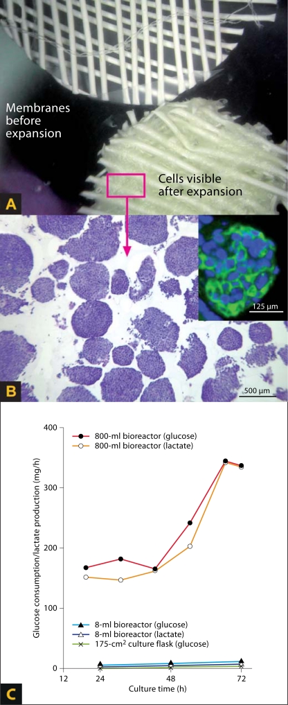
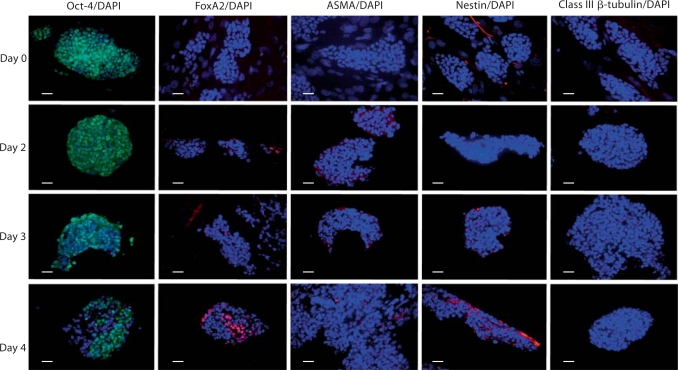
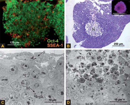
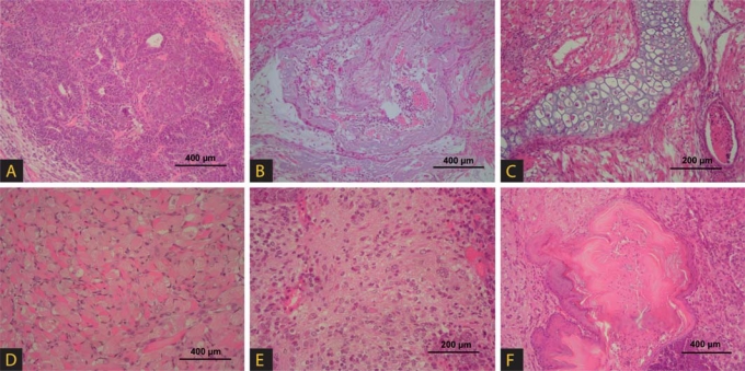
We describe hollow fiber-based three-dimensional (3D) dynamic perfusion bioreactor technology for embryonic stem cells (ESC) which is scalable for laboratory and potentially clinical translation applications. We added 2 more compartments to the typical 2-compartment devices, namely an additional media capillary compartment for countercurrent 'arteriovenous' flow and an oxygenation capillary compartment. Each capillary membrane compartment can be perfused independently. Interweaving the 3 capillary systems to form repetitive units allows bioreactor scalability by multiplying the capillary units and provides decentralized media perfusion while enhancing mass exchange and reducing gradient distances from decimeters to more physiologic lengths of <1 mm. The exterior of the resulting membrane network, the cell compartment, is used as a physically active scaffold for cell aggregation; adjusting intercapillary distances enables control of the size of cell aggregates. To demonstrate the technology, mouse ESC (mESC) were cultured in 8- or 800-ml cell compartment bioreactors. We were able to confirm the hypothesis that this bioreactor enables mESC expansion qualitatively comparable to that obtained with Petri dishes, but on a larger scale. To test this, we compared the growth of 129/SVEV mESC in static two-dimensional Petri dishes with that in 3D perfusion bioreactors. We then tested the feasibility of scaling up the culture. In an 800-ml prototype, we cultured approximately 5 x 10(9) cells, replacing up to 800 conventional 100-mm Petri dishes. Teratoma formation studies in mice confirmed protein expression and gene expression results with regard to maintaining 'stemness' markers during cell expansion.


Figures

Fig. 1
Fig. 1
Bioreactor technology. A Smallest repetitive capillary membrane unit with compartments for medium perfusion (II, IV), oxygenation (III) and cells (I). The cells are not shown; the dots represent mass exchange distribution. B Single units forming capillary bundles. C 3D arrangement of capillaries within the cell compartment. The cells are not shown between the capillaries (for cell position see fig. 2A). Compartments can be perfused independently as shown in B, addressing the reduction of gradient distances between capillary units and enhancing mass exchange. All membrane compartments are interwoven, forming a tight network (C). The capillaries of each compartment are bundled towards the inlet and outlet heads and connected to tube systems for perfusion (B). Cells are inoculated via 16 (8-ml prototype) or 48 (800-ml prototype) open-ended tubes, allowing cell distribution within the cell compartment. Interweaving the smallest capillary units (A) allows scale-up without changing the smallest units, as shown in C. D Processor-controlled perfusion device with a modular multichannel pump unit with medium perfusion and substitution pumps for one 800-ml bioreactor or up to four 8-ml bioreactors and an N2/CO2/O2/air gas supply unit. Bioreactors are kept at a constant temperature under the transparent 37°C hood (upper part); monitoring of flow, pressure, temperature and gas (N2/CO2/O2/air) within the perfusion circuit is provided. To demonstrate their size, we placed an additional 800-ml bioreactor (left) and 8-ml bioreactor (right) at the front.
Fig. 2
Fig. 2
A One layer of the capillary membrane network from an 8-ml bioreactor before cell inoculation (upper left) and after 6 days of culture (lower right); aggregates are visible between the fibers. B Toluidine blue staining of cells obtained from an 800-ml bioreactor after day 3. Inset: SSEA-1 immunoreactivity (green) on day 3 in the 800-ml bioreactor culture (blue: DAPI staining of cell nuclei). C Glucose consumption and lactate production in the 800- and 8-ml bioreactors versus glucose consumption in 2D control dishes over 3 days. Values are given as consumption or production rates per hour per bioreactor/culture flask. Thus, the extremely different scales of the bioreactors and cell numbers are reflected by these medium parameters.
Fig. 3
Fig. 3
Immunoreactivity and DAPI nuclei staining (blue) of 2D mESC cultures before inoculation into the bioreactors (day 0) or of cell aggregates after culture in 8-ml bioreactors over 2, 3 or 4 days. Each time point represents an independent culture. Similar marker expression and distribution are seen in 2D and early 3D cultures. In prolonged 3D culture, the number of stem cell marker Oct-4-positive cells decreased and cell differentiation immunoreactivity markers increased. Early differentiation towards the 3 different germ layer lineages, indicated by immunoreactivity detection of the transcription factor FoxA2 (endoderm) and intermediate filament nestin (early ectoderm) increased in 4-day bioreactor cultures. Class III β-tubulin (Tuj-1; late ectoderm, postmitotic neurons) and anti-α-smooth muscle actin (ASMA; mesoderm) immunoreactivity staining increased in bioreactor cultures maintained for more than 4 days (not shown). Scale bars = 25 μm.
Fig. 4
Fig. 4
Immunoreactive coexpression of stem cell markers (A), toluidine blue staining (B) and transmission electron microscopy (C, D) of cells cultured over 3 days in an 800-ml bioreactor. Cells formed mainly aggregates with a size of 530 ± 146 μm in diameter, corresponding to approximately 4,000 cells. SSEA-1 immunoreactivity was regularly distributed and showed partial coexpression with Oct-4, observed in the vast majority of aggregates (A). Transmission electron microscopy studies (C) showed that aggregates were partly lined by feeder cells (F) characterized by flat shapes and cytoplasmic extrusions, and regularly contained mitotic cells (M). Whereas the majority of aggregates showed an intact cell morphology and ultrastructure, a few aggregates of larger size showed central necrosis (N) as assessed by HE staining (B) and transmission electron microscopy (D). The necrotic areas lacked Oct-4 staining (B, inset). This necrosis may be addressed by changing the membrane weaving patterns and thus reducing the interfiber distances to mechanically limit the available space for ESC aggregate growth.
Fig. 5
Fig. 5
Cells harvested from the 800-ml bioreactor after 3 days of culture were able to form teratomas in vivo within 56 days after subcutaneous injection into 129/S6 mice. Tumors exhibited the formation of tissue structures belonging histologically to all 3 germ layers, including epithelium-like (A), bone-like (B), chondral-like (C), muscle-like (D), neural-like (E) and cornified squamous epithelium-like (F) structures.

Similar articles

Cited by

References

    1. Abbott A. Cell culture: biology's new dimension. Nature. 2003;424:870–872. - PubMed
    1. Abranches E., Bekman E., Henrique D., Cabral J.M.S. Expansion of mouse embryonic stem cells on microcarriers. Biotechnol Bioeng. 2007;96:1211–1221. - PubMed
    1. Adewumi O., Aflatoonian B., Ahrlund-Richter L., Amit A., Andrews P.W., Beighton G., Bello P.A., Benvenisty N., Berry L.S., Bevan S., Blum B., Brooking J., Chen K.G., Choo A.B.H., Churchill G.A., Corbel M., Damjanov I., Draper J.S., Dvorak P., Emanuelsson K., Fleck R.A., Ford A., Gertow K., Gertsenstein M., Gokhale P.J., Hamilton R.S., Hampl A., Healy L.E., Hovatta O., Hyllner J., Imreh M.P., Itskovitz-Eldor J., Jackson J., Johnson J.L., Jones M., Kee K., King B.L., Knowles B.B., Lako M., Lebrin F., Mallon B.S., Manning D., Mayshar Y., McKay R.D.G., Michalska A.E., Mikkola M., Mileikovsky M., Minger S.L., Moore H.D., Mummery C.L., Nagy A., Nakatsuji N., O'Brien C.M., Oh S.K.W., Olsson C., Otonkoski T., Park K.Y., Passier R., Patel H., Patel M., Pedersen R., Pera M.F., Piekarczyk M.S., Pera R.A.P., Reubinoff B.E., Robins A.J., Rossant J., Rugg-Gunn P., Schulz T.C., Semb H., Sherrer E.S., Siemen H., Stacey G.N., Stojkovic M., Suemori H., Szatkiewicz J., Turetsky T., Tuuri T., S. van den Brink, K. Vintersten, S. Vuoristo, D. Ward, T.A. Weaver, L.A. Young, W. Zhang. Characterization of human embryonic stem cell lines by the International Stem Cell Initiative. Nat Biotechnol. 2007;25:803–816. - PubMed
    1. Aleckovic M., C. Simó. Is teratoma formation in stem cell research a characterization tool or a window to developmental biology? Reprod Biomed Online. 2008;17:270–280. - PubMed
    1. Blum B., Benvenisty N. The tumorigenicity of human embryonic stem cells. Adv Cancer Res. 2008;100:133–158. - PubMed

Publication types