

Review
doi: 10.1159/000289222.
Epub 2010 Mar 3.
Early detection of Alzheimer's disease with PET imaging
Affiliations
- PMID: 20197691
- PMCID: PMC3214828
- DOI: 10.1159/000289222
Item in Clipboard
Review
Early detection of Alzheimer's disease with PET imaging
Neurodegener Dis.
2010.
Abstract
Preclinical diagnosis of Alzheimer's disease (AD) is one of the major challenges for the prevention of AD. AD biomarkers are needed not only to reveal preclinical pathologic changes, but also to monitor progression and therapeutics. PET neuroimaging can reliably assess aspects of the molecular biology and neuropathology of AD. The aim of this article is to review the use of FDG-PET and amyloid PET imaging in the early detection of AD.
Copyright 2010 S. Karger AG, Basel.
Figures

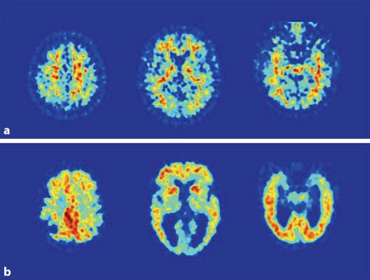
FDG-PET scans from two representative subjects: normal subject (a) and AD subject (b). PET scans are displayed in the axial plane, from the top to the bottom of the brain, at the level of the centrum-semiovale (left), basal ganglia (center), and MTL (right). Note the hypometabolism involving parietotemporal regions, posterior cingulate/precuneus, medial temporal cortex, and to a lesser extent FC in the AD patient as compared to the normal subject.

FDG-PET scan from a representative MCI subject. PET scan is displayed in the axial plane, from the top to the bottom of the brain, at the level of the centrum-semiovale (left), basal ganglia (center), and MTL (right). Note the moderate hypometabolism involving posterior cingulate/precuneus, parietal and medial temporal cortices.
PIB-PET scans from two representative subjects: normal subject (a) and AD subject (b). PET scans are displayed in the axial plane, from the top to the bottom of the brain, at the level of the centrum-semiovale (left), basal ganglia (center), and MTL (right). Note that in the normal subject PIB is distributed only in the white matter, reflecting nonspecific uptake, and in the AD patient PIB uptake is present in several cortical regions, such as frontal, parietal, posterior cingulate/precuneus and lateral temporal cortices, and in basal ganglia.
References
-
- Cummings JL. Toward a molecular neuropsychiatry of neurodegenerative diseases. Ann Neurol. 2003;54:147–154. - PubMed
-
- Mirra SS, Heyman A, McKeel D, Sumi SM, Crain BJ, Brownlee LM, et al. The consortium to establish a registry for Alzheimer's disease (CERAD). II. Standardization of the neuropathologic assessment of Alzheimer's disease. Neurology. 1991;41:479–486. - PubMed
-
- Braak H, Braak E. Neuropathological stageing of Alzheimer related changes. Acta Neuropathologica. 1991;82:239–259. - PubMed
-
- Berg D. Biomarkers for the early detection of Parkinson's and Alzheimer's disease. Neurodegener Dis. 2008;5:133–136. - PubMed
-
- Rusinek H, De Santi S, Frid D, Tsui W, Tarshish C, Convit A, et al. Regional brain atrophy rate predicts future cognitive decline: 6-year longitudinal MR imaging study of normal aging. Radiology. 2003;229:691–696. - PubMed
Publication types
MeSH terms
Substances
Grants and funding
LinkOut - more resources
Full Text Sources
Other Literature Sources
Medical
