. 2010 Aug;30(8):1477-86.
doi: 10.1038/jcbfm.2010.28. Epub 2010 Mar 3.

Inhibition of bradykinin receptor B1 protects mice from focal brain injury by reducing blood-brain barrier leakage and inflammation

Affiliations

Inhibition of bradykinin receptor B1 protects mice from focal brain injury by reducing blood-brain barrier leakage and inflammation

Furat Raslan et al. J Cereb Blood Flow Metab. 2010 Aug.

Abstract

Kinins are proinflammatory and vasoactive peptides that are released during tissue damage and may contribute to neuronal degeneration, inflammation, and edema formation after brain injury by acting on discrete bradykinin receptors, B1R and B2R. We studied the expression of B1R and B2R and the effect of their inhibition on lesion size, blood-brain barrier (BBB) disruption, and inflammatory processes after a focal cryolesion of the right parietal cortex in mice. B1R and B2R gene transcripts were significantly induced in the lesioned hemispheres of wild-type mice (P<0.05). The volume of the cortical lesions and neuronal damage at 24 h after injury in B1R(-/-) mice were significantly smaller than in wild-type controls (2.5+/-2.6 versus 11.5+/-3.9 mm(3), P<0.001). Treatment with the B1R antagonist R-715 1 h after lesion induction likewise reduced lesion volume in wild-type mice (2.6+/-1.4 versus 12.2+/-6.1 mm(3), P<0.001). This was accompanied by a remarkable reduction of BBB disruption and tissue inflammation. In contrast, genetic deletion or pharmacological inhibition of B2R had no significant impact on lesion formation or the development of brain edema. We conclude that B1R inhibition may offer a novel therapeutic strategy after acute brain injuries.


Figures

Figure 1
Figure 1
Bradykinin receptor B1 (B1R) and B2R are induced after focal brain damage. Relative gene expression of B1R and B2R in the lesioned cortex of wild-type mice 12, 24, and 48 h after cryogenic cortical injury or sham operation (n=5 per time point). *P<0.05, one-way ANOVA, Bonferroni post hoc test compared with sham-operated mice.
Figure 2
Figure 2
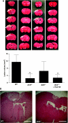
Bradykinin receptor B1 (B1R) deficiency protects from cortical brain damage. (A) (Top) Representative 2,3,5-triphenyltetrazolium chloride (TTC) stains of six corresponding coronal brain sections from wild-type (WT) mice, B1R−/− mice, B2R−/− mice, and B1R−/− mice treated with the B2R inhibitor Hoe140 (0.2 mg/kg) on day 1 after cryoinjury. Lesions appear to be smallest in B1R−/− mice (white arrows), which was confirmed by lesion volumetry (n=8 per group) (bottom) and (B) hematoxylin and eosin staining (broken black lines). Note that the pharmacological blockade of B2R in B1R−/− mice had no additive effect on lesion volume reduction. **P<0.001, one-way analysis of variance (ANOVA), Bonferroni post hoc test compared with WT mice. Scale bar represents 200 μm.
Figure 3
Figure 3
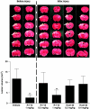
Pharmacological blockade of bradykinin receptor B1 (B1R) but not B2R protects from cortical brain damage. (top, left panel) Representative 2,3,5-triphenyltetrazolium chloride (TTC) stains of six corresponding coronal brain sections 24 h after cryogenic cortical injury from mice treated with the B1R inhibitor R-715 (1 mg/kg) or vehicle 1 h before the operation. (top, right panel) Representative TTC stains of six corresponding coronal brain sections 24 h after cryogenic cortical injury from mice treated with R-715 (1 mg/kg), R-715 (0.5 mg/kg), or the B2R inhibitor Hoe140 (0.2 and 0.4 mg/kg) 1 h after the operation. (bottom) Lesion volumes as assessed by volumetry (n=8 per group). Note that antagonism of B1R is effective both before or after cortical injury whereas the pharmacological blockade of the B2R is ineffective. **P<0.001, one-way analysis of variance (ANOVA), Bonferroni post hoc test compared with vehicle-treated mice.
Figure 4
Figure 4
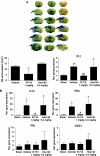
Bradykinin receptor B1 (B1R) blockade but not B2R blockade reduces blood–brain barrier (BBB) leakage and inflammation after focal brain trauma. (A) (Top) Representative corresponding coronal brain sections from R-715-treated (1 mg/kg), Hoe140-treated (0.2 mg/kg), or vehicle-treated mice 24 h after cryogenic cortical injury and injection of Evan's blue. Evan's blue extravasation (black arrows) appeared lowest in R-715-treated mice. (bottom, left) Volume of parenchymal Evan's blue extravasation determined by planimetry in the three animals groups 24 h after cryogenic cortical injury (n=6 per group). *P<0.05, one-way analysis of variance (ANOVA), Bonferroni post hoc test compared with vehicle-treated mice. (bottom, right) Relative gene expression of endothelin-1 (ET-1) in the lesioned cortex of R-715-treated (1 mg/kg), Hoe140-treated (0.2 mg/kg), or vehicle-treated mice 24 h after cryogenic cortical injury or sham operation (n=5 per group). Note that antagonism of B1R reverses the ET-1 mRNA expression peak observed in placebo-treated mice or after B2R blockade. *P<0.05, **P<0.01, ##P<0.01, one-way ANOVA, Bonferroni post hoc test compared with sham operation or vehicle-treated mice. (B) Relative gene expression of interleukin-1β (IL-1β), tumor necrosis factor-α (TNF-α), interferon-γ (IFNγ), and transforming growth factor-β (TGF-β)-1 in the lesioned cortex of R-715-treated (1 mg/kg), Hoe140-treated (0.2 mg/kg), and vehicle-treated mice 24 h after cryogenic cortical injury or sham operation (n=5/group). *P<0.05, #P<0.05, one-way ANOVA, Bonferroni post hoc test compared with sham operation or vehicle-treated mice.
Figure 5
Figure 5
Activated macrophages/microglia in the lesioned cortex (broken white lines) on day 1 after cryogenic brain trauma in wild-type (WT) mice, B1R−/− mice, and B2R−/− mice. Paraffin sections were stained by immunoperoxidase labeling with rat anti-mouse F4/80 antibodies for the detection of activated macrophages/microglia (black arrows). Note that macrophage/microglia invasion was markedly reduced in B1R−/− mice but not in B2R−/− mice compared with WT controls. Bar represents 100 μm (inset: magnification 2.5-fold).

Similar articles

Cited by

References

    1. Austinat M, Braeuninger S, Pesquero JB, Brede M, Bader M, Stoll G, Renne T, Kleinschnitz C. Blockade of bradykinin receptor B1 but not bradykinin receptor B2 provides protection from cerebral infarction and brain edema. Stroke. 2009;40:285–293. - PubMed
    1. Beauchamp K, Mutlak H, Smith WR, Shohami E, Stahel PF. Pharmacology of traumatic brain injury: where is the ‘golden bullet'. Mol Med. 2008;14:731–740. - PMC - PubMed
    1. Camargo AC, Shapanka R, Greene LJ. Preparation, assay, and partial characterization of a neutral endopeptidase from rabbit brain. Biochemistry. 1973;129:1838–1844. - PubMed
    1. Gobeil F, Neugebauer W, Filteau C, Jukic D, Allogho SN, Pheng LH, Nguyen-Le XK, Blouin D, Regoli D. Structure-activity studies of B1 receptor-related peptides. Antagonists. Hypertension. 1996;28:833–839. - PubMed
    1. Gorlach C, Hortobagyi T, Hortobagyi S, Benyo Z, Relton J, Whalley ET, Wahl M. Bradykinin B2, but not B1, receptor antagonism has a neuroprotective effect after brain injury. J Neurotrauma. 2001;18:833–838. - PubMed

Publication types

MeSH terms

Substances

LinkOut - more resources