. 2009;4(6):621-635.
doi: 10.2217/fvl.09.56.

Ebolavirus glycoprotein structure and mechanism of entry

Affiliations

Ebolavirus glycoprotein structure and mechanism of entry

Jeffrey E Lee et al. Future Virol. 2009.

Abstract

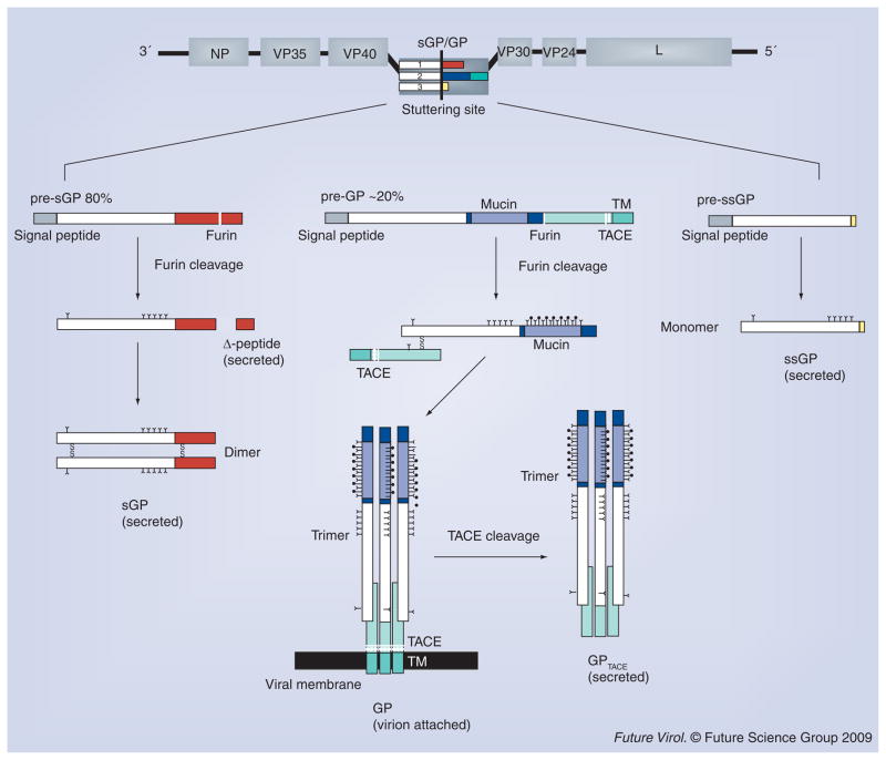
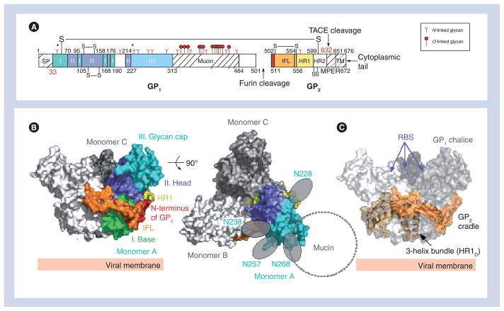
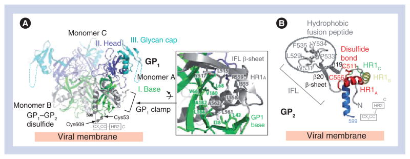
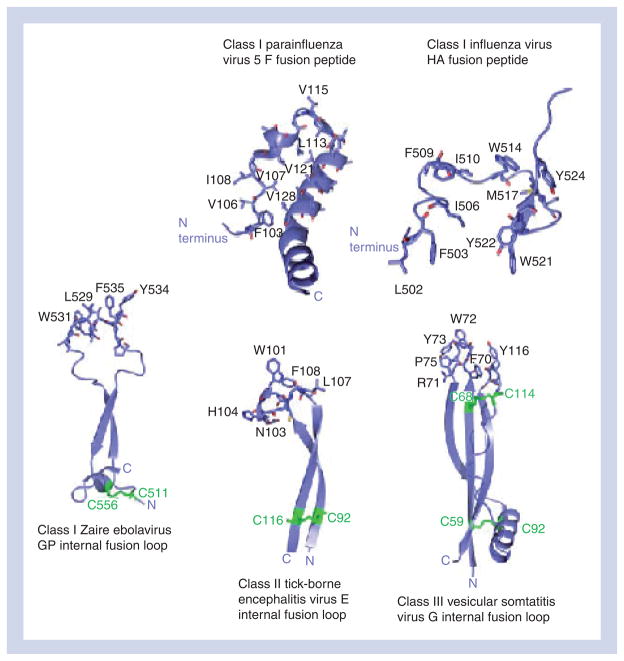
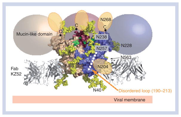
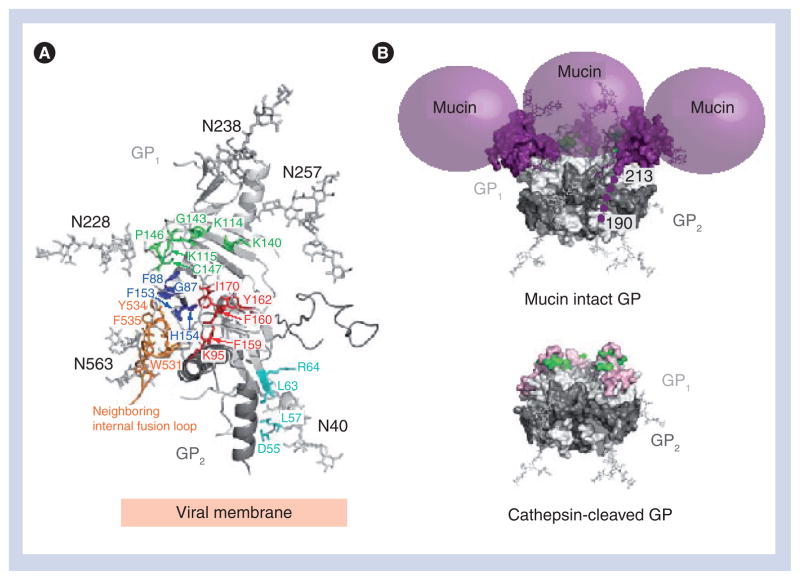
Ebolavirus (EBOV) is a highly virulent pathogen capable of causing a severe hemorrhagic fever with 50-90% lethality. The EBOV glycoprotein (GP) is the only virally expressed protein on the virion surface and is critical for attachment to host cells and catalysis of membrane fusion. Hence, the EBOV GP is a critical component of vaccines as well as a target of neutralizing antibodies and inhibitors of attachment and fusion. The crystal structure of the Zaire ebolavirus GP in its trimeric, prefusion conformation (3 GP(1) plus 3 GP(2)) in complex with a neutralizing antibody fragment, derived from a human survivor of the 1995 Kikwit outbreak, was recently determined. This is the first near-complete structure of any filovirus glycoprotein. The overall molecular architecture of the Zaire ebolavirus GP and its role in viral entry and membrane fusion are discussed in this article.


Figures

Figure 1
Figure 1. Transcription and processing of Zaire ebolavirus glycoproteins
The primary open reading frame of ebolavirus GP encodes a sGP (shown as white and red rectangles). Proteolytic cleavage of pre-sGP by furin results in the formation of the mature sGP and a small nonstructural fragment, termed Δ-peptide. Co-transcriptional stuttering of the GP gene results in two additional glycoproteins: GP and ssGP. GP is the virion-attached glycoprotein and proteolytic cleavage of its precursor (pre-GP) by furin results in two subunits, GP1 and GP2, that remain linked by a disulfide bond. The GP1 and GP2 heterodimer trimerizes and forms the viral surface peplomer. TNF-α-converting enzyme can also cleave envelope GP, at a site proximal to the GP2 transmembrane domain, thereby releasing a soluble trimeric GP. ssGP is a small secreted glycoprotein that shares the first 295 amino acids with sGP and GP, but has a different C-terminus (two nonshared residues, as colored in yellow). It has been reported that ssGP forms a monomer in solution. GP: Glycoprotein; sGP: Secreted glycoprotein; ssGP: Small, secreted glycoprotein; TACE: TNF-α-converting enzyme; TM: Transmembrane anchor.
Figure 2
Figure 2. Overall structure of Zaire ebolavirus GPΔmucΔtm
(A) Domain schematic of GP. The disulfide bridges (-S-S-), SP, IFL, HR1, HR2, MPER, TACE cleavage site, TM and cytoplasmic tail are labeled accordingly. White and hash-marked regions designate crystallographically disordered and construct-deleted regions, respectively. (B) Molecular surface of the GP trimer viewed on its side and down its threefold axis. Monomer A is colored according to its subdomains: GP1 base – green; GP1 head – purple; GP1 glycan cap – cyan; GP2 N-terminus – red; GP2 IFL – orange; and GP2 HR1 – yellow. (C) Molecular surface of the Zaire ebolavirus GPΔmucΔtm chalice and cradle. Three lobes of GP1, shown in shades of gray form the GP chalice, while three subunits of GP2 (orange) wrap around the base of the chalice to form the cradle. GP: Glycoprotein; HR: Heptad repeat region; IFL: Internal fusion loop; MPER: Membrane-proximal external region; RBS: Receptor-binding site; SP: Signal peptide; TACE: TNF-α-converting enzyme; TM: Transmembrane anchor. Adapted from [34].
Figure 3
Figure 3. Zaire ebolavirus GP1 and GP2
(A) Ribbon diagram of the Zaire ebolavirus GP trimer with each GP1 subdomain colored according to Figure 1B and the three GP2 subunits colored in gray. The GP1 base subdomain forms a hydrophobic, semicircular β-sheet surface that interacts with the hydrophobic face of the GP2 HR1A helix and the β-scaffold of the internal fusion loop (inset box). (B) Ribbon diagram of the prefusion conformation of Zaire ebolavirus GP2. Hydrophobic residues of the GP2 internal fusion loop are displayed on a β-scaffold (gray). The internal fusion loop is stabilized by a disulfide linkage at the base (Cys511–Cys556) and HR1 is separated into four segments (HR1A, HR1B, HR1C and HR1D). Note that the GP1–GP2 disulfide bridge (CX6CC motif) and HR2 region are disordered in the structure; their positions are marked by dashed lines. GP: Glycoprotein; HR: Heptad repeat region; IFL: Internal fusion loop. Adapted from [34].
Figure 4
Figure 4. Class I, II and III prefusion viral glycoprotein fusion peptides and internal fusion loops
Comparison of prefusion conformation fusion peptide and internal fusion loop structures from class I, II and III GPs. The Zaire ebolavirus GP internal fusion loop more closely resembles those observed in class II and class III GPs than those observed in other trimeric class I prefusion viral GPs. GP: Glycoprotein; HA: Hemagglutinin. Adapted from [34].
Figure 5
Figure 5. Zaire ebolavirus glycoprotein glycosylation
N-linked biantennary complex-type glycans (Gal2Man3GlcNAc4) are modeled as yellow space-filling spheres onto the Zaire ebolavirus GPΔmucΔtm structure at predicted glycosylation sites: Asn40, Asn204, Asn228, Asn238, Asn257, Asn268 and Asn563. The glycans at Asn204 and Asn268 reside in regions that are poorly ordered and, thus, their tentative locations are shown as orange ovals. The C-terminus of each last ordered residue of GP1, to which each mucin-like domain is linked, is marked with a ‘C’ (top of the chalice). Colored spheres (beige, pink and purple) outline the predicted location of the mucin-like domains. The putative receptor-binding site residues, recessed within the Zaire ebolavirus GP chalice bowl, are colored green. Adapted from [34].
Figure 6
Figure 6. Zaire ebolavirus glycoprotein site critical for viral entry
(A) Arrays of GP1 point mutations have identified at least 19 residues critical for viral entry [–80]. These residues map to four distinct regions (cyan, green, royal blue and red) on the ZEBOV GPΔmucΔtm structure (monomer shown). (B) Zaire ebolavirus GP cathepsin cleavage. A molecular surface representation of the GPΔmucΔtm trimer with N-linked glycans drawn as ball-and-sticks and the mucin-like domain shown as purple spheres. Residues identified by the mutagenesis to be important for viral entry and located on or near the surface of the GPΔmucΔtm structure are colored in green (Lys114, Lys115, Lys140, Gly143, Pro146 and Cys147). Cleavage by cathepsin on a disordered loop (around residue 190) removes the glycan cap and mucin-like domain, exposing additional residues (shown in pink) on the site critical for viral entry [82]. GP: Glycoprotein. Adapted from [34].
Figure 7
Figure 7. Zaire ebolavirus glycoprotein-mediated entryEbolavirus is thought to enter cells through an endocytic mechanism
(A) Initially, the metastable, prefusion Zaire ebolavirus GP may bind lectins [71] or an unidentified attachment factor at the cell surface (green ovals) via the mucin-like domains (grey spheres) or other sites on GP. (B) Subsequently, ebolavirus is internalized and trafficked to the endosome. Lectins may or may not remain bound to GP, depending on the nature of the individual lectin. In the endosome, host cathepsins cleave GP to remove the glycan cap and mucin-like domain, yielding an approximately 19-kDa GP1 core, disulfide bonded to GP2. (C) The newly exposed surface may allow either tighter binding to a receptor trafficked from the cell surface or binding to an alternate molecule in the endosome. Binding of this molecule, or perhaps further cathepsin cleavage, could then trigger conformational changes in the GP2 fusion subunit. (D) Structural rearrangements in GP2 allow HR1 to form a single 44-residue helix and position the IFL for insertion into the host-endosomal membrane. Upon insertion in the host membrane, the IFL adopts a 310 helix. This is the extended prehairpin intermediate. (E) Based on studies with the influenza virus, more than one trimer of GP2 may be required to complete the membrane fusion process. (F) The HR2 and MPER regions swing from the viral membrane towards the host membrane and HR1. Initial fold-back of the HR2 onto HR1 distorts the virus and host-cell bilayers and brings the two membranes into contact to form a hemifusion stalk. (G) The hemifusion stalk opens up to form the fusion pore and the low energy, postfusion 6HB is formed when three HR2 helices pack into the HR1 trimeric bundle. 6HB: Six helix bundle; CatL/B: Cathepsin L/B; GP: Glycoprotein; HR: Heptad repeat region; IFL: Internal fusion loop; MPER: Membrane-proximal external region; TACE: TNF-α-converting enzyme; TM: Transmembrane domain. Adapted from [34,51].

Similar articles

Cited by

References

Bibliography

    1. Sanchez A, Geisbert TW, Feldmann H. Filoviridae: Marburg and Ebola viruses. In: Knipe DM, Howley PM, Griffin DE, et al., editors. Fields Virology. Lippincott Williams and Wilkins; Philadelphia, PA, USA: 2007. pp. 1409–1448. Comprehensive review of Filoviruses.
    1. Towner JS, Sealy TK, Khristova ML, et al. Newly discovered ebola virus associated with hemorrhagic fever outbreak in Uganda. PLoS Pathog. 2008;4(11):e1000212. - PMC - PubMed
    1. Kuhn JH. History of filoviral disease outbreaks. In: Calisher CH, editor. Filoviruses. Springer Wien; NewYork, Wien, Austria: 2008. Comprehensive review of Filoviruses.
    1. Feldmann H, Geisbert TW, Jahrling PB, et al. Negative sense single stranded RNA viruses. In: Fauquet CM, Mayo MA, Maniloff J, Desselberger U, Ball LA, editors. Virus Taxonomy: Classification and Nomenclature of Viruses. Elsevier Academic Press; London, UK: 2005. pp. 645–653.
    1. Barrette RW, Metwally SA, Rowland JM, et al. Discovery of swine as a host for the Reston ebolavirus. Science. 2009;325(5937):204–206. - PubMed

Website

    1. Model of EBOV GP-mediated entry. www.nature.com/nature/journal/v454/n7201/index.html.

LinkOut - more resources